Multi-modal imaging of the subscapularis muscle
- PMID: 27752837
- PMCID: PMC5110480
- DOI: 10.1007/s13244-016-0526-1
Multi-modal imaging of the subscapularis muscle
Abstract
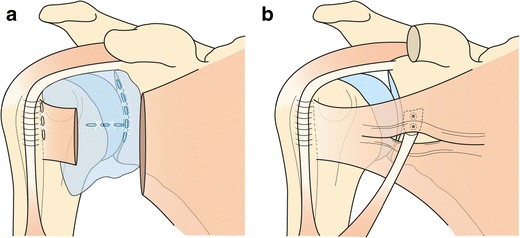
The subscapularis (SSC) muscle is the most powerful of the rotator cuff muscles, and plays an important role in shoulder motion and stabilization. SSC tendon tear is quite uncommon, compared to the supraspinatus (SSP) tendon, and, most of the time, part of a large rupture of the rotator cuff. Various complementary imaging techniques can be used to obtain an accurate diagnosis of SSC tendon lesions, as well as their extension and muscular impact. Pre-operative diagnosis by imaging is a key issue, since a lesion of the SSC tendon impacts on treatment, surgical approach, and post-operative functional prognosis of rotator cuff injuries. Radiologists should be aware of the SSC anatomy, variability in radiological presentation of muscle or tendon injury, and particular mechanisms that may lead to a SSC injury, such as coracoid impingement.
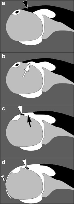
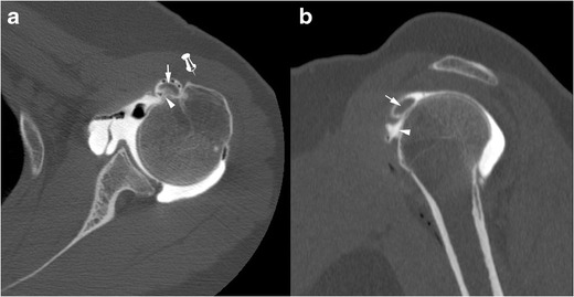
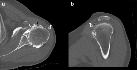
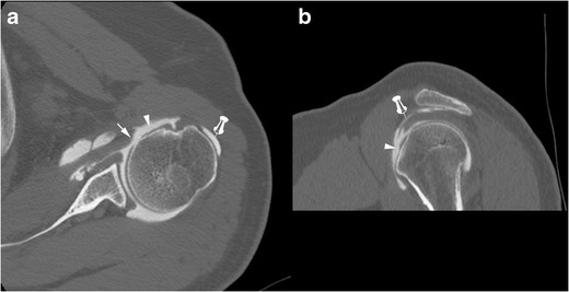
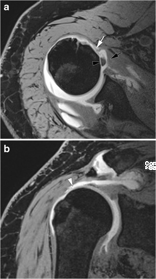
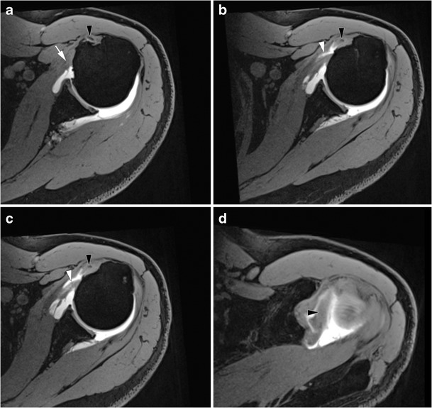
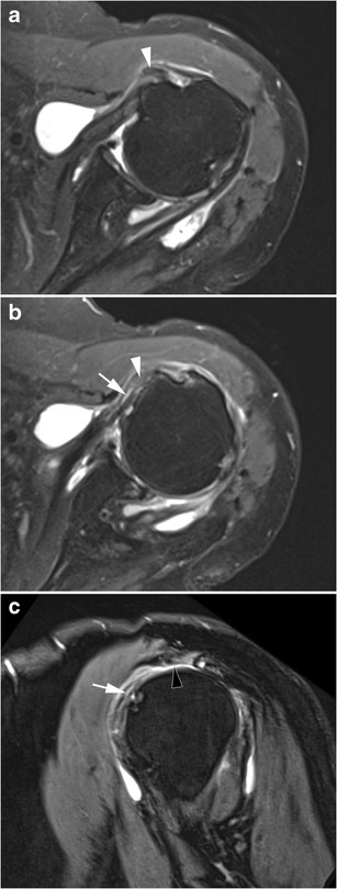
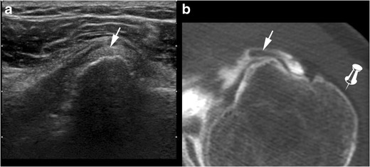
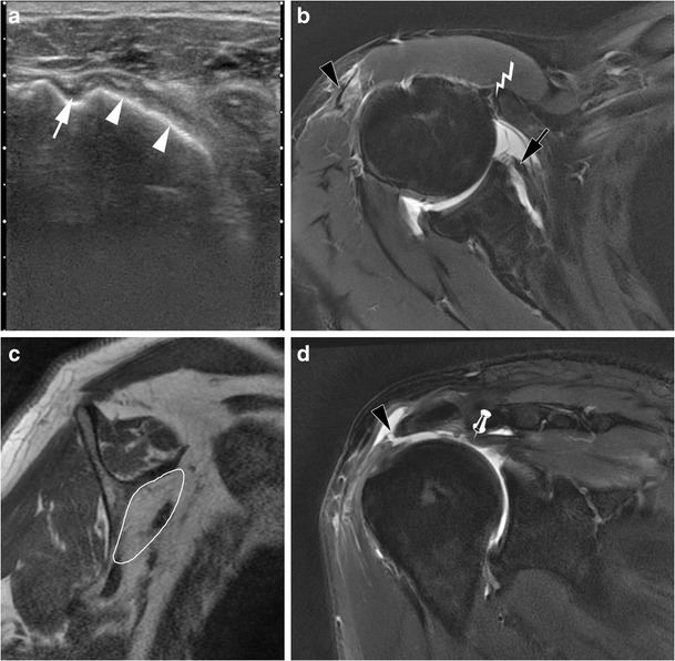
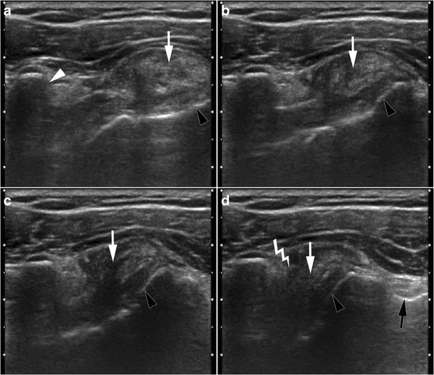
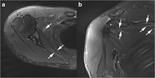
Teaching points: • Isolated subscapularis (SSC) tendon tears are uncommon. • Classically, partial thickness SSC tendon tears start superomedially and progress inferolaterally. • Long head of biceps tendon medial dislocation can indirectly signify SSC tendon tears. • SSC tendon injury is associated with anterior shoulder instability. • Dynamic ultrasound study of the SSC helps to diagnose coracoid impingement.
Keywords: Coracoid impingement; Magnetic resonance imaging; Rotator cuff; Subscapularis; Tendon injury.
Figures


Similar articles
-
Imaging Review of Subscapularis Tendon and Rotator Interval Pathology.Radiol Res Pract. 2022 Jan 11;2022:4009829. doi: 10.1155/2022/4009829. eCollection 2022. Radiol Res Pract. 2022. PMID: 35070451 Free PMC article. Review.
-
The "bridging sign": a MR finding for combined full-thickness tears of the subscapularis tendon and the supraspinatus tendon.Acta Radiol. 2013 Feb 1;54(1):83-8. doi: 10.1258/ar.2012.120353. Epub 2012 Oct 23. Acta Radiol. 2013. PMID: 23093726
-
Arthroscopic Repair of Massive Cuff Tears With Large Subscapularis Tendon Ruptures (Lafosse III/IV): A Prospective Magnetic Resonance Imaging-Controlled Case Series of 26 Cases With a Minimum Follow-up of 1 Year.Arthroscopy. 2015 Nov;31(11):2173-82. doi: 10.1016/j.arthro.2015.05.012. Epub 2015 Jul 15. Arthroscopy. 2015. PMID: 26188785
-
[Coraco-humeral space and rotator cuff tears].Rev Chir Orthop Reparatrice Appar Mot. 1999 Nov;85(7):677-83. Rev Chir Orthop Reparatrice Appar Mot. 1999. PMID: 10612131 French.
-
Pectoralis major and pectoralis minor transfer for irreparable subscapularis tendon tears.Oper Orthop Traumatol. 2022 Feb;34(1):45-54. doi: 10.1007/s00064-021-00760-5. Epub 2022 Feb 3. Oper Orthop Traumatol. 2022. PMID: 35113176 Review. English.
Cited by
-
Blood flow velocity in the anterior humeral circumflex artery and tear size can predict synovitis severity in patients with rotator cuff tears.Clin Shoulder Elb. 2024 Mar;27(1):11-17. doi: 10.5397/cise.2023.00752. Epub 2024 Jan 24. Clin Shoulder Elb. 2024. PMID: 38268319 Free PMC article.
-
Literature Review of Subscapularis Tear, Associated injuries, and the Available Treatment Options.Hawaii J Health Soc Welf. 2022 Mar;81(3 Suppl 1):2-7. Hawaii J Health Soc Welf. 2022. PMID: 35340936 Free PMC article. Review.
-
Subscapularis tendon tear detection using axial internal rotation MRI: semiquantitative and quantitative analysis.Quant Imaging Med Surg. 2023 Dec 1;13(12):8274-8289. doi: 10.21037/qims-23-273. Epub 2023 Nov 9. Quant Imaging Med Surg. 2023. PMID: 38106250 Free PMC article.
-
Neuromuscular compartmentation of the subscapularis muscle and its clinical implication for botulinum neurotoxin injection.Sci Rep. 2023 Jul 10;13(1):11167. doi: 10.1038/s41598-023-38406-0. Sci Rep. 2023. PMID: 37430018 Free PMC article.
-
Imaging Review of Subscapularis Tendon and Rotator Interval Pathology.Radiol Res Pract. 2022 Jan 11;2022:4009829. doi: 10.1155/2022/4009829. eCollection 2022. Radiol Res Pract. 2022. PMID: 35070451 Free PMC article. Review.
References
Publication types
LinkOut - more resources
Full Text Sources
Other Literature Sources