. 2017 Jun;28(4):400-408.
doi: 10.1080/09537104.2016.1235685. Epub 2016 Oct 18.

Golgi proteins in circulating human platelets are distributed across non-stacked, scattered structures

Affiliations

Golgi proteins in circulating human platelets are distributed across non-stacked, scattered structures

Shilpi Yadav et al. Platelets. 2017 Jun.

Abstract

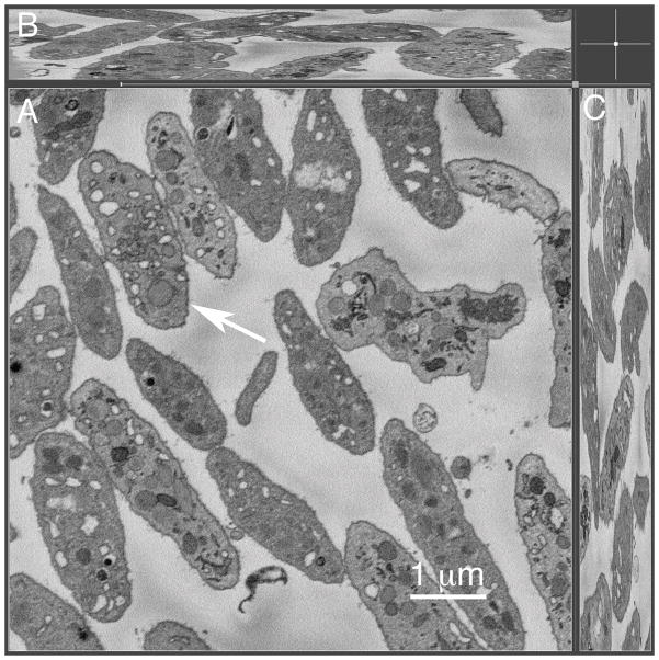
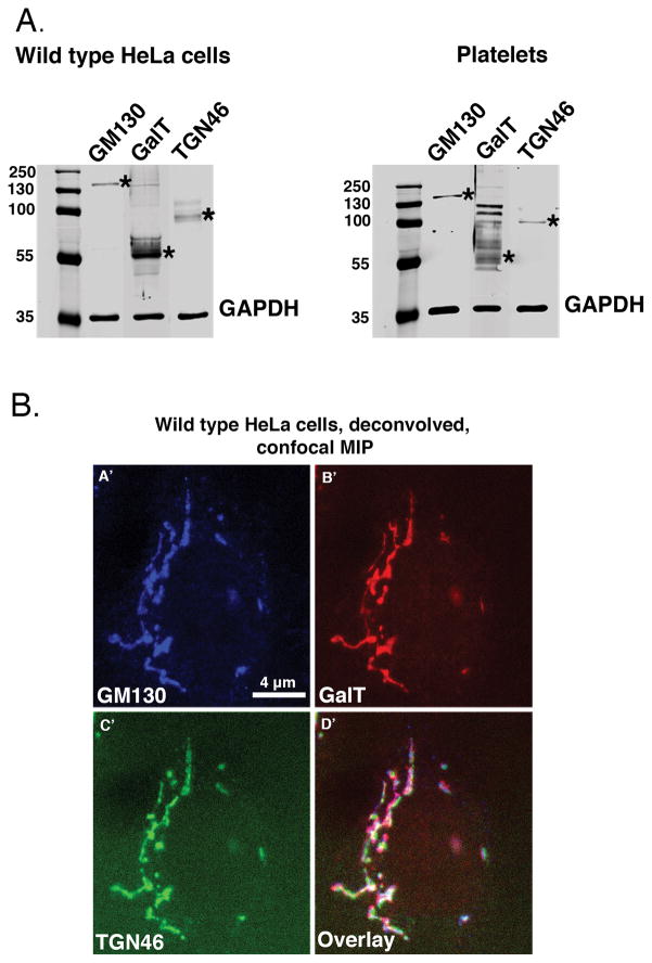
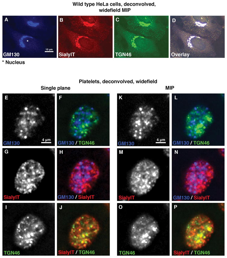
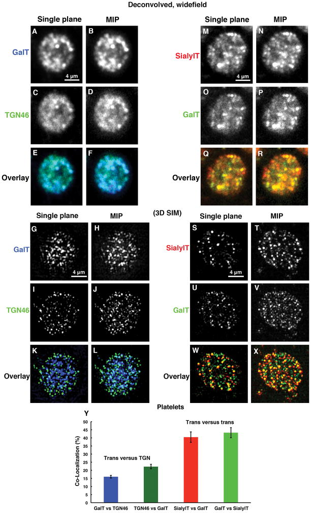
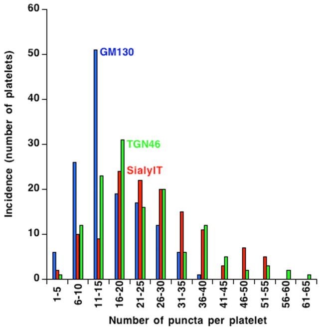
Platelets are small, anucleate cell fragments that are central to hemostasis, thrombosis, and inflammation. They are derived from megakaryocytes from which they inherit their organelles. As platelets can synthesize proteins and contain many of the enzymes of the secretory pathway, one might expect all mature human platelets to contain a stacked Golgi apparatus, the central organelle of the secretory pathway. By thin section electron microscopy, stacked membranes resembling the stacked Golgi compartment in megakaryocytes and other nucleated cells can be detected in both proplatelets and platelets. However, the incidence of such structures is low and whether each and every platelet contains such a structure remains an open question. By single-label, immunofluorescence staining, Golgi glycosyltransferases are found within each platelet and map to scattered structures. Whether these structures are positive for marker proteins from multiple Golgi subcompartments remains unknown. Here, we have applied state-of-the-art techniques to probe the organization state of the Golgi apparatus in resting human platelets. By the whole cell volume technique of serial-block-face scanning electron microscopy (SBF-SEM), we failed to observe stacked, Golgi-like structures in any of the 65 platelets scored. When antibodies directed against Golgi proteins were tested against HeLa cells, labeling was restricted to an elongated juxtanuclear ribbon characteristic of a stacked Golgi apparatus. By multi-label immunofluorescence microscopy, we found that each and every resting human platelet was positive for cis, trans, and trans Golgi network (TGN) proteins. However, in each case, the proteins were found in small puncta scattered about the platelet. At the resolution of deconvolved, widefield fluorescence microscopy, these proteins had limited tendency to map adjacent to one another. When the results of 3D structured illumination microscopy (3D SIM), a super resolution technique, were scored quantitatively, the Golgi marker proteins failed to map together indicating at the protein level considerable degeneration of the platelet Golgi apparatus relative to the layered stack as seen in the megakaryocyte. In conclusion, we suggest that these results have important implications for organelle structure/function relationships in the mature platelet and the extent to which Golgi apparatus organization is maintained in platelets. Our results suggest that Golgi proteins in circulating platelets are present within a series of scattered, separated structures. As separate elements, selective sets of Golgi enzymes or sugar nucleotides could be secreted during platelet activation. The establishment of the functional importance, if any, of these scattered structures in sequential protein modification in circulating platelets will require further research.

Keywords: 3D SIM; Golgi apparatus; fluorescence microscopy; platelets; serial-block-face scanning electron microscopy.


Conflict of interest statement

Declaration of interest

This work was supported in part by NIH grants S10 OD018065, R01 HL119393, and R01 GM092960. Work in the Leapman laboratory was supported by the NIBIB, NIH intramural program. The authors report no other declarations of interest.

Figures

Figure 1
Figure 1. Serial-block-face scanning electron microscopy of full platelet cell volume indicates that mature platelets do not contain stacked Golgi-like structures
Resting human platelets (n=65) were analyzed for full platelet cell volume using serial-block-face SEM technique. The representative micrograph showing high-resolution image of the full platelet cell volume on XY dimension (A) and Z dimension (B–C). Arrow in (A) points to the platelet shown in the full cell volume video (Supplemental Figure 1, 2460 nm depth). The overall depth of the SBF-SEM sample imaged was 7.5 μm.
Figure 2
Figure 2. Western blot and multilabel immunofluorescence analysis indicates that the marker antibodies recognize distinct protein species
(A.) Wild type HeLa cell and human platelet lysate was run on a 10% SDS-PAGE gel and blotted with GM130 (M.W. = 130 kDa), GalT (M.W. = ~55 kDa plus possible oligomers) and TGN46 (M.W. = predicted 51 kDa, typically runs as a dimer on SDS-PAGE) antibodies to identify cis, trans and trans Golgi network proteins, respectively. GAPDH is shown as a loading control. Asterisks indicate the position of the Golgi proteins. The blots were imaged with an Odyssey Infrared Imaging System (LI-COR Biosciences). (B.) A representative image of wild type HeLa cells showing a classic” stacked, ribbon-like juxtanuclear Golgi structure in which markers to the Golgi cisternae subcompartments cis, GM130 (blue); Trans, GalT (red); and trans Golgi network, TGN46 (green), overlay image map proximal to each other in deconvolved, spinning-disk confocal micrographs (A′–D′).
Figure 3
Figure 3. Validated Golgi proteins show a scattered distribution with limited to no overlapping in resting human platelets
(A–D) Representative deconvolved, widefield MIP fluorescence images of wild type HeLa cells stained with Golgi markers show a “classic” stacked ribbon-like juxtanuclear distribution. This provides validation of the antibodies as markers of Golgi subcompartments. (E–P) Immunofluorescence micrograph of resting human platelets show many puncta when stained for Golgi proteins marking cis, trans or TGN subcompartments using GM130 (blue), SialylT (red) or TGN46 (green) primary antibodies, respectively. Widefield Z-stack images were taken and the deconvolved images are presented as either single plane or maximum intensity projections (MIP).
Figure 4
Figure 4. Deconvolved, widefield microscopy and 3D SIM analysis reveals that trans and TGN Golgi markers show little tendency to map in an adjacent manner suggestive of mini-Golgi stacks while the co-localization of two trans markers with one another suggests that the trans Golgi is a distinctive subcompartment in platelets
(A–F) Trans, GalT (blue) and trans Golgi network, TGN46 (green) proteins showed apparent overlapping (light blue/Cyan color) with each other as seen by deconvolved, widefield microscopy where as (G–L) in three-dimensional structured illumination microscopy (3D SIM) of trans, GalT (blue) and trans Golgi network, TGN46 (green) proteins reveals little to no overlapping with each other. (M–R) Deconvolved, widefield microscopy suggests that two trans proteins SialylT (red) versus GalT (green), share a common compartment and show extensive overlapping (yellow color). (S–X) Furthermore considerable overlapping confirmed by 3D SIM analysis. (Y) Quantification of the limited overlapping. Seventeen individual platelets for trans versus (vs.) trans Golgi network and 9 individual platelets for trans versus trans are quantified in pairings. All pairings were averaged, and bars are presented as the mean ± SEM.
Figure 5
Figure 5. The incidence of Golgi-protein-positive puncta in resting human platelets suggest that circulating platelets behave as a single population with respect to Golgi organization
The incidence of Golgi-protein-positive puncta were manually calculated for 3 Golgi markers GM130 (blue), SialylT (red) and TGN46 (Green) using deconvolved, widefield images of 135 platelets. The frequency distribution of puncta per platelet is shown as histograms using KaleidaGraph software version 4.5.2. The histogram plots of each of the three markers are smooth and fairly similar suggesting that circulating platelets behave as a single population.

Similar articles

Cited by

References

    1. King S, Reed GL. Development of platelet secretory granules. Semin Cell Dev Biol. 2002;13:203–302. - PubMed
    1. Warshaw AL, Laster L, Shulman NR. Protein Synthesis by Human Platelets. J Biol Chem. 1967;242(9):2094–7. - PubMed
    1. Weyrich AS, Schwertz H, Kraiss LW, Zimmerman GA. Protein Synthesis by Platelets: Historical and New Perspectives. J Thromb Haemost. 2009;7(2):241–6. - PMC - PubMed
    1. Schubert P, Devine DV. De novo protein synthesis in mature platelets: a consideration for transfusion medicine. Vox Sang. 2010;99(2):112–22. - PubMed
    1. Schwertz H, Rowley JW, Tolley ND, Campbell RA, Weyrich AS. Assessing protein synthesis by platelets. Methods Mol Biol. 2012;788:141–53. - PubMed