

Autophagy-mediated catabolism of visual transduction proteins prevents retinal degeneration
- PMID: 27753525
- PMCID: PMC5173283
- DOI: 10.1080/15548627.2016.1238553
Autophagy-mediated catabolism of visual transduction proteins prevents retinal degeneration
Abstract
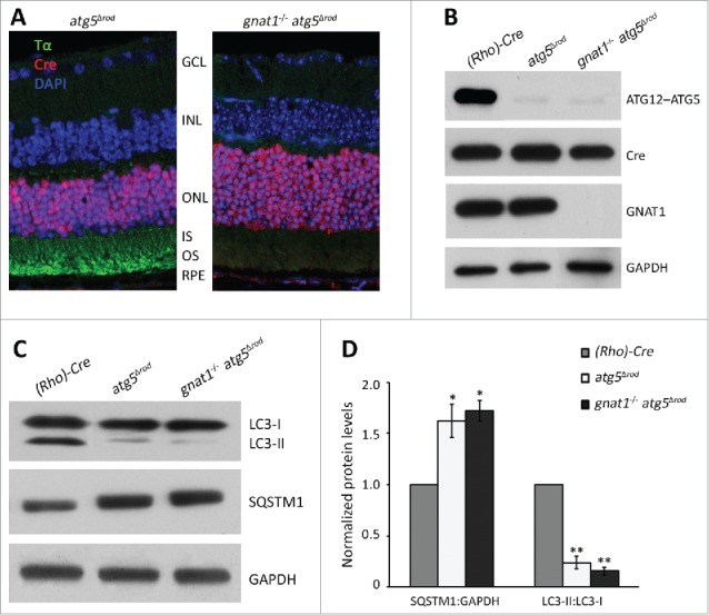
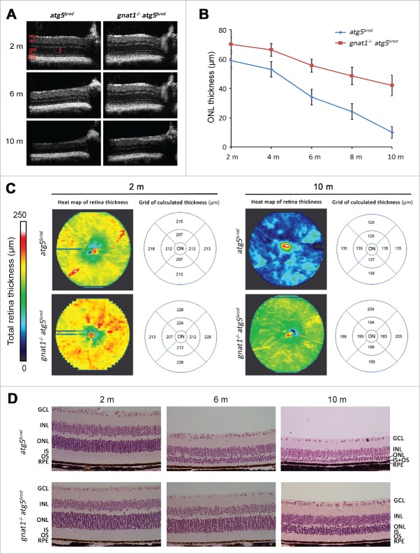
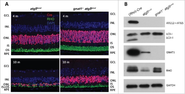
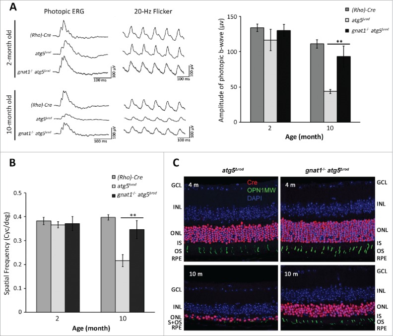
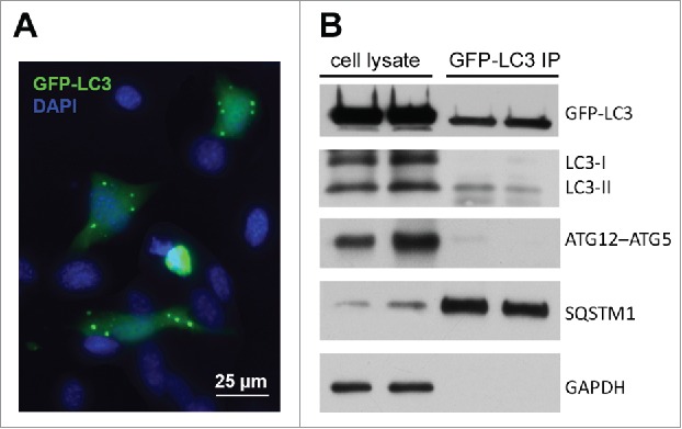
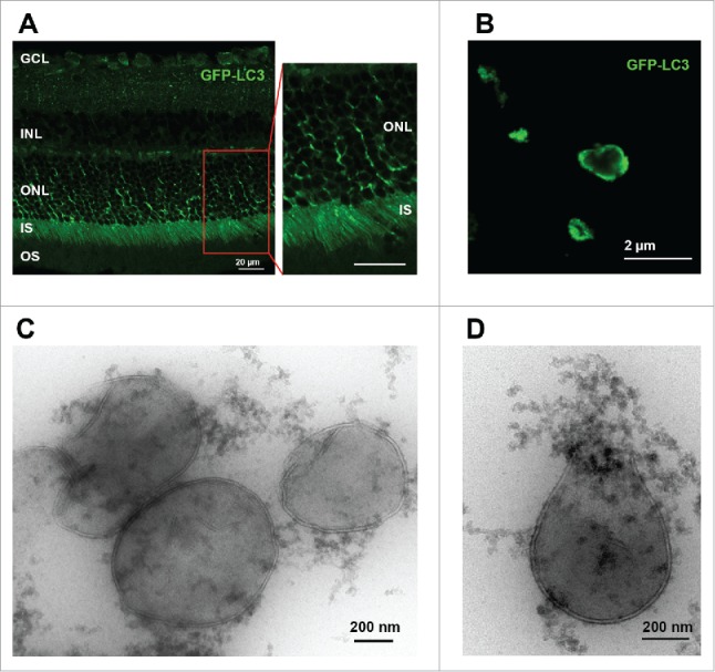
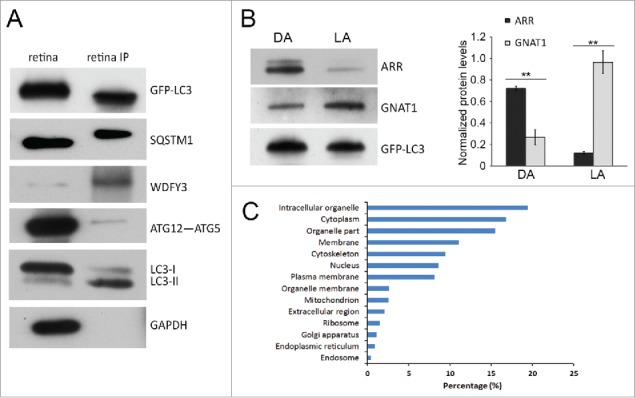
Autophagy is a lysosomal degradation pathway critical to preventing the accumulation of cytotoxic proteins. Deletion of the essential autophagy gene Atg5 from the rod photoreceptors of the retina (atg5Δrod mouse) results in the accumulation of the phototransduction protein transducin and the degeneration of these neurons. The purpose of this study is to test the hypothesis that autophagic degradation of visual transduction proteins prevents retinal degeneration. Targeted deletion of both Gnat1 (a gene encoding the α subunit of the heterotrimeric G-protein transducin) and Atg5 in the rod photoreceptors resulted in a significantly decreased rate of rod cell degeneration as compared to the atg5Δrod mouse retina, and considerable preservation of photoreceptors. Supporting this we used a novel technique to immunoprecipitate green fluorescent protein (GFP)-tagged autophagosomes from the retinas of the GFP-LC3 mice and demonstrated that the visual transduction proteins transducin and ARR/arrestin are associated with autophagosome-specific proteins. Altogether, this study shows that degradation of phototransduction proteins by autophagy is necessary to prevent retinal degeneration. In addition, we demonstrate a simple and easily reproducible immunoisolation technique for enrichment of autophagosomes from the GFP-LC3 mouse retina, providing a novel application to the study of autophagosome contents across different organs and specific cell types in vivo.
Keywords: autophagy; photoreceptor; retina; retinal degeneration; transducin.
Figures
Similar articles
-
Autophagy in Xenopus laevis rod photoreceptors is independently regulated by phototransduction and misfolded RHOP23H.Autophagy. 2019 Nov;15(11):1970-1989. doi: 10.1080/15548627.2019.1596487. Epub 2019 Apr 12. Autophagy. 2019. PMID: 30975014 Free PMC article.
-
Downregulation of cone-specific gene expression and degeneration of cone photoreceptors in the Rpe65-/- mouse at early ages.Invest Ophthalmol Vis Sci. 2005 Apr;46(4):1473-9. doi: 10.1167/iovs.04-0653. Invest Ophthalmol Vis Sci. 2005. PMID: 15790918
-
Autophagy supports survival and phototransduction protein levels in rod photoreceptors.Cell Death Differ. 2015 Mar;22(3):488-98. doi: 10.1038/cdd.2014.229. Epub 2015 Jan 9. Cell Death Differ. 2015. PMID: 25571975 Free PMC article.
-
New insights into the role of autophagy in retinal and eye diseases.Mol Aspects Med. 2021 Dec;82:101038. doi: 10.1016/j.mam.2021.101038. Epub 2021 Oct 5. Mol Aspects Med. 2021. PMID: 34620506 Review.
-
A Balancing Act.J Pediatr Ophthalmol Strabismus. 2018 Jul 1;55(4):216-217. doi: 10.3928/01913913-20180530-01. J Pediatr Ophthalmol Strabismus. 2018. PMID: 30024017 Review. No abstract available.
Cited by
-
Preservation of retinal structure and function in two mouse models of inherited retinal degeneration by ONL1204, an inhibitor of the Fas receptor.Cell Death Dis. 2024 Aug 8;15(8):576. doi: 10.1038/s41419-024-06970-6. Cell Death Dis. 2024. PMID: 39117629 Free PMC article.
-
Autophagy in Xenopus laevis rod photoreceptors is independently regulated by phototransduction and misfolded RHOP23H.Autophagy. 2019 Nov;15(11):1970-1989. doi: 10.1080/15548627.2019.1596487. Epub 2019 Apr 12. Autophagy. 2019. PMID: 30975014 Free PMC article.
-
Clinical and preclinical therapeutic outcome metrics for USH2A-related disease.Hum Mol Genet. 2020 Jul 21;29(11):1882-1899. doi: 10.1093/hmg/ddaa004. Hum Mol Genet. 2020. PMID: 31998945 Free PMC article.
-
Autophagy is a novel pathway for neurofilament protein degradation in vivo.Autophagy. 2023 Apr;19(4):1277-1292. doi: 10.1080/15548627.2022.2124500. Epub 2022 Sep 21. Autophagy. 2023. PMID: 36131358 Free PMC article.
-
Importance of Autoimmune Responses in Progression of Retinal Degeneration Initiated by Gene Mutations.Front Med (Lausanne). 2021 Dec 2;8:672444. doi: 10.3389/fmed.2021.672444. eCollection 2021. Front Med (Lausanne). 2021. PMID: 34926479 Free PMC article. Review.
References
-
- Mustafi D, Engel AH, Palczewski K. Structure of cone photoreceptors. Prog Ret Eye Res 2009; 28:289-302; http://dx.doi.org/10.1016/j.preteyeres.2009.05.003 - DOI - PMC - PubMed
-
- Calvert PD, Strissel KJ, Schiesser WE, Pugh EN Jr, Arshavsky VY. Light-driven translocation of signaling proteins in vertebrate photoreceptors. Trends Cell Biol 2006; 16:560-8; PMID:16996267; http://dx.doi.org/10.1016/j.tcb.2006.09.001 - DOI - PubMed
-
- Yang Z, Klionsky DJ. Mammalian autophagy: core molecular machinery and signaling regulation. Curr Opin Cell Biol 2010; 22:124-31; PMID:20034776; http://dx.doi.org/10.1016/j.ceb.2009.11.014 - DOI - PMC - PubMed
-
- Yoshimori T. Autophagy: a regulated bulk degradation process inside cells. Biochem Biophys Res Commun 2004; 313:453-8; PMID:14684184; http://dx.doi.org/10.1016/j.bbrc.2003.07.023 - DOI - PubMed
-
- Yao J, Jia L, Shelby SJ, Ganios AM, Feathers K, Thompson DA, Zacks DN. Circadian and noncircadian modulation of autophagy in photoreceptors and retinal pigment epithelium. Invest Ophthalmol Vis Sci 2014; 55:3237-46; PMID:24781939; http://dx.doi.org/10.1167/iovs.13-13336 - DOI - PMC - PubMed
Publication types
MeSH terms
Substances
Grants and funding
LinkOut - more resources
Full Text Sources
Other Literature Sources
Research Materials