

Doxycycline inhibits the cancer stem cell phenotype and epithelial-to-mesenchymal transition in breast cancer
- PMID: 27753527
- PMCID: PMC5405729
- DOI: 10.1080/15384101.2016.1241929
Doxycycline inhibits the cancer stem cell phenotype and epithelial-to-mesenchymal transition in breast cancer
Abstract
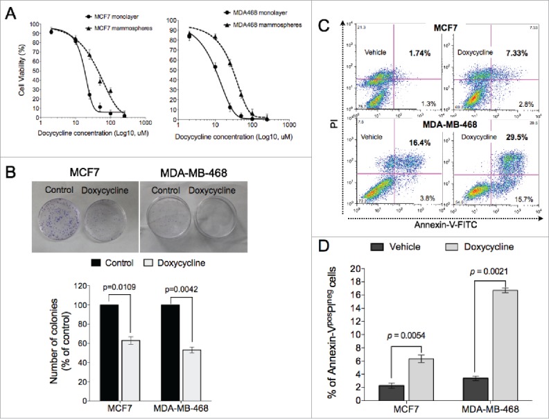
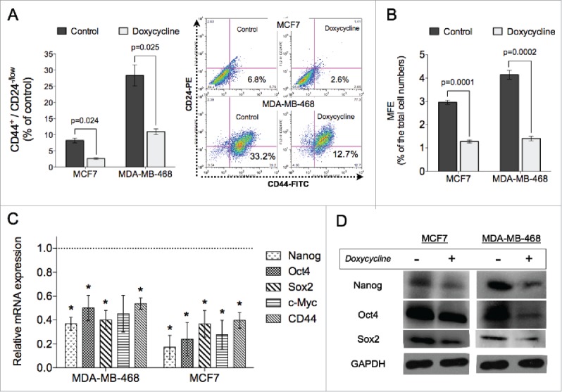
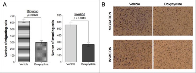
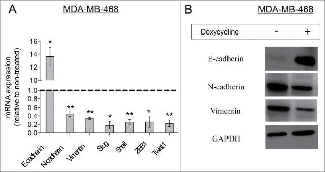
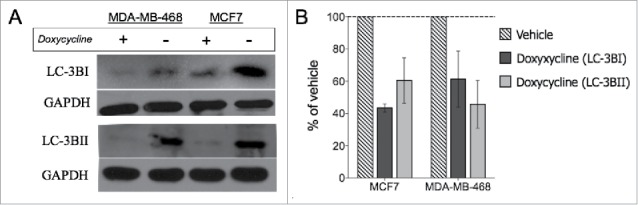
Experimental evidence suggest that breast tumors originate from breast cancer stem cells (BCSCs), and that mitochondrial biogenesis is essential for the anchorage-independent clonal expansion and survival of CSCs, thus rendering mitochondria a significant target for novel treatment approaches. One of the recognized side effects of the FDA-approved drug, doxycycline is the inhibition of mitochondrial biogenesis. Here we investigate the mechanism by which doxycycline exerts its inhibitory effects on the properties of breast cancer cells and BCSCs, such as mammosphere forming efficiency, invasion, migration, apoptosis, the expression of stem cell markers and epithelial-to-mesenchymal transition (EMT) related markers of breast cancer cells. In addition, we explored whether autophagy plays a role in the inhibitory effect of doxycycline on breast cancer cells. We find that doxycyline can inhibit the viability and proliferation of breast cancer cells and BCSCs, decrease mammosphere forming efficiency, migration and invasion, and EMT of breast cancer cells. Expression of stem cell factors Oct4, Sox2, Nanog and CD44 were also significantly downregulated after doxycycline treatment. Moreover, doxycycline could down-regulate the expression of the autophagy marker LC-3BI and LC-3BII, suggesting that inhibiting autophagy may be responsible in part for the observed effects on proliferation, EMT and stem cell markers. The potent inhibition of EMT and cancer stem-like characteristics in breast cancer cells by doxycycline treatment suggests that this drug can be repurposed as an anti-cancer drug in the treatment of breast cancer patients in the clinic.
Keywords: autophagy; breast cancer; cancer stem cells; doxycycline; epithelial-to-mesenchymal transition; mitochondria.
Figures
Similar articles
-
Effect of Melatonin in Epithelial Mesenchymal Transition Markers and Invasive Properties of Breast Cancer Stem Cells of Canine and Human Cell Lines.PLoS One. 2016 Mar 2;11(3):e0150407. doi: 10.1371/journal.pone.0150407. eCollection 2016. PLoS One. 2016. PMID: 26934679 Free PMC article.
-
Anti-metastasis activity of curcumin against breast cancer via the inhibition of stem cell-like properties and EMT.Phytomedicine. 2019 May;58:152740. doi: 10.1016/j.phymed.2018.11.001. Epub 2018 Nov 12. Phytomedicine. 2019. PMID: 31005718
-
Inhibitory effect of melatonin on hypoxia-induced vasculogenic mimicry via suppressing epithelial-mesenchymal transition (EMT) in breast cancer stem cells.Eur J Pharmacol. 2020 Aug 15;881:173282. doi: 10.1016/j.ejphar.2020.173282. Epub 2020 Jun 21. Eur J Pharmacol. 2020. PMID: 32580038
-
Metabolic reprogramming on breast cancer stem Cells: Proliferation and self-renewal, epithelial-mesenchymal transition (EMT), and drug resistance.Biochem Biophys Res Commun. 2025 Aug 8;774:152079. doi: 10.1016/j.bbrc.2025.152079. Epub 2025 May 23. Biochem Biophys Res Commun. 2025. PMID: 40446745 Review.
-
The role of the mTOR pathway in breast cancer stem cells (BCSCs): mechanisms and therapeutic potentials.Stem Cell Res Ther. 2025 Mar 29;16(1):156. doi: 10.1186/s13287-025-04218-4. Stem Cell Res Ther. 2025. PMID: 40158191 Free PMC article. Review.
Cited by
-
Modulation of Immune Components on Stem Cell and Dormancy in Cancer.Cells. 2021 Oct 21;10(11):2826. doi: 10.3390/cells10112826. Cells. 2021. PMID: 34831048 Free PMC article. Review.
-
Tumour Stem Cells in Breast Cancer.Int J Mol Sci. 2022 May 2;23(9):5058. doi: 10.3390/ijms23095058. Int J Mol Sci. 2022. PMID: 35563449 Free PMC article. Review.
-
1,2,4-Oxadiazole/2-Imidazoline Hybrids: Multi-target-directed Compounds for the Treatment of Infectious Diseases and Cancer.Int J Mol Sci. 2019 Apr 5;20(7):1699. doi: 10.3390/ijms20071699. Int J Mol Sci. 2019. PMID: 30959765 Free PMC article.
-
Targeting Mitochondria with ClpP Agonists as a Novel Therapeutic Opportunity in Breast Cancer.Cancers (Basel). 2023 Mar 23;15(7):1936. doi: 10.3390/cancers15071936. Cancers (Basel). 2023. PMID: 37046596 Free PMC article. Review.
-
Navigating the therapeutic landscape for breast cancer: targeting breast cancer stem cells.Naunyn Schmiedebergs Arch Pharmacol. 2025 Mar;398(3):2387-2406. doi: 10.1007/s00210-024-03542-5. Epub 2024 Oct 23. Naunyn Schmiedebergs Arch Pharmacol. 2025. PMID: 39441235 Review.
References
-
- Wang D, Lu P, Zhang H, Luo M, Zhang X, Wei X, Gao J, Zhao Z, Liu C. Oct-4 and Nanog promote the epithelial-mesenchymal transition of breast cancer stem cells and are associated with poor prognosis in breast cancer patients. Oncotarget 2014; 5(21):10803; PMID:25301732; http://dx.doi.org/10.18632/oncotarget.2506 - DOI - PMC - PubMed
-
- Siegel RL MK, Jemal A. Cancer statistics. CA Cancer J Clin 2016; 66(1):7–30; PMID:26742998; http://dx.doi.org/10.3322/caac.21332 - DOI - PubMed
-
- Weigelt B, Peterse JL, Van't Veer LJ. Breast cancer metastasis: markers and models. Nat Rev Cancer 2005; 5(8):591–602; PMID:16056258; http://dx.doi.org/10.1038/nrc1670 - DOI - PubMed
-
- Jemal A, Bray F, Center MM, Ferlay J, Ward E, Forman D. Global cancer statistics. CA: Cancer J Clin 2011; 61(2):69–90; PMID:21296855 - PubMed
-
- Jordan CT, Guzman ML, Noble M. Cancer stem cells. New Engl J Med 2006; 355(12):1253–61; PMID:16990388; http://dx.doi.org/10.1056/NEJMra061808 - DOI - PubMed
MeSH terms
Substances
LinkOut - more resources
Full Text Sources
Other Literature Sources
Medical
Research Materials
Miscellaneous