. 2016 Dec;15(23):3191-3202.
doi: 10.1080/15384101.2016.1241914. Epub 2016 Oct 18.

Histone chaperone ASF1B promotes human β-cell proliferation via recruitment of histone H3.3

Affiliations

Histone chaperone ASF1B promotes human β-cell proliferation via recruitment of histone H3.3

Pradyut K Paul et al. Cell Cycle. 2016 Dec.

Abstract

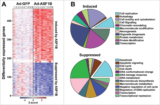
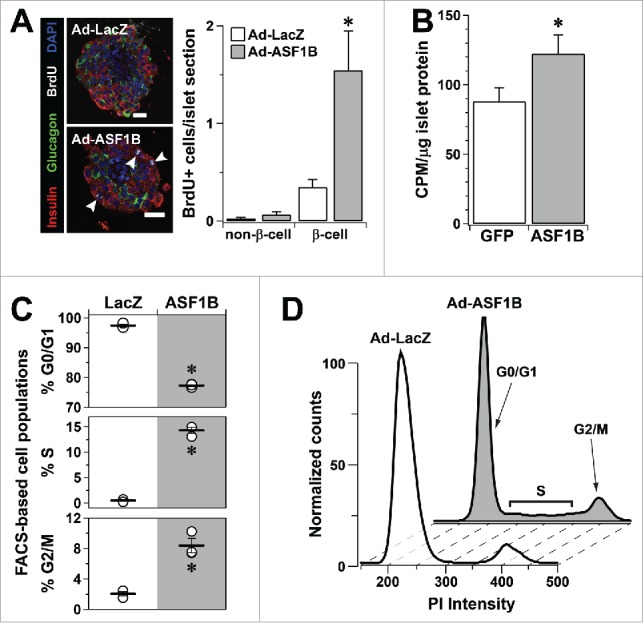
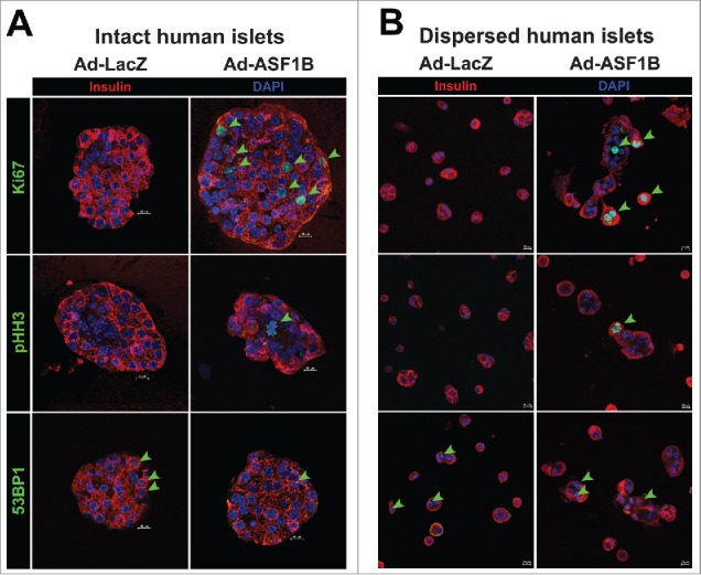
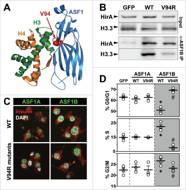
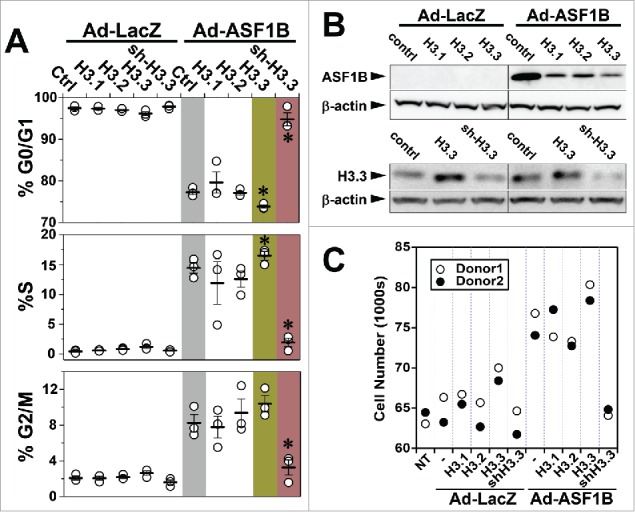
Anti-silencing function 1 (ASF1) is a histone H3-H4 chaperone involved in DNA replication and repair, and transcriptional regulation. Here, we identify ASF1B, the mammalian paralog to ASF1, as a proliferation-inducing histone chaperone in human β-cells. Overexpression of ASF1B led to distinct transcriptional signatures consistent with increased cellular proliferation and reduced cellular death. Using multiple methods of monitoring proliferation and mitotic progression, we show that overexpression of ASF1B is sufficient to induce human β-cell proliferation. Co-expression of histone H3.3 further augmented β-cell proliferation, whereas suppression of endogenous H3.3 attenuated the stimulatory effect of ASF1B. Using the histone binding-deficient mutant of ASF1B (V94R), we show that histone binding to ASF1B is required for the induction of β-cell proliferation. In contrast to H3.3, overexpression of histone H3 variants H3.1 and H3.2 did not have an impact on ASF1B-mediated induction of proliferation. Our findings reveal a novel role of ASF1B in human β-cell replication and show that ASF1B and histone H3.3A synergistically stimulate human β-cell proliferation.

Keywords: ASF1B; cell cycle; histone H3.3; replication-independent histone deposition; β-cell proliferation.


Figures

Figure 1.
Figure 1.
Expression of Asf1b, but not Asf1a, strongly correlates with Mki67 in mouse islets. Obesity-dependent changes in the expression of Asf1a, Asf1b, and Mki67 in islets from B6 or BTBR mice at 4 or 10 weeks of age (male, N = 5 each). Gene expression values are log10-transformed ratios of individual measurements vs. a strain-specific reference pool. *, P ≤ 0.05 for obese (Leptinob/ob) vs. lean.
Figure 2.
Figure 2.
ASF1B modulates genes regulating cell proliferation and apoptosis in human β-cells. (A) Heat map illustrating differentially expressed (DE) genes in response to Ad-ASF1B overexpression in 10 human islet samples. Color corresponds to Z-score (decreased, blue; increased, red) relative to Ad-GFP treated islets. (B) Gene Ontology (GO) analysis of DE genes in response to ASF1B. Pie charts represent the number of genes for significant categories (Z > 2.0). A complete listing of all enriched terms is provided in Table S3.
Figure 3.
Figure 3.
ASF1B overexpression induces proliferation of human β-cells. (A) Representative confocal images (60X) of intact human islets (N = 400) treated with Ad-LacZ or Ad-ASF1B, and labeled with BrdU (white) to mark proliferating cells (48 h). β-cells are identified by insulin (red), α-cells by glucagon (green) and nuclei by DAPI (blue). (B) [3H]-thymidine incorporation into human islet DNA measured 48 h after Ad-LacZ or Ad-ASF1B treatment (N = 10). (C) Flow cytometry quantitation of the % cells in G0/G1, S and G2/M phases 48 h after Ad-LacZ or Ad-ASF1B treatment of dispersed human islet cells (N = 4). (D) Representative histograms of FACS-analyzed cell cycle profiles of disrupted human islets. Data represent means ± SEM, N = 3. *, P ≤ 0.05. All comparisons are for Ad-LacZ or Ad-GFP vs. Ad-ASF1B.
Figure 4.
Figure 4.
ASF1B overexpression induces mitotic progression, but not DNA damage, in intact and dispersed human islets. Representative confocal images (60X) of (A) intact human islets (N = 20) and (B) dispersed human islet cells treated with Ad-LacZ or Ad-ASF1B, and labeled with the proliferation markers Ki67 and phospho-histone H3 (pHH3 S10) that mark cells through mitotic progression, and with the DNA damage marker, 53BP1 to mark nuclei undergoing DNA damage response (48 h). β-cells are identified by insulin (red), nuclei by DAPI (blue).
Figure 5.
Figure 5.
Histone-binding mutant of ASF1B fails to promote β-cell proliferation. (A) Crystal structure of yeast ASF1-H3-H4 complex (PDB 2HUE); V94 of ASF1 is highlighted red. (B) Binding of HIRA and histone H3.3 was detected in anti-Twin-strep-tag (ASF1B) immunoprecipitates and total cell lysates from MIN6 cells transiently expressing GFP, WT-ASF1B or ASF1B-V94R mutant proteins. (C) Representative images (100X) of MIN6 β-cells expressing ASF1A or ASF1B (green) for WT (top panels) or the V94R mutants (bottom panels). Cells are co-stained for DAPI (nuclei, white), and insulin (red). (D) Flow cytometry analysis of cell cycle progression in MIN6 β-cells 48 h post-transfection with indicated plasmid-based constructs. Data represent means ± SEM, N = 3. *, GFP vs. ASF1B-WT and #, ASF1B-WT vs. ASF1B-V94R, P ≤ 0.05.
Figure 6.
Figure 6.
An interaction with H3.3 is necessary for ASF1B to induce human β-cell proliferation. (A) Flow cytometry analysis of cell cycle progression in disrupted human islets 48 h after indicated viral-mediated transductions. (B) Western blot illustrating relative protein abundance of ASF1B in islet cells expressing histone H3.1, H3.2 or H3.3 alone or in combination with Ad-LacZ (control) or ASF1B (upper panel); reduced protein abundance of histone H3.3 in response to sh-RNA mediated suppression (lower panel) N = 4 human donors. (C) Quantitation of change in cell number after ASF1B-mediated induction in human β-cell proliferation. Changes are measured using CyQuant assay and compared to control (no treatment, NT), N = 2 human donors.

Comment in

Similar articles

Cited by

References

    1. Butler AE, Janson J, Bonner-Weir S, Ritzel R, Rizza RA, Butler PC. Beta-cell deficit and increased β-cell apoptosis in humans with type 2 diabetes. Diabetes 2003; 52:102-10; PMID:12502499; http://dx.doi.org/ 10.2337/diabetes.52.1.102 - DOI - PubMed
    1. Rankin MM, Kushner JA. Adaptive β-cell proliferation is severely restricted with advanced age. Diabetes 2009; 58:1365-72; PMID:19265026; http://dx.doi.org/ 10.2337/db08-1198 - DOI - PMC - PubMed
    1. Salpeter SJ, Khalaileh A, Weinberg-Corem N, Ziv O, Glaser B, Dor Y. Systemic regulation of the age-related decline of pancreatic β-cell replication. Diabetes 2013; 62:2843-8; PMID:23630298; http://dx.doi.org/ 10.2337/db13-0160 - DOI - PMC - PubMed
    1. Butler AE, Cao-Minh L, Galasso R, Rizza RA, Corradin A, Cobelli C, Butler PC. Adaptive changes in pancreatic β cell fractional area and β cell turnover in human pregnancy. Diabetologia 2010; 53:2167-76; PMID:20523966; http://dx.doi.org/ 10.1007/s00125-010-1809-6 - DOI - PMC - PubMed
    1. Georgia S, Bhushan A. Beta cell replication is the primary mechanism for maintaining postnatal β cell mass. J Clin Invest 2004; 114:963-8; PMID:15467835; http://dx.doi.org/ 10.1172/JCI22098 - DOI - PMC - PubMed