. 2016 Oct 12;8(10):275.
doi: 10.3390/v8100275.

The Role of the Equine Herpesvirus Type 1 (EHV-1) US3-Encoded Protein Kinase in Actin Reorganization and Nuclear Egress

Affiliations

The Role of the Equine Herpesvirus Type 1 (EHV-1) US3-Encoded Protein Kinase in Actin Reorganization and Nuclear Egress

Alexandra Proft et al. Viruses. .

Abstract

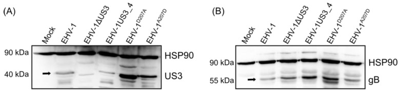
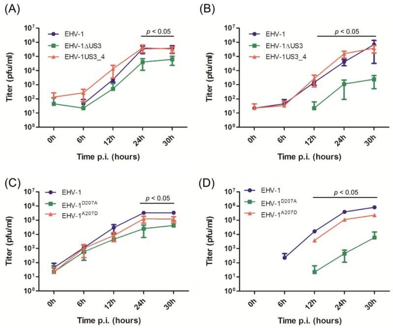
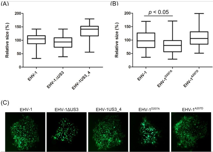
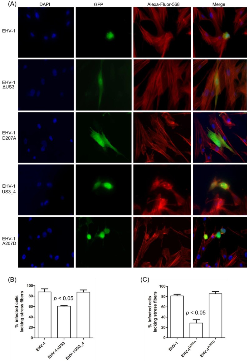
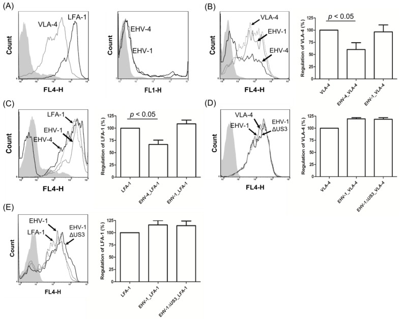
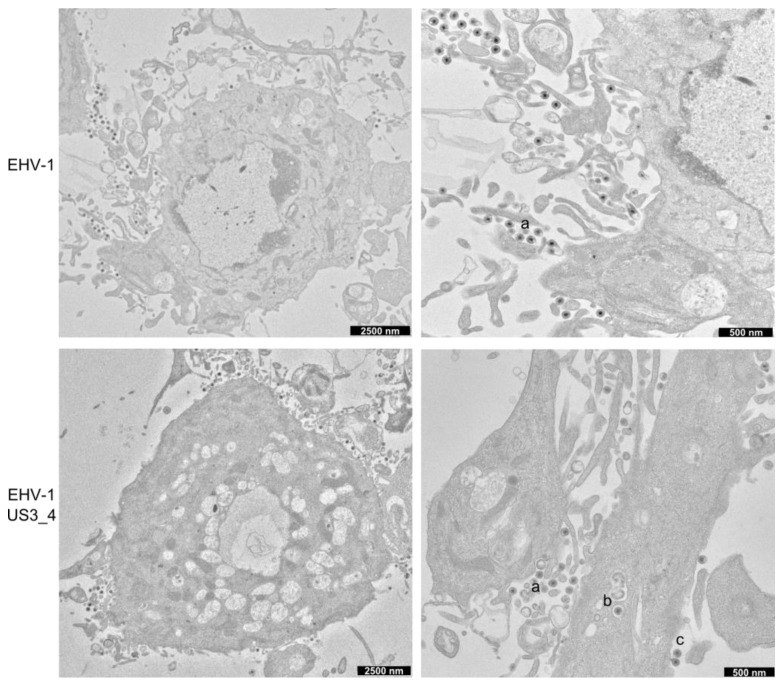
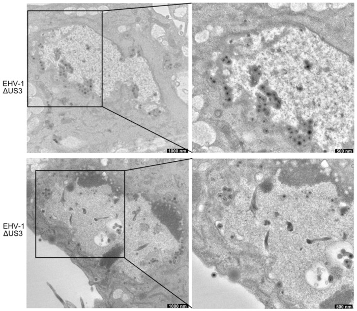
The serine-threonine protein kinase encoded by US3 gene (pUS3) of alphaherpesviruses was shown to modulate actin reorganization, cell-to-cell spread, and virus egress in a number of virus species. However, the role of the US3 orthologues of equine herpesvirus type 1 and 4 (EHV-1 and EHV-4) has not yet been studied. Here, we show that US3 is not essential for virus replication in vitro. However, growth rates and plaque diameters of a US3-deleted EHV-1 and a mutant in which the catalytic active site was destroyed were significantly reduced when compared with parental and revertant viruses or a virus in which EHV-1 US3 was replaced with the corresponding EHV-4 gene. The reduced plaque sizes were consistent with accumulation of primarily enveloped virions in the perinuclear space of the US3-negative EHV-1, a phenotype that was also rescued by the EHV-4 orthologue. Furthermore, actin stress fiber disassembly was significantly more pronounced in cells infected with parental EHV-1, revertant, or the recombinant EHV-1 expressing EHV-4 US3. Finally, we observed that deletion of US3 in EHV-1 did not affect the expression of adhesion molecules on the surface of infected cells.

Keywords: EHV-1; EHV-4; EM; US3; actin cytoskeleton; adhesion molecules.


Conflict of interest statement

The authors declare no conflict of interest.

Figures

Figure 1
Figure 1
Analysing US3 expression by western blot. Cells were either infected with parental, mutant, recombinant, or revertant viruses. Cell lysates were prepared with radioimmunoprecipitation assay (RIPA) buffer. The pUS3 (A) and gB (B) proteins were separated by 12% sodium dodecyl sulfate-polyacrylamide gel electrophoresis (SDS-PAGE). The expression of pUS3 and gB (black arrows) were detected with anti-US3 and anti-gB antibodies, respectively. Heat shock protein 90 α/β (HSP90α/β, 1:1000 dilution) was used as a loading control
Figure 2
Figure 2
Growth properties of mutant and recombinant viruses. Confluent equine dermal (ED) cells were infected with the different viruses as indicated in the figure. Infected cells (A and C) and supernatant (B and D) were collected separately at different time points post-infection (p.i.; 0, 6, 12, 24 and 30 h) and virus titers were determined. The data presented are means ± standard deviations (SD) of three independent experiments. Significance levels (p < 0.05) were determined for US3-deleted or EHV-1D207A viruses when compared to parental, mutant, recombinant and revertant viruses (Friedman test-Dunn’s multiple comparison test). EHV-1: equine herpesvirus type 1; EHV-1ΔUS3: EHV-1 where US3 has been deleted; EHV-1US3_4: EHV-1 where US3_4 replaces US3_1; EHV-1D207A: EHV-1 with a point mutation in the catalytic active site of pUS3 where aspartic acid was replaced with alanine; EHV-1A207D: EHV-1 revertant virus where the original aspartic acid was restored.
Figure 3
Figure 3
Determination of cell-to-cell spread by plaque size assay. ED cells were infected with EHV-1∆US3 (A) or EHV-1D207A (B) viruses at a multiplicity of infection (MOI) of 0.001. After 72 h, 50 plaques were measured for each virus. The central line in the box plot indicates the median of the data, while the edges of the box indicate the 25th and 75th percentiles. Extending from the box are whiskers, the top whisker expands to the 95th percentile and the bottom whisker to the 5th percentile. The plaque diameter of parental viruses was set to 100%. A significant reduction (one-way ANOVA; p < 0.05) of plaque size for EHV-1D207A was seen when compared to parental, mutant, recombinant or revertant viruses. (C) plaques produced by different green fluorescent protein (GFP)-expressing viruses. Pictures were taken using a Zeiss Axiovert fluorescence microscope.
Figure 4
Figure 4
Rearrangement of actin cytoskeleton in infected ED cells. (A) ED cells were infected with parental, US3_1-deleted, EHV-1D207A, US3_4-recombinant, or revertant viruses and imaged by immunofluorescence microscopy using a Zeiss Axiovert fluorescence microscope. Pictures were taken with a 63x oil objective. The nucleus was stained with 4′,6-diamidino-2-phenylindole (DAPI, blue), the actin cytoskeleton was stained with phalloidin-Alexa 568 (red), and the virus-infected cells were visualized through enhanced GFP (eGFP) expression (green). (B and C) A total of 200 infected cells for each virus in three independent experiments were inspected and the percentage of infected cells with or without changes in actin cytoskeleton was calculated. A significant change in actin cytoskeleton rearrangement was detected for either EHV-1ΔUS3 or EHV-1D207A when compared to parental, recombinant, or revertant viruses (one-way ANOVA; p < 0.05).
Figure 5
Figure 5
EHV-4 downregulates cell surface adhesion molecules on peripheral blood mononuclear cells (PBMC). (A) surface expression of very late antigen-4 (VLA-4) and lymphocyte function-associated antigen-1 (LFA-1) on the surface of mock-infected PBMC and virus infection (black line: EHV-1; dotted line: EHV-4) levels of PBMC were determined by flow cytometry. (B and C) Equine PBMC were either mock-infected or infected with EHV-4 or EHV-1 and subjected to flow cytometric analysis. Histograms and bars of VLA-4 (B) and LFA-1 (C) expression after detection with the relevant antibodies are shown. Viable cells (10,000) were analyzed for each sample. Grey filled: mock-infected cells; dotted line: mock-infected cells with the relevant antibody; black line: EHV-4-infected cells; grey line: EHV-1-infected cells. For the bars, the expression level of adhesion molecules (VLA-4 or LFA-1) was set to 100%. All data represent the mean ± SD of three independent experiments. A significant downregulation (one-way ANOVA; p < 0.05) of VLA-4 and LFA-1 for EHV-4-infected cells was seen when compared to mock- or EHV-1-infected cells. (D and E) PBMC were either mock-infected or infected with EHV-1ΔUS3 and subjected to flow cytometric analysis. Histograms and bars of VLA-4 (D) and LFA-1 (E) expression are shown. Grey filled: mock-infected cells; dotted line: mock-infected cells with the relevant antibody; grey line: EHV-1-infected cells; black line: EHV-1ΔUS3-infected cells. Data are from one representative experiment out of three. FL1 and FL4: fluorescence detector.
Figure 6
Figure 6
Electron microscopy of ED cells infected with parental and recombinant EHV-1. ED cells were infected for 14 h and analysed by transmission electron microscopy. Cells show different stages of virion maturation, including extracellular virions (a), enveloped virions within cytoplasmic vesicles (b) and primary enveloped virions in the nucleus (c).
Figure 7
Figure 7
Ultrastructural analysis of ED cells infected with US3-deleted EHV-1. ED cells were infected for 14 h and analysed by transmission electron microscopy. Cells show accumulation of enveloped virions in the perinuclear space.

Similar articles

Cited by

References

    1. Calton C.M., Randall J.A., Adkins M.W., Banfield B.W. The pseudorabies virus serine/threonine kinase Us3 contains mitochondrial, nuclear and membrane localization signals. Virus Genes. 2004;29:131–145. doi: 10.1023/B:VIRU.0000032796.27878.7f. - DOI - PubMed
    1. Del Medico Zajac M.P., Ladelfa M.F., Kotsias F., Muylkens B., Thiry J., Thiry E., Romera S.A. Biology of bovine herpesvirus 5. Vet. J. 2010;184:138–145. doi: 10.1016/j.tvjl.2009.03.035. - DOI - PubMed
    1. Colle C.F., 3rd, Flowers C.C., O’Callaghan D.J. Open reading frames encoding a protein kinase, homolog of glycoprotein gX of pseudorabies virus, and a novel glycoprotein map within the unique short segment of equine herpesvirus type 1. Virology. 1992;188:545–557. doi: 10.1016/0042-6822(92)90509-N. - DOI - PubMed
    1. Telford E.A., Watson M.S., Perry J., Cullinane A.A., Davison A.J. The DNA sequence of equine herpesvirus-4. J. Gen. Virol. 1998;79:1197–1203. doi: 10.1099/0022-1317-79-5-1197. - DOI - PubMed
    1. Jacob T., Van den Broeke C., Favoreel H.W. Viral serine/threonine protein kinases. J. Virol. 2011;85:1158–1173. doi: 10.1128/JVI.01369-10. - DOI - PMC - PubMed

MeSH terms

LinkOut - more resources