Review
. 2016 Oct 15;17(10):1732.
doi: 10.3390/ijms17101732.

Glycosphingolipid-Protein Interaction in Signal Transduction

Affiliations
Review

Glycosphingolipid-Protein Interaction in Signal Transduction

Domenico Russo et al. Int J Mol Sci. .

Abstract

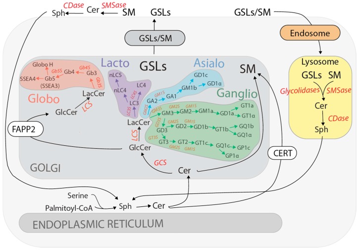
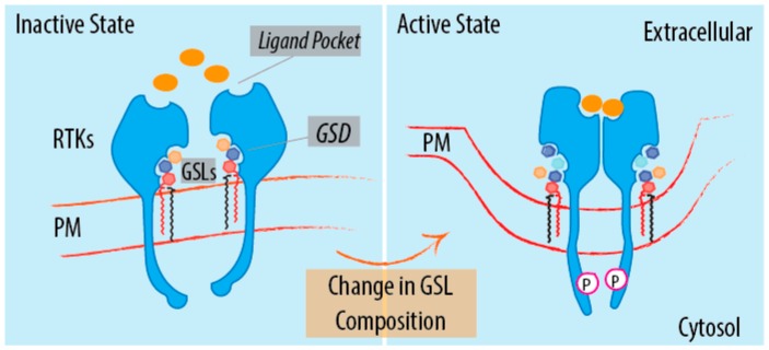
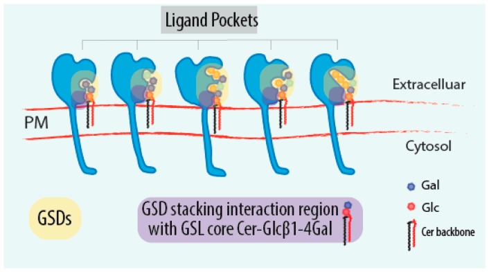
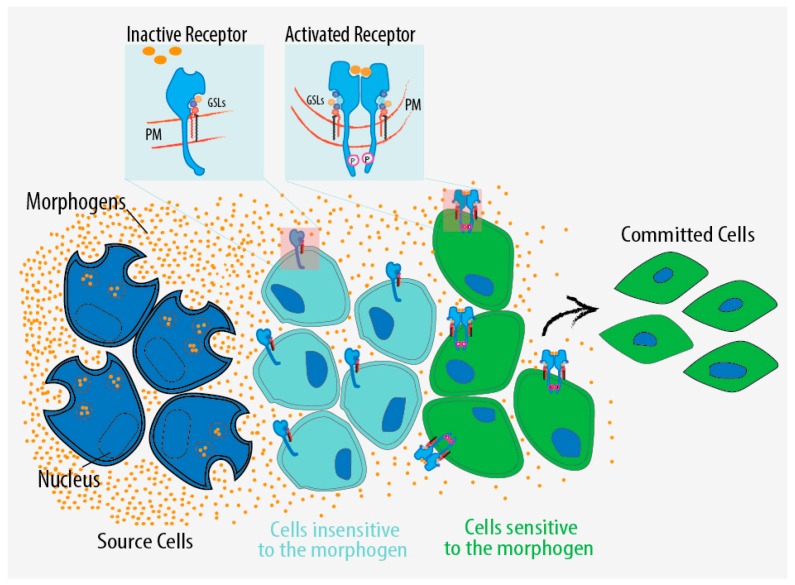
Glycosphingolipids (GSLs) are a class of ceramide-based glycolipids essential for embryo development in mammals. The synthesis of specific GSLs depends on the expression of distinctive sets of GSL synthesizing enzymes that is tightly regulated during development. Several reports have described how cell surface receptors can be kept in a resting state or activate alternative signalling events as a consequence of their interaction with GSLs. Specific GSLs, indeed, interface with specific protein domains that are found in signalling molecules and which act as GSL sensors to modify signalling responses. The regulation exerted by GSLs on signal transduction is orthogonal to the ligand-receptor axis, as it usually does not directly interfere with the ligand binding to receptors. Due to their properties of adjustable production and orthogonal action on receptors, GSLs add a new dimension to the control of the signalling in development. GSLs can, indeed, dynamically influence progenitor cell response to morphogenetic stimuli, resulting in alternative differentiation fates. Here, we review the available literature on GSL-protein interactions and their effects on cell signalling and development.

Keywords: glycan–protein interaction; glycosphingolipid; signalling.


Conflict of interest statement

The authors declare no conflict of interest.

Figures

Figure 1
Figure 1
GSL synthesis and turnover. Schematic representation of the metabolism of the major GSL classes synthesized along the secretory and endocytic pathways. For details refer to the text. GSEs catalysing the major synthetic reactions and the enzymes involved in GSLs/SM dismantling are reported in red. GSLs, glycosphingolipids; Sph, sphinganine from the de novo pathway or sphingosine from the salvage pathway; Cer, ceramide; SM, sphingomyelin; GCS, GlcCer synthase; LCS, LacCer synthase; CERT, ceramide transfer protein; FAPP2, four-phosphate adaptor protein 2; SMSase, sphingomyelinase; CDase, ceramidase.
Figure 2
Figure 2
GSL influence on signalling. Schematic representation of the regulatory role of the GSLs on signal transduction. The regulation of RTKs by GSLs is provided as an example to illustrate the signalling role of GSLs. A specific GSL profile on plasma membrane can keep a RTK in an inactive state (left panel); A change in GSLs composition can regulate receptor activation/inactivation. GSLs act as allosteric regulators of receptor capability to recognize the ligand, to undergo multimerization and to auto-phosphorylate (right panel). GSLs, glycosphingolipids; Orange spheres represent ligands; Colored hexagons represent the different sugar residues of GSL-glycan moieties; Black-Red parallelogram, Cer backbone. Pink circle surrounding P represent phosphate groups; GSD, glycosphingolipid sensing domain; PM, plasma membrane; RTK, receptor tyrosine kinase.
Figure 3
Figure 3
GSDs as sensors of the GSL-glycocode. Schematic representation of receptors containing different GSDs that recognize specificGSL-glycan portions. In light orange are highlighted GSDs in the receptors. In violet are indicated the conserved GSD portions recognizing the basic GSL core motif Cer-Glcβ1-4Gal present in the most GSLs. Coloured hexagons represent the different sugar residues of GSL-glycan moieties. Blue hexagon, Gal residue; Red hexagon, Glc residue; Black-Red parallelogram, Cer backbone; GSD, glycosphingolipid sensing domain; PM, plasma membrane.
Figure 4
Figure 4
GSL regulation of signalling in development. Schematic representation of GSL-dependent regulation affecting the environmental sensing of the cell and the commitment to alternative differentiation fates. This regulatory layer might act in parallel to known mechanisms of tissue patterning such as morphogen gradients that induce the differentiation of specific cell types in a distinct spatial order. The same differentiation factor might be invisible to some cells and at the same time perceived by others depending on the fact that the target receptor is in a dormant or active state in relation to the GSL composition on the plasma membrane. Orange spheres represent morphogens; PM, plasma membrane.

Similar articles

Cited by

References

    1. Merrill A.H., Jr. Sphingolipid and glycosphingolipid metabolic pathways in the era of sphingolipidomics. Chem. Rev. 2011;111:6387–6422. doi: 10.1021/cr2002917. - DOI - PMC - PubMed
    1. Coskun U., Grzybek M., Drechsel D., Simons K. Regulation of human EGF receptor by lipids. Proc. Natl. Acad. Sci. USA. 2011;108:9044–9048. doi: 10.1073/pnas.1105666108. - DOI - PMC - PubMed
    1. Vyas K.A., Patel H.V., Vyas A.A., Schnaar R.L. Segregation of gangliosides GM1 and GD3 on cell membranes, isolated membrane rafts, and defined supported lipid monolayers. Biol. Chem. 2001;382:241–250. doi: 10.1515/BC.2001.031. - DOI - PubMed
    1. Kiyokawa E., Baba T., Otsuka N., Makino A., Ohno S., Kobayashi T. Spatial and functional heterogeneity of sphingolipid-rich membrane domains. J. Biol. Chem. 2005;280:24072–24084. doi: 10.1074/jbc.M502244200. - DOI - PubMed
    1. Lindquist S., Karitkina D., Langnaese K., Posevitz-Fejfar A., Schraven B., Xavier R., Seed B., Lindquist J.A. Phosphoprotein associated with glycosphingolipid-enriched microdomains differentially modulates SRC kinase activity in brain maturation. PLoS ONE. 2011;6:1732. doi: 10.1371/journal.pone.0023978. - DOI - PMC - PubMed

LinkOut - more resources