. 2017 Jan;108(1):32-41.
doi: 10.1111/cas.13105. Epub 2016 Dec 1.

A novel prognostic factor TRIM44 promotes cell proliferation and migration, and inhibits apoptosis in testicular germ cell tumor

Affiliations

A novel prognostic factor TRIM44 promotes cell proliferation and migration, and inhibits apoptosis in testicular germ cell tumor

Yuta Yamada et al. Cancer Sci. 2017 Jan.

Abstract

Tripartite motif 44 (TRIM44) is one of the TRIM family proteins that are involved in ubiquitination and degradation of target proteins by modulating E3 ubiquitin ligases. TRIM44 overexpression has been observed in various cancers. However, its association with testicular germ cell tumor (TGCT) is unknown. We aimed to investigate the clinical significance of TRIM44 and its function in TGCT. High expression of TRIM44 was significantly associated with α feto-protein levels, clinical stage, nonseminomatous germ cell tumor (NSGCT), and cancer-specific survival (P = 0.0009, P = 0.0035, P = 0.0004, and P = 0.0140, respectively). Multivariate analysis showed that positive TRIM44 IR was an independent predictor of cancer-specific mortality (P = 0.046). Gain-of-function study revealed that overexpression of TRIM44 promoted cell proliferation and migration of NTERA2 and NEC8 cells. Knockdown of TRIM44 using siRNA promoted apoptosis and repressed cell proliferation and migration in these cells. Microarray analysis of NTERA2 cells revealed that tumor suppressor genes such as CADM1, CDK19, and PRKACB were upregulated in TRIM44-knockdown cells compared to control cells. In contrast, oncogenic genes including C3AR1, ST3GAL5, and NT5E were downregulated in those cells. These results suggest that high expression of TRIM44 is associated with poor prognosis and that TRIM44 plays significant role in cell proliferation, migration, and anti-apoptosis in TGCT.

Keywords: Apoptosis; TRIM family; immunohistochemistry; microarray; testicular germ cell tumor.


Figures

Figure 1
Figure 1
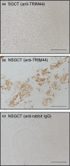
TRIM44 was strongly expressed in nonseminomatous germ cell tumor (NSGCT). (a–c) Representative images of immunohistochemistry. (a) anti‐TRIM44 in seminomatous germ cell tumor (SGCT), (b) anti‐TRIM44 in nonseminomatous germ cell tumor (NSGCT), (c) negative control (NSGCT immunostained with rabbit IgG antibody). Scale bar = 100 μm.
Figure 2
Figure 2
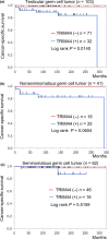
Overexpression of TRIM44 was associated with cancer‐specific survival in patients with testicular germ cell tumor (TGCT). (a) Cancer‐specific survival of patients with TGCT according to TRIM44 immunoreactivity (IR); n = 103. Patients with positive TRIM44 IR showed worse prognosis (P = 0.0140, log‐rank test). (b) There was a trend towards lower rate of cancer‐specific survival in positive TRIM44 IR in patients with NSGCT (P = 0.0604, log‐rank test). (c) There was no significant difference between positive and negative TRIM44 IR in terms of cancer‐specific survival rate in SGCT patients (P = 0.5159, log‐rank test).
Figure 3
Figure 3
Overexpression of TRIM44 promoted cell proliferation and migration in NTERA2 cells. (a) TRIM44 protein levels were analyzed in NTERA2 cells. 293T cells were transfected with TRIM44 DNA plasmid to obtain positive control for TRIM44. NTERA2 cells were transfected with pcDNA3‐FLAGTRIM44 or an empty vector. TRIM44 is overexpressed in NTERA2‐TRIM44 cells. β‐actin was used as loading control. (b) MTS assay of NTERA2‐TRIM44 cells. NTERA2‐TRIM44 cells promoted cell growth 48 h after transient transfection of TRIM44 (*P < 0.05). Results are presented as means and SD of five wells for NTERA2‐Vector and NTERA2‐TRIM44 cells. (c) Representative images of migration assay of NTERA‐Vector and NTERA2‐TRIM44 cells. Cell migration assay was performed in NTERA‐Vector and NTERA2‐TRIM44 cells. Average numbers of migrated cells were counted in five representative fields. (d) NTERA2‐TRIM44 cells had higher motility (*P < 0.05; Student's t‐test) than NETRA2‐Vector cells. Migrated cells were counted in five randomly selected fields. Data are presented as mean value ± SD. Scale bar = 200 μm.
Figure 4
Figure 4
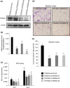
Inhibition of TRIM44 showed an anticancer effect in vitro in NTERA2 cells. Three TRIM44‐specific siRNAs (siTRIM44 #1, siTRIM44 #2, siTRIM44 #3) were used for TRIM44 knockdown. (a) TRIM44 protein levels were analyzed by Western blot analysis. NTERA2 cells treated with siControl showed expression of TRIM44 protein, whereas NTERA2‐siTRIM44 cells showed reduced expression levels of TRIM44 protein. (b) Measurement of TRIM44 mRNA levels of NTERA2‐siControl and NTERA2‐siTRIM44 cells was performed. The results are presented as the average of three wells ± SD (***P < 0.0001). (c) Cell proliferation was measured by MTS assay at the indicated time points after siTRIM44 or siControl transfection (*P < 0.05, **P < 0.005, Student's t‐test). Results are presented as means and SD of five wells for NTERA2‐siControl and each type of NTERA2‐siTRIM44 cells. (d) Representative images of cell migration assay of NTERA2‐siControl and NTERA2‐siTRIM44 cells. Scale bar = 200 μm. (e) Migrated cells of NTERA2‐siControl and NTERA2‐siTRIM44 were counted in five randomly selected fields. NTERA2‐siTRIM44 cells had significantly lower motility compared to that of NTERA2‐siControl (***P < 0.0001). Data are presented as mean value ± SD.
Figure 5
Figure 5
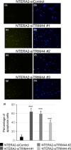
Inhibition of TRIM44 promoted apoptosis in NTERA2 cells. TUNEL (TdT‐mediated dUTP Nick‐End Labeling) assay was performed to investigate apoptosis in TRIM44 knockdown NTERA2 (NTERA2‐siTRIM44) and NTERA2‐siControl cells. Cells showing blue light (stained with DAPI) were counted as total number of cells, and cells showing green light (stained with TdT) were counted as apoptotic cells. Percentage of apoptotic cells to total number of cells were calculated in five randomly selected microfields (×100). Significantly more apoptotic cells were observed in NTERA2‐siTRIM44 cells than in NTERA2‐siControl cells (***P < 0.0001, versus control, Student's t‐test).
Figure 6
Figure 6
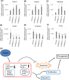
mRNA levels of oncogenic and tumor suppressive genes regulated by siTRIM44 in NTERA2 cells and a proposed model of TRIM44 action in TGCT. (a–f): qRTPCR was used for measuring mRNA levels of the six tumorigenesis‐related candidate genes that were highly regulated by siTRIM44. mRNA levels of these tumorigenesis‐related candidate genes were in line with the microarray results. P‐values are presented versus control; *P < 0.05, **P < 0.005, ***P < 0.0001, Student's t‐test. (g): Schematic figure of proposed mechanism of TRIM44 involvement in tumorigenesis. TRIM44 may promote cell proliferation, migration, and inhibit apoptosis by regulating oncogenic (C3AR1, ST3GAL5, NT5E) and tumor suppressive (CDK19, CADM1, PRKACB) genes.

References

    1. Albers P, Albrecht W, Algaba F et al European Association of Urology. Guidelines on testicular cancer. [Cited 13 June 2015.] Available from URL: http://uroweb.org/guideline/testicular-cancer.
    1. Hatakeyama S. TRIM proteins and cancer. Nat Rev 2011; 11: 792–804. - PubMed
    1. Ikeda K, Inoue S. TRIM proteins as RING finger E3 ubiquitin ligases. Adv Exp Med Biol 2012; 770: 27–37. - PubMed
    1. Liu C, Huang X, Hou S, Hu B, Li H. Silencing of tripartite motif (TRIM) 29 inhibits proliferation and invasion and increases chemosensitivity to cisplatin in human squamous cancer NCI‐H520 cells. Thorac Cancer 2015; 6(1): 31–7. - PMC - PubMed
    1. Wang Y, He D, Yang L et al TRIM26 functions as a novel tumor suppressor of hepatocellular carcinoma and its down regulation contributes to worse prognosis. Biochem Biophys Res Commun 2015; 463: 458–65. - PubMed

MeSH terms

Substances

Supplementary concepts