. 2017 Jan;23(1):47-57.
doi: 10.1261/rna.058065.116. Epub 2016 Oct 17.

Global analysis of pre-mRNA subcellular localization following splicing inhibition by spliceostatin A

Affiliations

Global analysis of pre-mRNA subcellular localization following splicing inhibition by spliceostatin A

Rei Yoshimoto et al. RNA. 2017 Jan.

Abstract

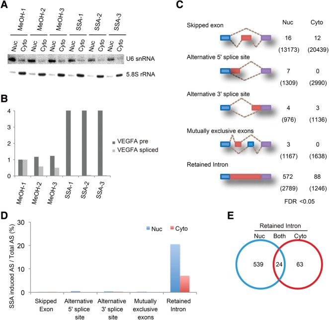
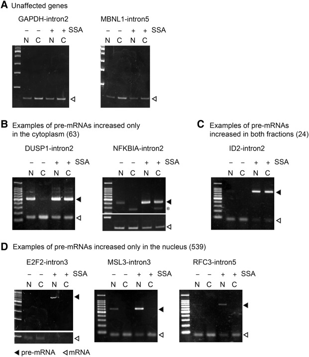
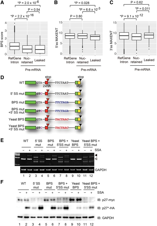
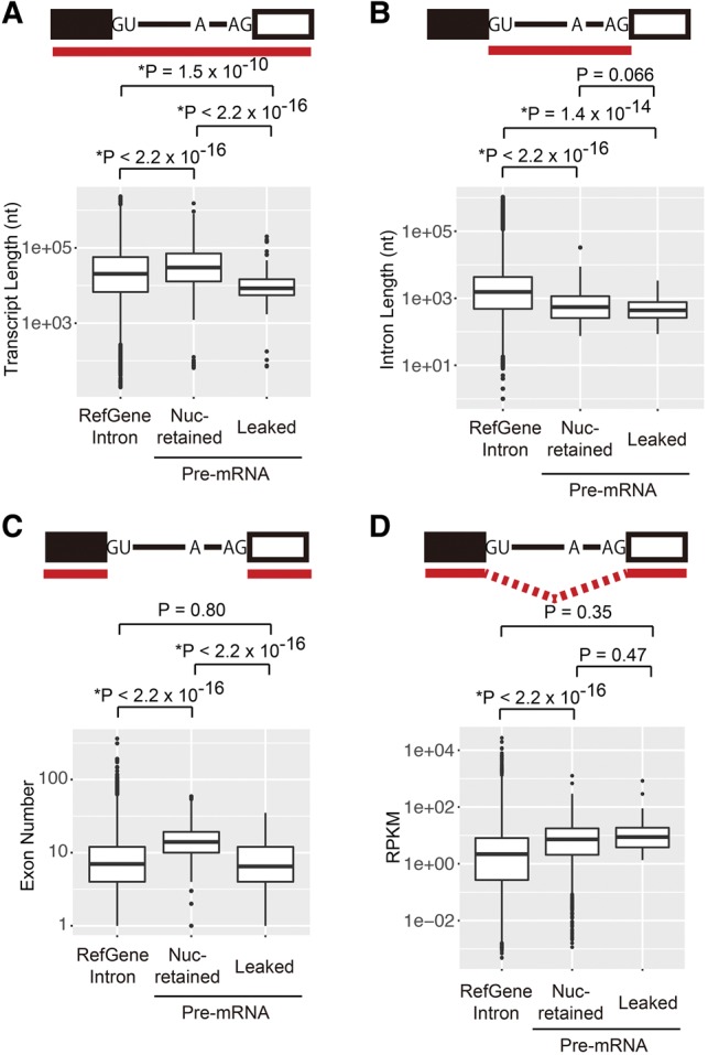
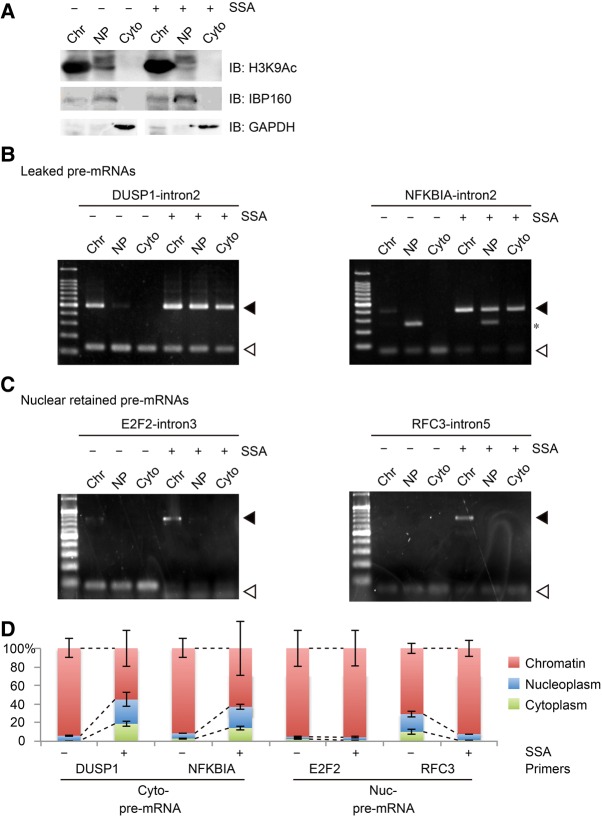
Spliceostatin A (SSA) is a methyl ketal derivative of FR901464, a potent antitumor compound isolated from a culture broth of Pseudomonas sp no. 2663. These compounds selectively bind to the essential spliceosome component SF3b, a subcomplex of the U2 snRNP, to inhibit pre-mRNA splicing. However, the mechanism of SSA's antitumor activity is unknown. It is noteworthy that SSA causes accumulation of a truncated form of the CDK inhibitor protein p27 translated from CDKN1B pre-mRNA, which is involved in SSA-induced cell-cycle arrest. However, it is still unclear whether pre-mRNAs are uniformly exported from the nucleus following SSA treatment. We performed RNA-seq analysis on nuclear and cytoplasmic fractions of SSA-treated cells. Our statistical analyses showed that intron retention is the major consequence of SSA treatment, and a small number of intron-containing pre-mRNAs leak into the cytoplasm. Using a series of reporter plasmids to investigate the roles of intronic sequences in the pre-mRNA leakage, we showed that the strength of the 5' splice site affects pre-mRNA leakage. Additionally, we found that the level of pre-mRNA leakage is related to transcript length. These results suggest that the strength of the 5' splice site and the length of the transcripts are determinants of the pre-mRNA leakage induced by SF3b inhibitors.

Keywords: RNA-seq; pre-mRNA nuclear retention; pre-mRNA splicing; spliceostatin A.


Figures

FIGURE 1.
FIGURE 1.
SSA treatment causes intron retention. (A) Northern blots performed on RNA isolated from nuclear and cytoplasmic fractions of SSA-treated cells (100 ng mL−1, 6 h). MeOH-treated cells were used as an experimental control. Probes against U6 snRNA and 5.8S RNA were used specifically to detect endogenous RNA. (B) RT-PCR analysis of VEGFA pre-mRNA from SSA-treated cells (100 ng mL−1, 6 h). RT-PCR analysis was performed using specific primers against VEGFA pre-mRNA and spliced mRNA. (C) The summary of RNA-seq analysis for alternative splicing. Five representative alternative splicing patterns are depicted. The numbers of statistically significant (FDR < 0.05) splicing pattern changes after SSA treatment are indicated on the right. The numbers of total splicing events are indicated in parenthesis. (D) Ratio of the SSA-induced change in splicing pattern to total alternative splicing events of the data in C. (E) Venn diagram shows the number of introns, the level of which was increased by SSA. The numbers of introns that accumulated only in the nuclear fraction (539 events), only in the cytoplasmic fraction (63 events), and in both (24 events) were all counted.
FIGURE 2.
FIGURE 2.
SSA causes pre-mRNA leakage in a gene-specific manner. (AD) HeLa S3 cells were treated with SSA (100 ng mL−1, 6 h) and the cells were disrupted and separated into cytoplasmic and nuclear fractions. RNA samples were purified from the fractions. RT-PCR analysis was performed using the RNA samples to detect spliced and unspliced forms of the genes, of which pre-mRNAs were not affected by SSA (A), accumulated only in the cytoplasm (B), accumulated in both fractions (C), and accumulated only in the nucleus (D). Open triangles indicate spliced mRNA, while the filled triangles indicate pre-mRNA. Asterisk indicates nonspecific bands.
FIGURE 3.
FIGURE 3.
Effects of BPS and splice sites on splicing and leakage of pre-mRNA. (AC) Boxplots of the strength of BPS (BPS scores), 5′ splice site (5′ss MAXENT), and 3′ splice site (3′ss MAXENT). All introns of human RefGenes, nuclear-retained pre-mRNAs that contain introns in the Nuc category in Figure 1E, and leaked pre-mRNAs that contain introns in the Cyto category in Figure 1E were analyzed. (D) Schematic representation of CDKN1B-derived reporters. Each box shows an exon and a line shows an intron. (E) HEK293T cells were transfected with the reporter plasmids, then treated with SSA (100 ng mL−1, 6 h). RNA samples were purified from the cells. RT-PCR analysis was performed for detecting the spliced and unspliced forms of the reporter constructs. Open triangle and filled triangle indicate spliced mRNA and pre-mRNA, respectively. (F) HEK293T cells were transfected with the reporter plasmids and then treated with SSA (100 ng mL−1, 6 h). Total cell extracts were analyzed by immunoblotting. The antibodies used are indicated to the right of the respective blots.
FIGURE 4.
FIGURE 4.
Short pre-mRNAs are prone to leak from the nucleus. (A) Boxplots of the transcript length of human RefGenes, nuclear retained pre-mRNAs, and leaked pre-mRNAs. (B) Boxplots of the intron length of human RefGenes, nuclear retained pre-mRNAs, and leaked pre-mRNAs. (C) Boxplots of the exon number of human RefGenes, nuclear retained pre-mRNAs, and leaked pre-mRNAs. (D) Boxplots of the RPKM of human RefGenes, nuclear retained pre-mRNAs, and leaked pre-mRNAs.
FIGURE 5.
FIGURE 5.
Nuclear-retained pre-mRNAs are localized in the chromatin fraction. (A) Validation of cellular fractionation into chromatin, nucleoplasm, and cytoplasmic fractions. H3K9Ac, IBP160, and GAPDH were used as markers for chromatin, nucleoplasm, and cytoplasmic fractionation, respectively. (B,C) HeLa cells were treated with SSA (100 ng mL−1, 6 h) before they were disrupted and separated into chromatin, nucleoplasmic, and cytoplasmic fractions. RNA samples were purified from the fractions. RT-PCR analysis was performed using RNA samples to detect the spliced and unspliced forms of the leaked pre-mRNA (B) and nuclear retained pre-mRNA (C). Open triangles indicate the spliced mRNA, while the filled triangles indicate the pre-mRNA. Asterisk indicates a nonspecific band. (D) The amount of pre-mRNA in the three fractions from control and SSA-treated cells was measured by quantitative RT-PCR using the same sample as B and C.

References

    1. Bentley DL. 2014. Coupling mRNA processing with transcription in time and space. Nat Rev Genet 15: 163–175. - PMC - PubMed
    1. Berglund JA, Chua K, Abovich N, Reed R, Rosbash M. 1997. The splicing factor BBP interacts specifically with the pre-mRNA branchpoint sequence UACUAAC. Cell 89: 781–787. - PubMed
    1. Brody Y, Neufeld N, Bieberstein N, Causse SZ, Bohnlein EM, Neugebauer KM, Darzacq X, Shav-Tal Y. 2011. The in vivo kinetics of RNA polymerase II elongation during co-transcriptional splicing. PLoS Biol 9: e1000573. - PMC - PubMed
    1. Corrionero A, Minana B, Valcarcel J. 2011. Reduced fidelity of branch point recognition and alternative splicing induced by the anti-tumor drug spliceostatin A. Genes Dev 25: 445–459. - PMC - PubMed
    1. Corvelo A, Hallegger M, Smith CW, Eyras E. 2010. Genome-wide association between branch point properties and alternative splicing. PLoS Comput Biol 6: e1001016. - PMC - PubMed

Publication types