. 2016 Dec;15(12):3653-3664.
doi: 10.1074/mcp.M116.063206. Epub 2016 Oct 17.

Salmonella Typhimurium Enzymatically Landscapes the Host Intestinal Epithelial Cell (IEC) Surface Glycome to Increase Invasion

Affiliations

Salmonella Typhimurium Enzymatically Landscapes the Host Intestinal Epithelial Cell (IEC) Surface Glycome to Increase Invasion

Dayoung Park et al. Mol Cell Proteomics. 2016 Dec.

Abstract

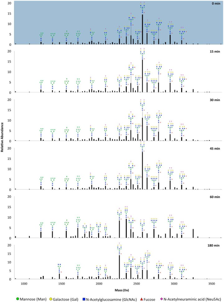
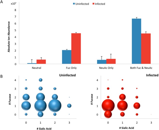
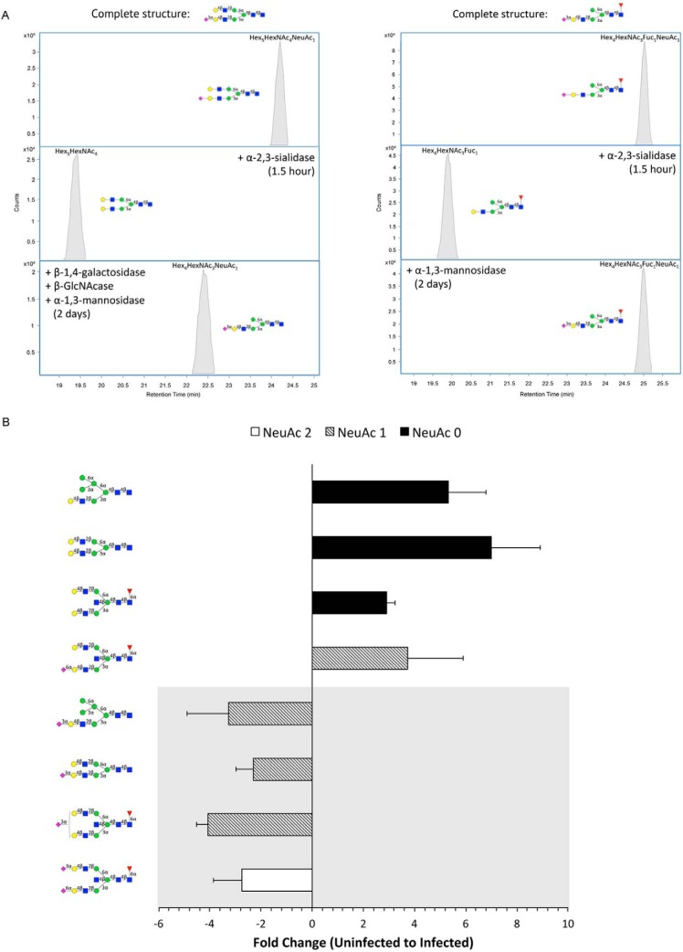
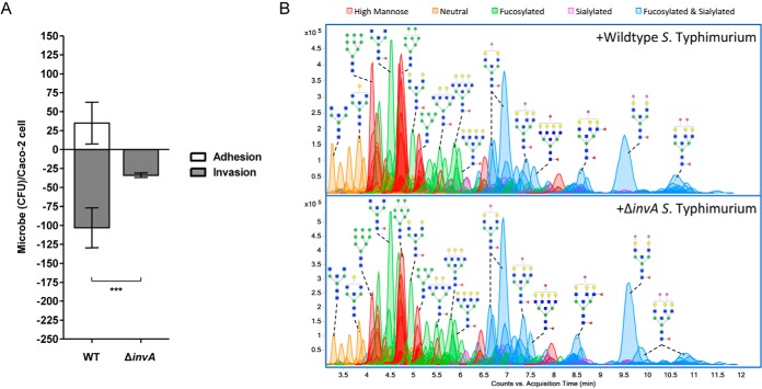
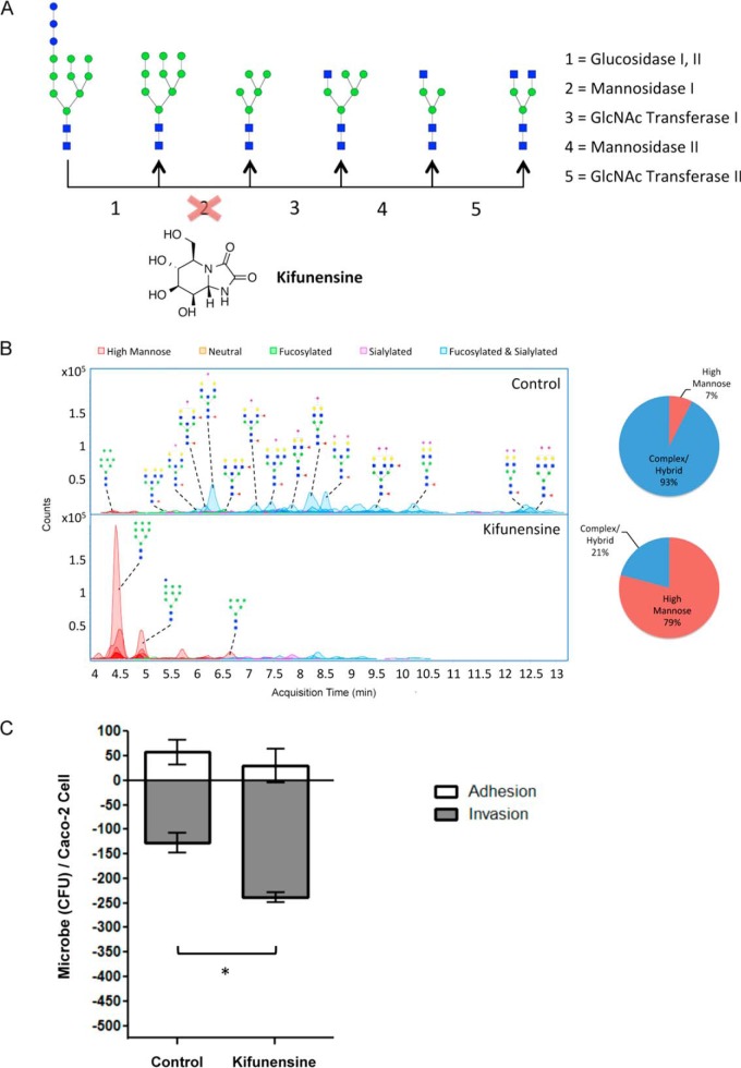
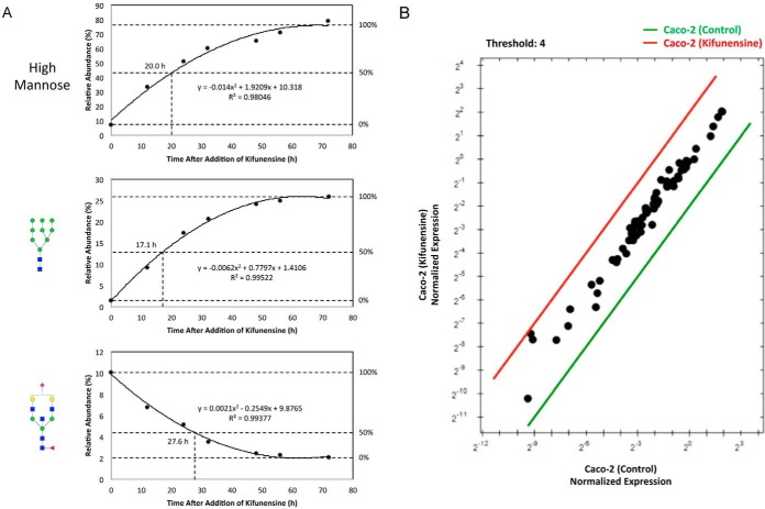
Although gut host-pathogen interactions are glycan-mediated processes, few details are known about the participating structures. Here we employ high-resolution mass spectrometric profiling to comprehensively identify and quantitatively measure the exact modifications of native intestinal epithelial cell surface N-glycans induced by S. typhimurium infection. Sixty minutes postinfection, select sialylated structures showed decreases in terms of total number and abundances. To assess the effect of cell surface mannosylation, we selectively rerouted glycan expression on the host using the alpha-mannosidase inhibitor, kifunensine, toward overexpression of high mannose. Under these conditions, internalization of S. typhimurium significantly increased, demonstrating that bacteria show preference for particular structures. Finally, we developed a novel assay to measure membrane glycoprotein turnover rates, which revealed that glycan modifications occur by bacterial enzyme activity rather than by host-derived restructuring strategies. This study is the first to provide precise structural information on how host N-glycans are altered to support S. typhimurium invasion.


Figures

Fig. 1.
Fig. 1.
Relative abundances of glycan compositions found in the glycocalyx of Caco-2 after interactions with S. typhimurium for varying time durations. Values were averaged for replicate experiments. Putative structures are drawn for abundant peaks. Symbol nomenclature is used for representing glycan structures (52).
Fig. 2.
Fig. 2.
Global compositional profiling of complex/hybrid (C/H) signals in uninfected (blue) versus infected (red) cells. A, Absolute ion abundances summed together for each type of decorated and nondecorated C/H structures. Error bars represent S.D. (n = 3). B, Distribution of C/H type glycans containing fucose and/or sialic acid. The size of the dots represents the relative abundances of the indicated groups of glycans.
Fig. 3.
Fig. 3.
Exoglycosidase digestion of sialyl glycans. A, Extracted compound chromatograms (ECCs) of HPLC fractionated glycan compounds before and after digestion with a mixture of exoglycosidases. Location and linkage determination of sialic acid is shown for Hex5HexNAc4NeuAc1 (m/z 1931.69) (left) and Hex4HexNAc3Fuc1NeuAc1 (m/z 1712.61) (right). Elucidated structures are displayed at the top of each panel. B, Fold changes of representative isomeric-specific forms of sialylated and desialylated structures of different subtypes (bisecting complex, biantennary complex, and monosialylated hybrid type glycans). Negative values indicate decreases in abundance and positive values indicate increases from uninfected to infected cell samples. Sequentially lower orders of sialylation are distinguished by color. Glycans with an α-2,3-sialyl feature are displayed in the gray panel. Symbol nomenclature is used for representing glycan structures (52).
Fig. 4.
Fig. 4.
Infection of Caco-2 with ΔinvA S. typhimurium. A, Comparison of the CFU of wildtype and mutant S. typhimurium that adhered (white bars) and invaded (gray bars) per Caco-2 cell. Error bars show S.E. between 3 biological replicates. Astericks indicate statistical significance, where ***, p < 0.001. B, Chromatograms of identified glycan compounds on Caco-2 after co-incubation with WT and ΔinvA S. typhimurium. Abundant peaks are annotated with putative structures. Symbol nomenclature is used for representing glycan structures (52).
Fig. 5.
Fig. 5.
Effects of kifunensine treatment on cell surface glycosylation and bacterial infection. A, Kifunensine inhibits the mannosidase I enzyme that participates in N-glycan biosynthesis. B, Chromatograms display each compound at its column retention time. Pie charts show the summed abundances of high mannose and complex/hybrid type glycans before and after treatment. C, S. typhimurium adhesion (white bars) and invasion (gray bars) plot of untreated versus kifunensine-treated Caco-2 cells. S.E. is represented by error bars (n = 3). Astericks indicate statistical significance, where *, p < 0.05. Symbol nomenclature is used for representing glycan structures (52).
Fig. 6.
Fig. 6.
Changes in glycan expression over time after addition of kifunensine. A, Rate lines are interpolated based on the average abundances of high mannose, Man 9, and Hex5HexNAc5Fuc1NeuAc1 following kifunensine treatment. B, Expression level comparison of total RNA extracted from untreated and kifunensine-treated cells. Data are shown as fold changes relative to reference genes. Symbol nomenclature is used for representing glycan structures (52).

References

    1. Holgersson J., Gustafsson A., and Breimer M. E. (2005) Characteristics of protein-carbohydrate interactions as a basis for developing novel carbohydrate-based antirejection therapies. Immunol. Cell Biol. 83, 694–708 - PubMed
    1. Marcobal A., Southwick A. M., Earle K. A., and Sonnenburg J. L. (2013) A refined palate: bacterial consumption of host glycans in the gut. Glycobiology 23, 1038–1046 - PMC - PubMed
    1. Karlsson K. A. (2001) Pathogen-host protein-carbohydrate interactions as the basis of important infections. Adv. Exp. Med. Biol. 491, 431–443 - PubMed
    1. Ohtsubo K., and Marth J. D. (2006) Glycosylation in cellular mechanisms of health and disease. Cell 126, 855–867 - PubMed
    1. Dube D. H., and Bertozzi C. R. (2005) Glycans in cancer and inflammation–potential for therapeutics and diagnostics. Nat. Rev. Drug Discov. 4, 477–488 - PubMed