. 2017 Aug;101(8):1793-1800.
doi: 10.1097/TP.0000000000001529.

C-C Chemokine Receptor Type 2-Dependent Migration of Myeloid-Derived Suppressor Cells in Protection of Islet Transplants

Affiliations

C-C Chemokine Receptor Type 2-Dependent Migration of Myeloid-Derived Suppressor Cells in Protection of Islet Transplants

Jie Qin et al. Transplantation. 2017 Aug.

Abstract

Background: Islet transplantation is a promising therapeutic approach to restore the physical response to blood glucose in type 1 diabetes. Current chronic use of immunosuppressive reagents for preventing islet allograft rejection is associated with severe complications. In addition, many of the immunosuppressive drugs are diabetogenic. The induction of transplant tolerance to eliminate the dependency on immunosuppression is ideal, but remains challenging.

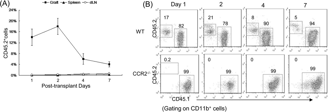
Methods: Addition of hepatic stellate cells allowed generation of myeloid-derived suppressor cells (MDSC) from precursors in mouse bone marrow. Migration of MDSC was examined in an islet allograft transplant model by tracking the systemic administered MDSC from CD45.1 congenic mice.

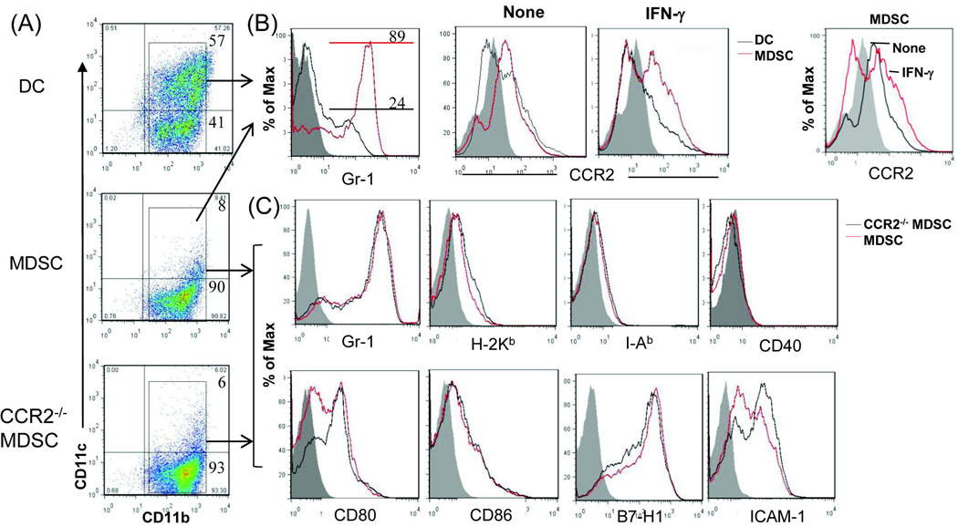
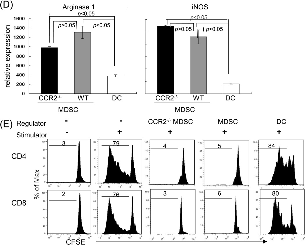
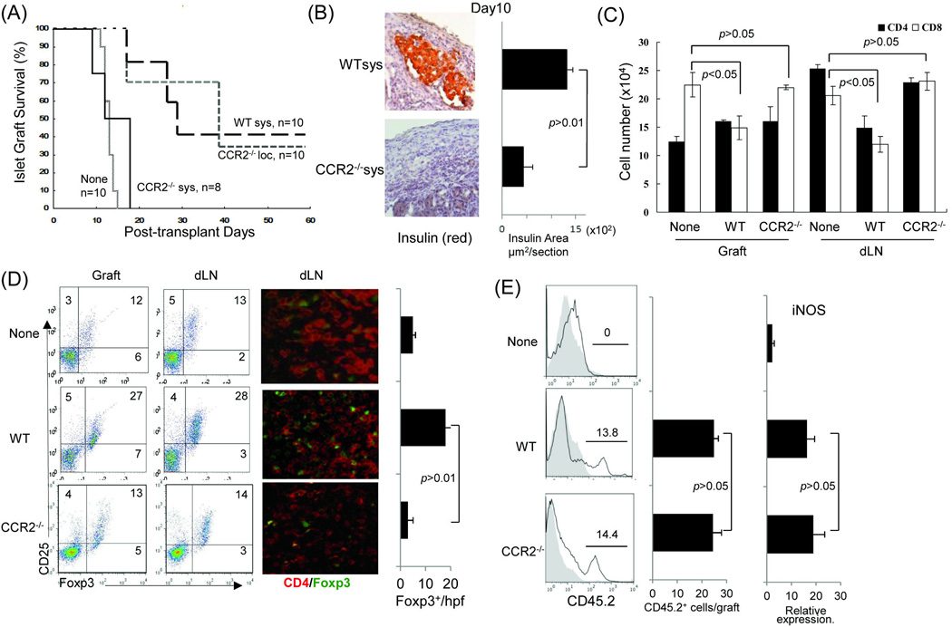
Results: The generated MDSC were expressed C-C chemokine receptor type 2 (CCR2), which was enhanced by exposure to interferon-γ. A single systemic administration of MDSC markedly prolonged survival of islet allografts without requirement of immunosuppression. Tracking the administered MDSC showed that they promptly migrated to the islet graft sites, at which point they exerted potent immune suppressive activity by inhibiting CD8 T cells, enhancing regulatory T cell activity. MDSC generated from CCR2 mice failed to be mobilized and lost tolerogenic activity in vivo, but sustained suppressive activity in vitro.

Conclusions: MDSC migration was dependent on expression of CCR2, whereas CCR2 does not directly participate in immune suppression. Expression of CCR2 needs to be closely monitored for quality control purpose when MDSC are generated in vitro for immune therapy.


Conflict of interest statement

Disclosures

The authors declare no conflict of interest.

Figures

Figure 1
Figure 1. Deficiency in CCR2 expression does not alter MDSC phenotype and immunosuppressive activity in vitro
The MDSC used in this study were generated in vitro by addition of HpSC (B6) at the beginning into the culture of BM cells that were isolated from WT or CCR2−/− B6 mice at a ratio of 1:50 in the presence of GM-CSF and IL-4 for 5 days. The floating cells were harvested. The BM cells cultured in the absence of served as controls (DC). Cells were stained for CD11b, CD11c and the indicated key surface molecules, and analyzed by flow cytometry. (A) Expression of CD11b and CD11c on DC, MDSC and CCR2−/− MDSC. The number is percentage of positive cells in whole cell populations. (B) Expression of Gr-1 and CCR2 on DC and MDSC. MDSC expressed high Gr-1 (left panel). For IFN-γ stimulation, the cells were exposed to IFN-γ (100U/ml) for last 18 hours of the cultures. Right panel shows an overlay of CCR2 expression on MDSC with or without exposure to IFN-γ. All data are displayed as histograms gated on CD11b+ populations. (C) Null of CCR2 expression in MDSC does not affect expression of the key surface molecules. Expression of the indicated molecules was analyzed on WT and CCR2−/− MDSC. The data are displayed as histograms gated on CD11b+ cells. (D) Deficiency in expression of CCR2 does not alter expression of arginase-1 and iNOS in MDSC. RNA was isolated from the magnetic beads purified CD11b+ cells. Expression of mRNA was determined by q-PCR, displayed as mean (n=3) relative expression ± SD, and analyzed by two-way t test with Bonferroni correction. (E) CCR2−/− MDSC demonstrate comparable T cell inhibitory activity in vitro. DC and MDSC generated from WT or CCR2−/− mice (B6) were used as regulators, and added into a 3-day MLR culture in which allogeneic T cells (BALB/c) were elicited by B6 DC as stimulators (T:DC = 20:1). T cell proliferation was determined by CFSE dilution assay, and expressed as histograms gated on CD4 and CD8 populations. The number is percentage of dividing CD4+ or CD8+ T cells. The data are representative of three separate experiments.
Figure 2
Figure 2. Systemically administered CCR2−/− MDSC lose ability to prolong survival of islet allografts
Immediately after transplantation of 300 islets (BALB/c) under renal capsule of B6 diabetic STZ induced) recipient, 2 × 10⁶ WT or CCR2−/− MDSC (B6) were intravenously injected (sys). For comparison purpose in a separate group, CCR2−/− MDSC were locally delivered (loc) by being mixed with islets, and then transplanted, as previously described (5). Islet transplantation alone (without MDSC treatment) served as control (None). For mechanistic studies, the recipients treated with systemic administration of WT or CCR2−/− MDSC were sacrificed on POD 10. The islet grafts and draining lymph node were harvested for sections and isolation of cells. (A) Survival of islet allografts. Systemic administration of WT MDSC or local treatment of CCR2−/− MDSC markedly prolonged survival of islet allografts (p<0.05, WT Sys or CCR2−/− Loc vs. None). Systemic administration of CCR2−/− MDSC failed to prolong islet allograft survival (p>0.05, CCR2−/− vs. none; p<0.05, CCR2−/− Sys vs. CCR2−/− Loc). (B) Islet allografts sections were stained with anti-insulin mAb (red). The pictures (left panels) show the presence of functional islets in the recipients receiving systemic administration of WT MDSC, but not in an animal (CCR2−/− MDSC group) with rejected islet grafts. Right panel shows the quantitative data for insulin areas analysis (n=10 in each group). The data were expressed as mean µm2/section ± SD. (C) Poor protection of islet allograft by systemic administration of CCR2−/− MDSC is associated with increased CD8+ T cells. Lymphocytes isolated from islet allografts and draining lymph node (dLN) from the islet allograft recipients receiving systemic administration of WT or CCR2−/− MDSC were stained with anti-CD4, -CD8 mAbs. Lymphocytes isolated from naïve animals served as the controls (None). CD4+ and CD8+ cell number was calculated based on flow analysis, and expressed as mean cell number ± SD (n=3). CD8+ cells, WT sys vs. CCDR2−/− sys, p<0.05 for two-way t test with Bonferroni correction. (D) Systemic administration of CCR2−/− MDSC is not associated with enhanced Treg cell activity. Treg cell activity was examined by flow cytometry for expression of CD25 and Foxp3 gated on CD4+ cells (left panels) or by immunohistochemistry where the cell suspensions were stained with anti-CD4 (red) and -Foxp3 (green) mAbs using fluorescent immunochemical protocol and examined by a microscope. The Foxp3+ cells were counted and expressed as mean Foxp3+ cells/high power field ± SD (n=3). p<0.05 for 2-way t test with Bonferroni correction. (E) Null of CCR2 does not affect MDSC stability in vivo. In a separate experiment, 2 × 10⁶ MDSC propagated from WT (B6) or CCR2−/− mice (both CD45.2+) were mixed with allogeneic islets (BALB/c), and transplanted under the renal capsule of congenic B6 recipients (CD45.1+) (n=3). Islet allograft transplantation alone served as controls (None). On POD7, the grafts were peeled off under a microscope for leukocyte isolation. Cells were double stained with anti-CD11b and -CD45.2, analyzed by flow cytmetry gated on CD11b+ cells, and displayed as hystograms. The number is percentage of CD45.2+ cells (left panels). The absolute number of CD45.2+ cells were calculated based on the flow analysis (middle panel). The myeloid cells were purified using CD11b+ beads for isolation of mRNA. Expression of iNOS was determined by qPCR (right panel). The data were analyzed by t test (two-tailed) with Bonferroni correction.
Figure 3
Figure 3. Migration pattern of the systemically administered MDSC
(A) MDSC preferably migrate to islet allografts. 2 × 10⁶ MDSC generated from normal B6 mice (CD45.2) mice were i.v. injected into the congenic mice (CD45.1) immediately after transplantation of BALB/c islets. Leukocytes were isolated on POD 1, 2, 4 and 7 from islet allografts, draining lymph node (dLN) and spleen, and stained for CD11b and CD45.2 for flow analysis gated on CD11b+ population. The data show mean percentage of CD45.2+ cells ± SD (n=3) (p<0.05, graft vs. spleen or dLN at all time points). (B) Migration of MDSC to islet allografts requires CCR2. In a separate study, MDSC were generated from CCR2−/− mice, instead of WT mice, and i.v. injected into the congenic mice (CD45.1) immediately after transplantation of BALB/c islets. Leukocytes were isolated on POD 1, 2, 4 and 7 from islet allografts, and stained with anti-CD11b, CD45.1 and CD45.2 mAbs for low analysis. The number is percentage of CD45.1+ or CD45.2+ cells gated on CD11b+ cell population. The data are representative of three separate experiments.

Similar articles

Cited by

References

    1. Barr CC. Retinopathy and nephropathy in patients with type 1 diabetes four years after a trial of intensive insulin therapy, by The Diabetes Control and Complications Trial/Epidemiology of Diabetes Interventions and Complications Research Group. N. Engl. J. Med. 2000;342:381–389. - PubMed
    1. Harlan DM, Kenyon NS, Korsgren O, Roep BO. Current advances and travails in islet transplantation. Diabetes. 2009;58:2175–2184. - PMC - PubMed
    1. Senior P, Kin T, Shapior J, Koh A. Islet transplantation at the university of alberta: Status update and review of progress over the last decade. Can J Diabetes. 2012;36:32–37.
    1. Pepper AR, Gala-Lopez B, Ziff O, Shapiro AJ. Current status of clinical islet transplantation. World J Transplant. 2013;3:48–53. - PMC - PubMed
    1. Gala-Lopez B, Pepper AR, Shapiro AM. Biologic agents in islet transplantation. Curr Diab Rep. 2013;13:713–722. - PubMed

Publication types

MeSH terms