. 2016 Oct 18;11(10):e0165004.
doi: 10.1371/journal.pone.0165004. eCollection 2016.

Monocytes/Macrophages Upregulate the Hyaluronidase HYAL1 and Adapt Its Subcellular Trafficking to Promote Extracellular Residency upon Differentiation into Osteoclasts

Affiliations

Monocytes/Macrophages Upregulate the Hyaluronidase HYAL1 and Adapt Its Subcellular Trafficking to Promote Extracellular Residency upon Differentiation into Osteoclasts

Emeline Puissant et al. PLoS One. .

Abstract

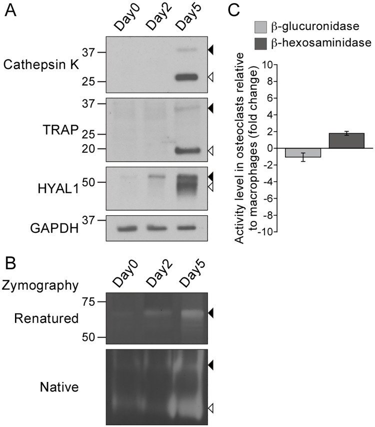
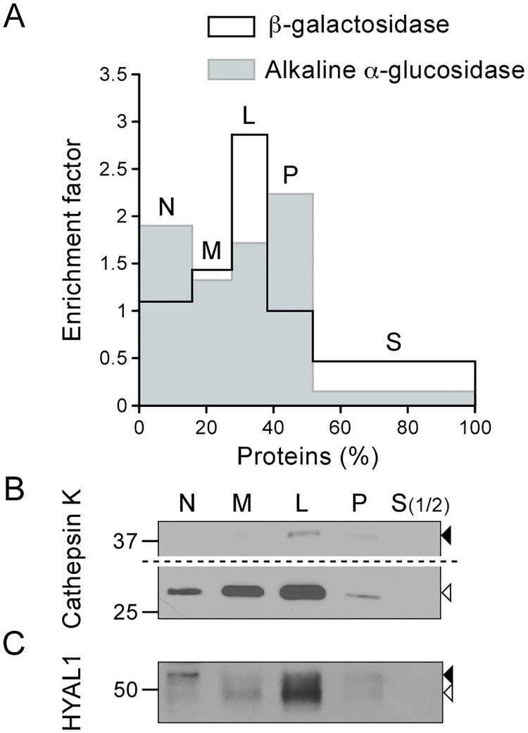
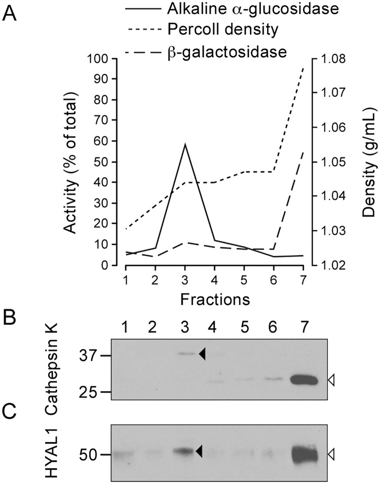
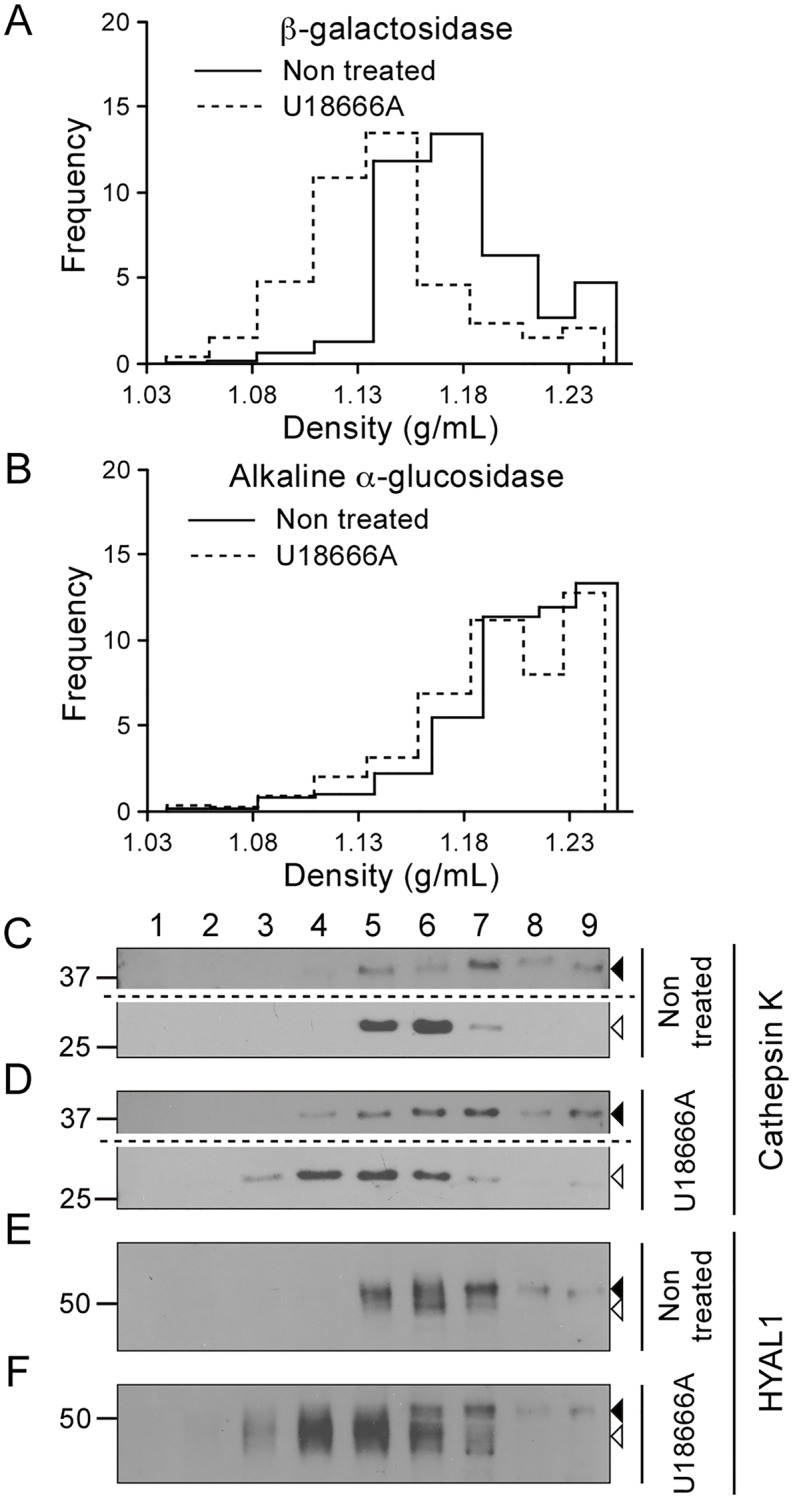
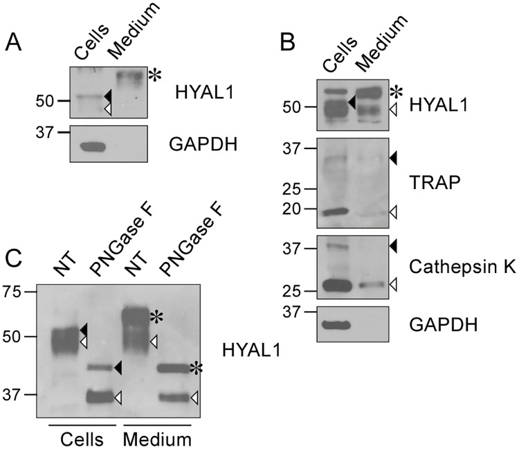
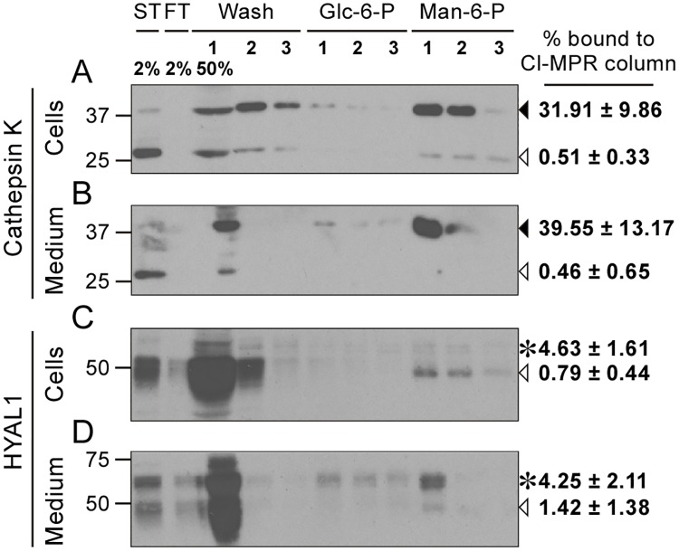
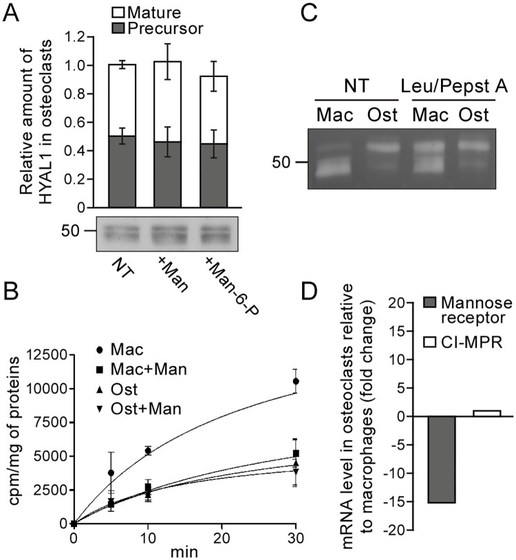
Osteoclasts are giant bone-resorbing cells originating from monocytes/macrophages. During their differentiation, they overexpress two lysosomal enzymes, cathepsin K and TRAP, which are secreted into the resorption lacuna, an acidified sealed area in contact with bone matrix where bone degradation takes place. Here we report that the acid hydrolase HYAL1, a hyaluronidase able to degrade the glycosaminoglycans hyaluronic acid (HA) and chondroitin sulfate, is also upregulated upon osteoclastogenesis. The mRNA expression and protein level of HYAL1 are markedly increased in osteoclasts differentiated from RAW264.7 mouse macrophages or primary mouse bone marrow monocytes compared to these precursor cells. As a result, the HYAL1-mediated HA hydrolysis ability of osteoclasts is strongly enhanced. Using subcellular fractionation, we demonstrate that HYAL1 proteins are sorted to the osteoclast lysosomes even though, in contrast to cathepsin K and TRAP, HYAL1 is poorly mannose 6-phosphorylated. We reported previously that macrophages secrete HYAL1 proforms by constitutive secretion, and that these are recaptured by the cell surface mannose receptor, processed in endosomes and sorted to lysosomes. Present work highlights that osteoclasts secrete HYAL1 in two ways, through lysosomal exocytosis and constitutive secretion, and that these cells promote the extracellular residency of HYAL1 through downregulation of the mannose receptor. Interestingly, the expression of the other main hyaluronidase, HYAL2, and of lysosomal exoglycosidases involved in HA degradation, does not increase similarly to HYAL1 upon osteoclastogenesis. Taken together, these findings point out the predominant involvement of HYAL1 in bone HA metabolism and perhaps bone remodeling via the resorption lacuna.


Conflict of interest statement

The authors have declared that no competing interests exist.

Figures

Fig 1
Fig 1. The protein and activity levels of HYAL1 are upregulated during osteoclastogenesis.
RAW264.7 macrophages were treated for 2 or 5 days with RANKL to induce osteoclastogenesis. (A) Detection of cathepsin K, TRAP, HYAL1 and GAPDH by western blotting (under reducing conditions). Closed and open arrowheads point to each hydrolase precursor and mature form, respectively. (B) Visualization of the HA-degrading activity of HYAL1 by renatured protein zymography (“Renatured”) and native protein zymography (“Native”) (non-reducing conditions). Precursor and mature forms are pointed as described in A. (C) Measurement of the activity of β-hexosaminidase and β-glucuronidase in RAW264.7 macrophages and in osteoclasts derived from these cells. The graph shows the fold change (mean ± SD of n = 3 independent experiments) in osteoclasts relative to the precursor cells.
Fig 2
Fig 2. The mature form of HYAL1 co-distributes with lysosomal markers after osteoclast fractionation by differential centrifugation.
Osteoclasts differentiated from RAW264.7 cells were fractionated into five fractions (N, M, L, P and S) following de Duve's fractionation scheme. (A) The activities of β-galactosidase and alkaline α-glucosidase were detected by fluorometric assay to establish the distribution of lysosomes and of the ER, respectively. The graph shows the relative specific activity (ratio of the percentage of activity of the enzyme in a given fraction to the percentage of proteins in this fraction), which is indicative of the enrichment factor of the enzyme in the fractions, plotted against the percentage of proteins in each fraction. (B-C) The distribution of cathepsin K and HYAL1 was analyzed by western blotting (reducing conditions). Equal amounts of proteins were loaded for each fraction, except for S, which was diluted 1:2 compared to the other fractions. The mature and precursor forms are highlighted by open and closed arrowheads, respectively. Of note, in panel B, a longer exposure time is shown for the upper part of the blot to help visualization of cathepsin K proforms.
Fig 3
Fig 3. HYAL1 and cathepsin K co-distribute in a self-forming Percoll density gradient.
An M+L+P pooled fraction of osteoclasts was centrifuged in a self-forming Percoll density gradient and 7 fractions were collected from top to bottom. (A) The distribution of lysosomes and of the ER was established based on the activities of β-galactosidase and alkaline α-glucosidase, respectively. The graph shows the percentage of β-galactosidase and alkaline α-glucosidase in each fraction of the Percoll gradient and the density of these fractions. (B-C) The distribution of the precursor and mature forms of cathepsin K and HYAL1 (pointed by closed and open arrowheads, respectively) was analyzed by western blotting (reducing conditions).
Fig 4
Fig 4. U18666A modifies the distribution of mature HYAL1 in a sucrose gradient, similarly to lysosomal markers.
An M+L+P fraction was prepared from control or U18666A treated osteoclasts and centrifuged in a linear sucrose density gradient extending from 1.04 g/mL to 1.26 g/mL. Nine fractions were collected from top to bottom. (A-B) The activities of the markers β-galactosidase (A) and alkaline α-glucosidase (B) were measured using fluorometric assay in the different fractions. The ordinate of the graphs corresponds to the frequency (Q/SQ.r, where Q represents the activity found in the fraction, SQ, the total activity recovered in the sum of the fractions, and r, the increment of density from top to bottom of the fraction). (C-F) Cathepsin K and HYAL1 were detected by western blotting (reducing conditions) in the fractions collected after centrifugation. The precursor and mature forms of these proteins are shown by closed and open arrowheads, respectively. Of note, a longer exposure time is shown for the upper part of the blots shown in panels C and D, to visualize cathepsin K proforms.
Fig 5
Fig 5. Osteoclasts secrete HYAL1 through both the constitutive secretory pathway and lysosomal exocytosis.
(A) Detection of HYAL1 by western blotting, under reducing conditions, in RAW264.7 cell lysate and medium samples collected after a 5 h incubation in serum-free conditions. The cytosolic protein GAPDH was used as a control of the cell integrity. The medium was concentrated 4-fold compared to the cell sample. The closed arrowhead, open arrowhead and asterisk mark the 52 kDa intracellular precursor form, 48 kDa intracellular mature form and 65 kDa secreted form of HYAL1, respectively. (B) Detection of cathepsin K, TRAP, GAPDH and HYAL1 by western blotting in RAW264.7-derived osteoclast cell lysate and concentrated medium (4 x). The precursor and mature forms of TRAP and cathepsin K are highlighted by closed and open arrowheads, respectively. The different forms of HYAL1 are marked as described in A. (C) Aliquots of osteoclast cell lysate and medium (concentrated 4-fold) were treated with PNGase F prior to the detection of HYAL1 by western blotting. The different forms of HYAL1 are pointed as described in A. Note that the extracellular precursor form (asterisk) exhibited the same MM as the intracellular precursor (closed arrowhead) after deglycosylation, and that osteoclasts secreted mature forms of HYAL1 (detected at ~35 kDa after deglycosylation, open arrowhead).
Fig 6
Fig 6. HYAL1 is poorly mannose 6-phosphorylated in osteoclasts.
Cell lysate (A, C) or culture medium (B, D) of osteoclasts differentiated from RAW264.7 cells were collected after a 5 h culture period in serum-free conditions and loaded on a CI-MPR affinity column. The presence of cathepsin K (A-B) and HYAL1 (C-D) was analyzed, using western blotting (reducing conditions), in the starting sample (ST), flow-through (FT), washes (Wash) and elution fractions (trichloroacetic acid-precipitated). Proteins non specifically bound to the column were eluted using 5 mM of Glucose 6-Phosphate (Glc-6-P) while proteins that bound specifically to CI-MPRs were eluted with 5 mM of mannose 6-phosphate (Man-6-P). Cathepsin K precursor and cleaved forms are indicated by closed and open arrowheads, respectively. The 65 kDa precursor and 48 kDa mature forms of HYAL1 are indicated by asterisks and open arrowheads, respectively. One representative experiment is shown. Specific binding of each cathepsin K or HYAL1 forms to the column was calculated in n = 3 independent experiments and expressed as a percentage of the total signal detected for the corresponding form in the starting sample (mean ± SD of the 3 independent experiments are shown).
Fig 7
Fig 7. Osteoclasts downregulate the mannose receptor-dependent recapture mechanism that targets HYAL1 to lysosomes in macrophages.
(A) Osteoclasts derived from RAW264.7 cells were treated for 24 h with 15 mg/mL of mannan (Man) or 5 mM of Man-6-P, and the intracellular amount of HYAL1 was subsequently visualized using western blotting. The graph shows the quantifications of 52 kDa precursor (grey squares) and 48 kDa mature (white squares) HYAL1 forms in n = 3 independent experiments (mean ± SD). (B) Osteoclasts (Ost) and their RAW264.7 precursor macrophages (Mac) were incubated for 5, 10 or 30 min with iodinated rhHYAL1. When indicated, 10 mg/mL of mannan (Man) were added on the cells 5 min prior to the assay and 20 mg/mL during the pulse period. After washing, the proteins bound to the surface were stripped prior to cell lysis and counting of the internalized radioactivity. Three independent experiments were quantified (mean ± SD). (C) After a 2 h incubation at 37°C with rhHYAL1 (± leupeptin [Leu]/pepstatin A [Pepst A]) followed by a 15 min chase, the amount of rhHYAL1 endocytosed by macrophages (Mac) and osteoclasts (Ost) was detected using renatured protein zymography. Of note, only low amounts of proteins are loaded on the gel in these assays; therefore, the endogenous protein is below detection level. One representative experiment is shown (total independent experiments n = 3). (D) The mRNA expression levels of the mannose receptor and CI-MPR were measured by qPCR in macrophages and osteoclasts. The ΔCT values obtained in 3 independent experiments were averaged and used to calculate fold changes in osteoclasts relative to precursor macrophages (2-ΔΔCT).

Similar articles

Cited by

References

    1. Väänänen HK, Zhao H, Mulari M, Halleen JM. The cell biology of osteoclast function. J Cell Sci. 2000;113: 377–381. - PubMed
    1. Teitelbaum SL. Bone resorption by osteoclasts. Science. 2000;289: 1504–1508. 10.1126/science.289.5484.1504 - DOI - PubMed
    1. DeSelm CJ, Miller BC, Zou W, Beatty WL, van Meel E, Takahata Y, et al. Autophagy proteins regulate the secretory component of osteoclastic bone resorption. Dev Cell. 2011;21: 966–974. 10.1016/j.devcel.2011.08.016 - DOI - PMC - PubMed
    1. Zhao H, Ito Y, Chappel J, Andrews NW, Teitelbaum SL, Ross FP. Synaptotagmin VII regulates bone remodeling by modulating osteoclast and osteoblast secretion. Dev Cell. 2008;14: 914–925. 10.1016/j.devcel.2008.03.022 - DOI - PMC - PubMed
    1. Väänänen HK, Karhukorpi EK, Sundquist K, Wallmark B, Roininen I, Hentunen T, et al. Evidence for the presence of a proton pump of the vacuolar H(+)-ATPase type in the ruffled borders of osteoclasts. J Cell Biol. 1990;111: 1305–1311. 10.1083/jcb.111.3.1305 - DOI - PMC - PubMed

MeSH terms

LinkOut - more resources