. 2016 Oct 19;9(1):64.
doi: 10.1186/s12920-016-0226-1.

Pitfalls of improperly procured adjacent non-neoplastic tissue for somatic mutation analysis using next-generation sequencing

Affiliations

Pitfalls of improperly procured adjacent non-neoplastic tissue for somatic mutation analysis using next-generation sequencing

Lei Wei et al. BMC Med Genomics. .

Abstract

Background: The rapid adoption of next-generation sequencing provides an efficient system for detecting somatic alterations in neoplasms. The detection of such alterations requires a matched non-neoplastic sample for adequate filtering of non-somatic events such as germline polymorphisms. Non-neoplastic tissue adjacent to the excised neoplasm is often used for this purpose as it is simultaneously collected and generally contains the same tissue type as the neoplasm. Following NGS analysis, we and others have frequently observed low-level somatic mutations in these non-neoplastic tissues, which may impose additional challenges to somatic mutation detection as it complicates germline variant filtering.

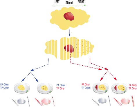
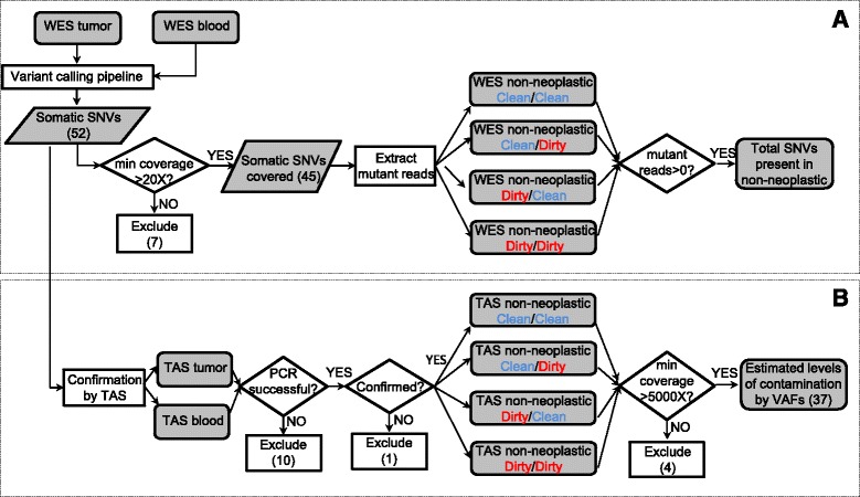
Methods: We hypothesized that the low-level somatic mutation observed in non-neoplastic tissues may be entirely or partially caused by inadvertent contamination by neoplastic cells during the surgical pathology gross assessment or tissue procurement process. To test this hypothesis, we applied a systematic protocol designed to collect multiple grossly non-neoplastic tissues using different methods surrounding each single neoplasm. The procedure was applied in two breast cancer lumpectomy specimens. In each case, all samples were first sequenced by whole-exome sequencing to identify somatic mutations in the neoplasm and determine their presence in the adjacent non-neoplastic tissues. We then generated ultra-deep coverage using targeted sequencing to assess the levels of contamination in non-neoplastic tissue samples collected under different conditions.

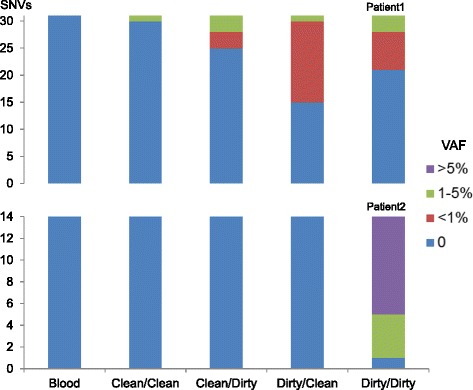
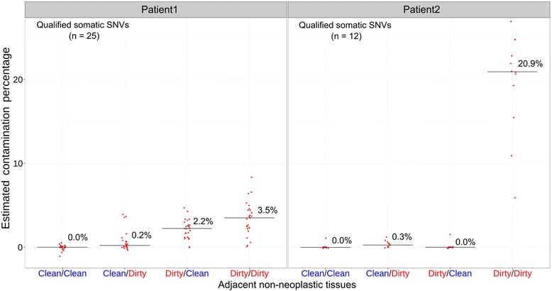
Results: Contamination levels in non-neoplastic tissues ranged up to 3.5 and 20.9 % respectively in the two cases tested, with consistent pattern correlated with the manner of grossing and procurement. By carefully controlling the conditions of various steps during this process, we were able to eliminate any detectable contamination in both patients.

Conclusion: The results demonstrated that the process of tissue procurement contributes to the level of contamination in non-neoplastic tissue, and contamination can be reduced to below detectable levels by using a carefully designed collection method. A standard protocol dedicated for acquiring adjacent non-neoplastic tissue that minimizes neoplasm contamination should be implemented for all somatic mutation detection studies.

Keywords: Adjacent normal tissues; Somatic mutations; Tumor contamination.


Figures

Fig. 1
Fig. 1
Design of the special collection of non-neoplastic breast tissue. After orienting, the lumpectomies were bread-loafed from left to right using one blade for the entire specimen. Non-neoplastic tissue sectioned before the neoplasm was designated “PA Clean”. Non-neoplastic tissue sectioned after the neoplasm was designated “PA Dirty”. The forceps, scalpel, and Petri dish used to cut the neoplasm were referred to as “dirty” tools (red). The forceps, scalpel, and Petri dish that had no contact with the neoplasm were referred to as “clean” tools (blue). During the tissue procurement process, samples collected with “clean” tools and “dirty” tools were designated “TP Clean” and “TP Dirty”, respectively. Except for a section of neoplasm, four samples were collected from each lumpectomy: “PA Clean” and “TP Clean” (Clean/Clean), “PA Clean” and “TP Dirty” (Clean/Dirty), “PA Dirty” and “TP Clean” (Dirty/Clean), “PA Dirty” and “TP Dirty” (Dirty/Dirty). Grossly non-neoplastic tissue fragments that had contact with neoplastic tissue have specs of red which are reflective of theoretical contamination
Fig. 2
Fig. 2
Overall flow chart of the two-stage analysis strategy. a, de-novo mutation calling and initial assessment of neoplasm contamination by using WES; b, targeted validation and ultra-deep sequencing to assess neoplasm contamination level in each adjacent non-neoplastic tissue. WES: whole exome sequencing; TAS: targeted amplicon sequencing; SNV: single nucleotide variant; VAF: variant allele fraction
Fig. 3
Fig. 3
Presence of somatic SNVs in adjacent non-neoplastic tissues as the first indication of contamination. Distribution of somatic SNVs’ allele fractions (VAF) in WES data of the blood and four types of adjacent non-neoplastic tissues, classified by four categories: zero (no mutant reads detected), less than one percent, one to five percent, and greater than five percent. Y axis values represent the numbers of SNVs in each category
Fig. 4
Fig. 4
The estimated contamination levels in adjacent non-neoplastic tissues by targeted amplicon sequencing. Each red dot represents a previously identified somatic SNV. The contamination level was estimated by: (VAF_in_non-neoplastic_tissue - VAF_in_blood)/VAF_in_neoplasm. The median contamination levels for each non-neoplastic sample are plotted as a horizontal bar with the percentage displayed in numeric value

References

    1. Stratton MR. Exploring the genomes of cancer cells: progress and promise. Science. 2011;331(6024):1553–8. doi: 10.1126/science.1204040. - DOI - PubMed
    1. Jones S, Anagnostou V, Lytle K, Parpart-Li S, Nesselbush M, Riley DR, Shukla M, Chesnick B, Kadan M, Papp E, et al. Personalized genomic analyses for cancer mutation discovery and interpretation. Sci Transl Med. 2015;7(283):283ra253. doi: 10.1126/scitranslmed.aaa7161. - DOI - PMC - PubMed
    1. Ding L, Getz G, Wheeler DA, Mardis ER, McLellan MD, Cibulskis K, Sougnez C, Greulich H, Muzny DM, Morgan MB, et al. Somatic mutations affect key pathways in lung adenocarcinoma. Nature. 2008;455(7216):1069–75. doi: 10.1038/nature07423. - DOI - PMC - PubMed
    1. Cancer Genome Atlas Research N Comprehensive genomic characterization defines human glioblastoma genes and core pathways. Nature. 2008;455(7216):1061–8. doi: 10.1038/nature07385. - DOI - PMC - PubMed
    1. Network TCGAR Corrigendum: Comprehensive genomic characterization defines human glioblastoma genes and core pathways. Nature. 2013;494(7438):506. doi: 10.1038/nature11903. - DOI - PubMed

Publication types