. 2016 Oct 18;17(1):433.
doi: 10.1186/s12891-016-1294-2.

Pre-surgical CT-assessment of neurogenic myositis ossificans of the hip and risk factors of recurrence: a series of 101 consecutive patients

Affiliations

Pre-surgical CT-assessment of neurogenic myositis ossificans of the hip and risk factors of recurrence: a series of 101 consecutive patients

Bruno Law-Ye et al. BMC Musculoskelet Disord. .

Abstract

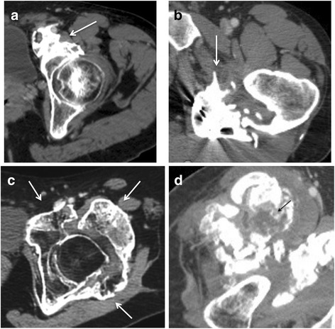
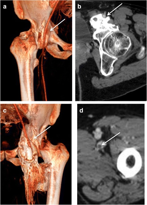
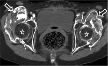
Background: Neurogenic Myositis Ossificans (NMO) is a rare disabling pathology characterized by peri-articular heterotopic ossifications following severe peripheral or central nervous system injuries. It results in ankylosis and vessels or nerves compressions. Our study aimed to describe the pre-operative findings of patients with NMO of the hip using biphasic computerized tomography (CT).

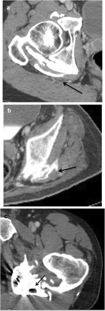
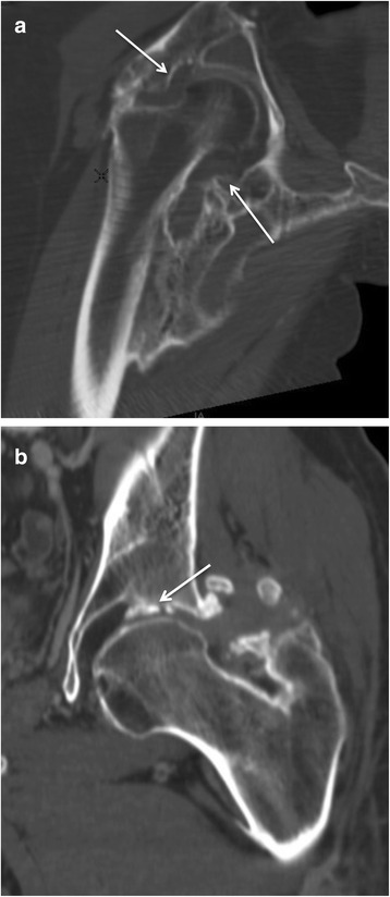
Methods: Between 2006 and 2012, we retrospectively analyzed 101 consecutive patients with hip NMO. We analyzed all CTs and surgical reports following a standardized grid depicting the osteoma and its relations with joint capsule, vessels and nerves and bone mineralization. We studied surgical complications and recurrence during follow-up. Chi2-test and Fischer's test were performed to compare qualitative values with respectively normal and non-normal distribution. Quantitative values were analyzed with a one factor analysis of variance (ANOVA) test. Agreement between pre-surgical CT and surgical observations was evaluated with Cohen's kappa test.

Results: Correlation between pre-operative CT and surgical findings was excellent regarding relationships with vessels (0,82) and was good concerning relationships with sciatic nerves (0.62) and with joint capsule (0.68). Close contact or disruption of joint capsule (p = 0.005), joint space narrowing (p = 0.007) and bone demineralization (p < 0.001) were correlated with NMO recurrence.

Conclusions: Biphasic enhanced-CT allows pre-operative assessment of NMO with good correlation to surgical observations and helps prevent surgical complications.

Keywords: Brain trauma; Neurogenic myositis ossificans; Osteoma; Paraplegia; Spinal cord injury.


Figures

Fig. 1
Fig. 1
Axial CT images in four different patients. a anterior osteoma (arrow). b posterior osteoma (arrow). c circumferential osteoma. d anterior osteoma in a 31 year-old male patient. Central hypodensity with thin peripheral enhancement corresponding to an immature portion (black arrow)
Fig. 2
Fig. 2
Enhanced-CT in volume rendering reconstruction (a and c) and axial images (b and d) illustrating the different types of relationships between osteomas and femoral arteries. a displacement of right femoral artery with a moderate compression (arrow). b The osteoma forms a groove surrounding the artery partially, less than 180°(arrow). c The osteoma forms a complete tunnel around the artery (30 year-old male patient) (arrow). d Endoluminal defect inside the femoral vein indicating thrombosis (arrow)
Fig. 3
Fig. 3
Axial images of a biphasic enhanced CT in three different patients with posterior osteomas with various types of relationships with sciatic nerves. a slight contact and compression of the right sciatic nerve by the osteoma (arrow). b the osteoma forms a groove around the sciatic nerve, surrounding it less than 180° (arrow). c the osteoma forms a complete bony tunnel around the sciatic nerve (19 year-old male patient) (arrow)
Fig. 4
Fig. 4
Coronal reconstruction of CT scan: neurogenic osteoarthropathy of the right hip (a) and left hip (b, other patient). While the osteoma only touches the joint capsule on image A (arrows), we can observe a disruption of joint capsule with an involvement of joint space (27 year-old male patient) (arrow)
Fig. 5
Fig. 5
Large bilateral osteomas of the hips on axial image (hollow arrows). Advanced demineralization classified M4 (evanescent bone) (stars) and collapsed joint space (arrows)

Similar articles

Cited by

References

    1. Carlier RY, Safa DML, Parva P, Mompoint D, Judet T, Denormandie P, et al. Ankylosing neurogenic myositis ossificans of the hip. An enhanced volumetric CT study. J Bone Joint Surg Br. 2005;87:301–5. doi: 10.1302/0301-620X.87B3.14737. - DOI - PubMed
    1. Pape HC, Marsh S, Morley JR, Krettek C, Giannoudis PV. Current concepts in the development of heterotopic ossification. J Bone Joint Surg Br. 2004;86:783–7. doi: 10.1302/0301-620X.86B6.15356. - DOI - PubMed
    1. Cassar-Pullicino VN, McClelland M, Badwan DA, McCall IW, Pringle RG, el Masry W. Sonographic diagnosis of heterotopic bone formation in spinal injury patients. Paraplegia. 1993;31:40–50. doi: 10.1038/sc.1993.7. - DOI - PubMed
    1. Singer BR. Heterotopic ossification. Br J Hosp Med. 1993;49:247–51. - PubMed
    1. Ebinger T, Roesch M, Kiefer H, Kinzl L, Schulte M. Influence of etiology in heterotopic bone formation of the hip. J Trauma. 2000;48:1058–62. doi: 10.1097/00005373-200006000-00010. - DOI - PubMed

MeSH terms

LinkOut - more resources