. 2016 Oct 19;35(1):164.
doi: 10.1186/s13046-016-0441-9.

RARγ-induced E-cadherin downregulation promotes hepatocellular carcinoma invasion and metastasis

Affiliations

RARγ-induced E-cadherin downregulation promotes hepatocellular carcinoma invasion and metastasis

Wen-Juan Gan et al. J Exp Clin Cancer Res. .

Abstract

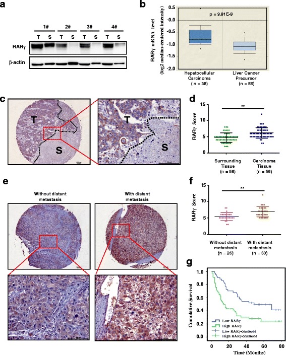
Background: Aberrant expression of Retinoic acid receptor γ (RARγ) is implicated in cancer development. Our previous study identified that RARγ functions as a tumor promoter to drive hepatocellular carcinoma (HCC) growth. However, its contribution to HCC invasion and metastasis remains unclear.

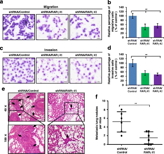
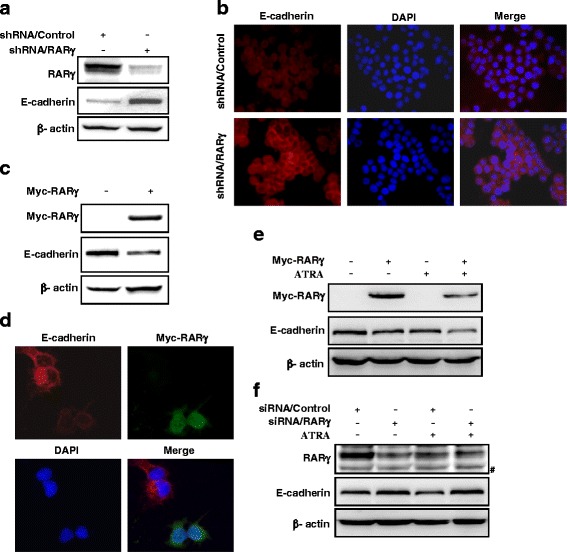
Methods: RARγ expression in clinical HCC samples was detected by western blot and immunohistochemistry. The relationship between RARγ expression levels and the clinical characteristics were evaluated. HCC cell line MHCC-97H were stably knocked down RARγ using a lentivirus vector-based shRNA technique. The cells were analyzed by migration and invasion assays, and injected into nude mice to assess tumor metastasis. E-cadherin expression regulated by RARγ was examined by qPCR, western blot and immunofluorescence staining.

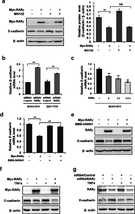
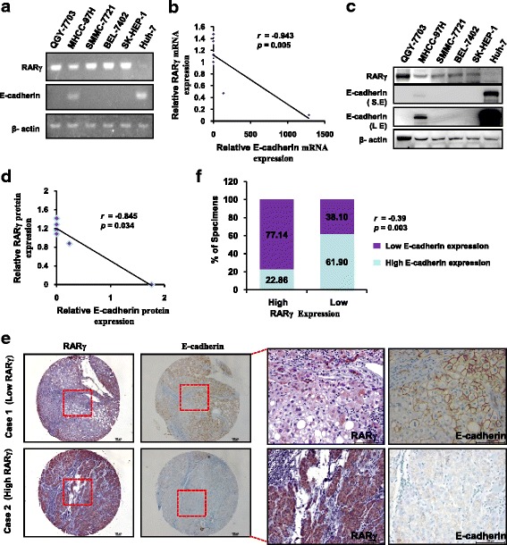
Results: The expression of RARγ is significantly upregulated in human HCC tissues. Moreover, its expression positively correlates with tumor size, distant metastasis and TNM stage, and negatively correlates with length of survival of HCC patients. Knockdown of RARγ markedly inhibits HCC cell invasion and metastasis both in vitro and in vivo. Mechanistic investigations reveal that RARγ functions through regulation of NF-κB-mediated E-cadherin downregulation to promote HCC invasion and metastasis. Notably, RARγ expression status negatively correlates with E-cadherin expression in HCC cell lines and clinical HCC samples.

Conclusions: These findings demonstrate that RARγ could promote HCC invasion and metastasis by regulating E-cadherin reduction, and implicate new strategies to aggressively treat HCC through targeting RARγ/E-cadherin signaling axis.

Keywords: E-cadherin; Hepatocellular carcinoma; Metastasis; RARγ.


Figures

Fig. 1
Fig. 1
Increased levels of RARγ in HCC correlates with distant metastasis and predicts poor clinical outcome. a RARγ expression in HCC samples was evaluated by western blotting. Four randomly selected pairs of HCC tumors (T) and matched surrounding tissues (S) are presented. b Box plot shows the mRNA levels of RARγ expression in 38 human HCC and 58 human liver cancer precursor tissues. Statistical significance was determined by a two-tailed, unpaired Student's t-test. c Representative bright-field images showing RARγ staining (brown) in human HCC sections. Nuclei (blue) were marked by hematoxylin staining. Scale bar: 100 μm. T, tumor. S, surrounding tissue. d Scatter plot analysis of RARγ levels in 56 HCC tissue samples and their surrounding tissues. Statistical significance was determined by a two-tailed, paired Student's t-test. **p < 0.01. e Representative bright-field images showing RARγ staining (brown) in human HCC tissues with distant metastasis (N = 30) and without distant metastasis (N = 26). Nuclei (blue) were marked by hematoxylin staining. Scale bar: 100 μm. f Scatter plot analysis of RARγ levels in human HCC tissues with distant metastasis (N = 30) and without distant metastasis (N = 26). Statistical significance was determined by a two-tailed, unpaired Student's t-test. **p < 0.01. g Kaplan-Meier survival curve of HCC patients with low (n = 44) and high (n = 46) RARγ expression
Fig. 2
Fig. 2
Silencing RARγ impaires HCC migration and invasion in vitro and metastasis in vivo. ad Migration (a) and invasion (c) assays were performed in wild-type MHCC-97H cells (shRNA/Control) and in the MHCC-97H cells with stable knock down of RARγ (shRNA/ RARγ), and the relative number of migratory (b) and invasive (d) cells were calculated with Wright-Giemsa staining. e Knockdown of RARγ inhibits HCC metastasis. Representative lung tissue sections from each group were shown by hematoxylin and eosin staining (magnification: × 40). Black arrows indicate lung tissues with matastatic nodules. f The number of lung metastatic foci in each group (n = 6 per group) was counted under the microscope. Statistical significance was determined by a two-tailed, unpaired Student's t-test. **p < 0.01
Fig. 3
Fig. 3
RARγ regulates E-cadherin expression. a, b silencing RARγ increases endogenous levels of E-cadherin proteins. MHCC-97H cells stably expressed shRNA/Control or shRNA/RARγ, then (a) total cell lysates were subjected to immunoblotting to determine E-cadherin levels, or (b) the cells were subjected to immunofluorescent staining of E-cadherin (red). Nuclei was stained with DAPI (blue). Representative images are shown. c, d RARγ overexpression decrease endogenous E-cadherin protein levels. Huh-7 cells were transiently transfected with vector or Myc-tagged RARγ, then endogeous protein levels of E-cadherin were detected by immunoblotting (c) or immunofluorescent staining (d). e, f The role of ATRA in RARγ-induced E-cadherin downregulation. Immunoblotting of E-cadherin in RARγ-transfected Huh-7 cells (e) or in RARγ-silenced MHCC-97H cells (f) treated with vehicle or 1 μM ATRA. #, no specific band
Fig. 4
Fig. 4
NF-κB is indispensable for RARγ-driven E-cadherin reduction. a RARγ-driven E-cadherin reduction does not depend on proteasome pathway. Immunoblotting (left) or qPCR (right) analysis of the E-cadherin expression in RARγ-transfected Huh-7 cells treated with vehicle or 10 μM MG132. b, c RARγ regulates E-cadherin at transcriptional level. qPCR analysis of the E-cadherin expression in RARγ siRNA-transduced MHCC-97H and QGY-7703 cells (b) or RARγ-transfected MHCC-97H cells (c). d, e BMS-345541 inhibits RARγ-driven E-cadherin reduction. qPCR (d) or immunoblotting (e) analysis of the E-cadherin expression in RARγ-transfected Huh-7 cells treated with vehicle or 10 μM BMS-345541. f, g TNFα promotes RARγ-driven E-cadherin reduction. Immunoblotting analysis of the levels of E-cadherin expression in RARγ-transfected Huh-7 cells (f) or RARγ siRNA-transduced MHCC-97H cells (g) treated with vehicle or 20 nM TNFα. Statistical significance was determined by a two-tailed, unpaired Student's t-test. **p < 0.01. ns, no significance
Fig. 5
Fig. 5
The expression levels of RARγ and E-cadherin in HCC cell lines and clinical HCC tissues. a The expression of RARγ and E-cadherin were evaluated by RT-PCR in the indicated cell lines. b Dot plot correlates the mRNA levels of RARγ and E-cadherin in HCC cell lines. The dotted line shows the negative correlation of RARγ and E-cadherin at the mRNA levels. c Immunoblotting analysis of RARγ and E-cadherin expression in the indicated cell lines. d The dot plot correlates RARγ and E-cadherin protein levels in six HCC cell lines. The dotted line shows the negative corralation of RARγ and E-cadherin at the protein levels. e Immunohistochemical staining of RARγ and E-cadherin in human CRC tissues. Representative bright-field images showing RARγ and E-cadherin staining in human HCC sections. Scale bar: 100 μm. f Spearman’s correlation analysis between RARγ and E-cadherin in 56 cases of HCC tissues

Similar articles

Cited by

References

    1. He J, Gu D, Wu X, Reynolds K, Duan X, Yao C, Wang J, Chen CS, Chen J, Wildman RP, et al. Major causes of death among men and women in China. N Engl J Med. 2005;353(11):1124–1134. doi: 10.1056/NEJMsa050467. - DOI - PubMed
    1. Parkin DM, Bray FI, Devesa SS. Cancer burden in the year 2000. The global picture. Eur J Cancer. 2001;37(Suppl 8):S4–66. doi: 10.1016/S0959-8049(01)00267-2. - DOI - PubMed
    1. Yeo W, Mok TS, Zee B, Leung TW, Lai PB, Lau WY, Koh J, Mo FK, Yu SC, Chan AT, et al. A randomized phase III study of doxorubicin versus cisplatin/interferon alpha-2b/doxorubicin/fluorouracil (PIAF) combination chemotherapy for unresectable hepatocellular carcinoma. J Natl Cancer Inst. 2005;97(20):1532–1538. doi: 10.1093/jnci/dji315. - DOI - PubMed
    1. Portolani N, Coniglio A, Ghidoni S, Giovanelli M, Benetti A, Tiberio GA, Giulini SM. Early and late recurrence after liver resection for hepatocellular carcinoma: prognostic and therapeutic implications. Ann Surg. 2006;243(2):229–235. doi: 10.1097/01.sla.0000197706.21803.a1. - DOI - PMC - PubMed
    1. Du ZG, Wei YG, Chen KF, Li B. Risk factors associated with early and late recurrence after curative resection of hepatocellular carcinoma: a single institution's experience with 398 consecutive patients. Hepatobiliary Pancreat Dis Int. 2014;13(2):153–161. doi: 10.1016/S1499-3872(14)60025-4. - DOI - PubMed

MeSH terms