. 2016 Nov:100:72-82.
doi: 10.1016/j.yjmcc.2016.10.006. Epub 2016 Oct 15.

Growth and maturation of heart valves leads to changes in endothelial cell distribution, impaired function, decreased metabolism and reduced cell proliferation

Affiliations

Growth and maturation of heart valves leads to changes in endothelial cell distribution, impaired function, decreased metabolism and reduced cell proliferation

Lindsey J Anstine et al. J Mol Cell Cardiol. 2016 Nov.

Abstract

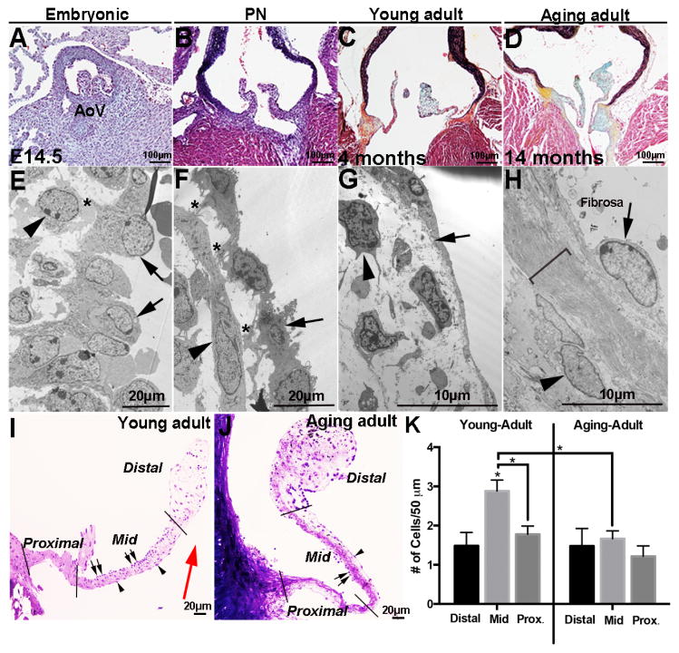
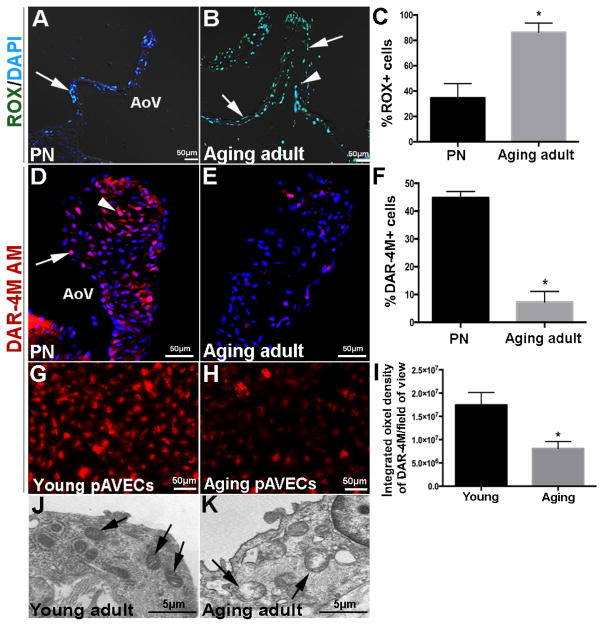
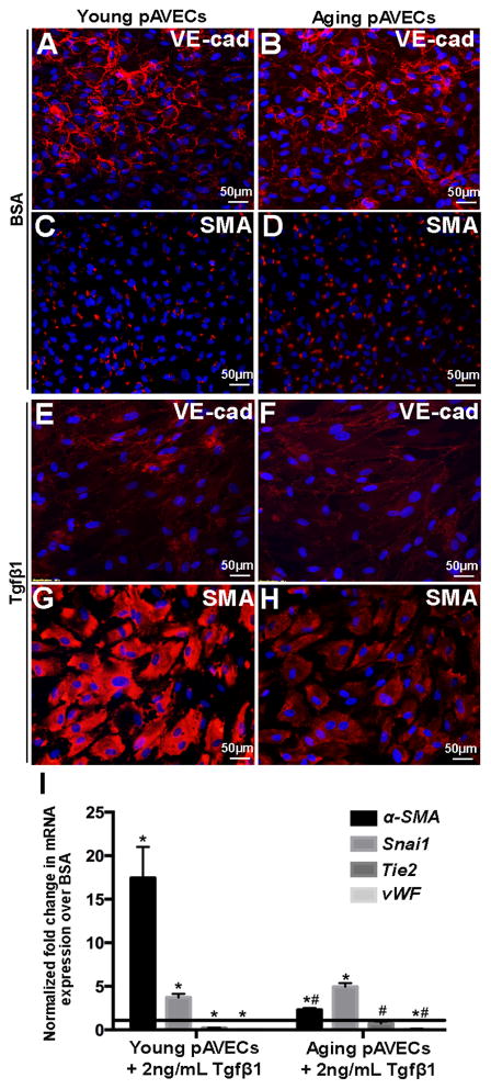
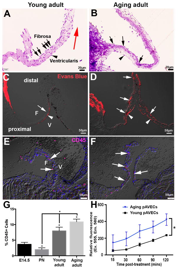
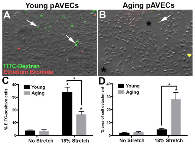
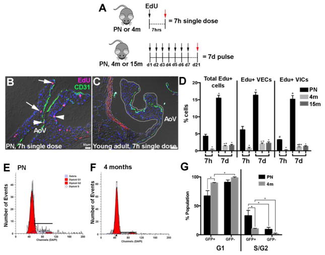
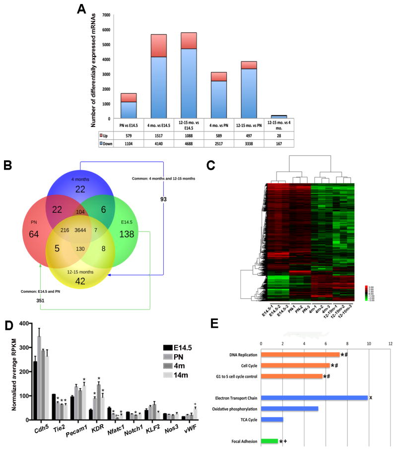
Risk factors of heart valve disease are well defined and prolonged exposure throughout life leads to degeneration and dysfunction in up to 33% of the population. While aortic valve replacement remains the most common need for cardiovascular surgery particularly in those aged over 65, the underlying mechanisms of progressive deterioration are unknown. In other cardiovascular systems, a decline in endothelial cell integrity and function play a major role in promoting pathological changes, and while similar mechanisms have been speculated in the valves, studies to support this are lacking. The goal of this study was to examine age-related changes in valve endothelial cell (VEC) distribution, morphology, function and transcriptomes during critical stages of valve development (embryonic), growth (postnatal (PN)), maintenance (young adult) and aging (aging adult). Using a combination of in vivo mouse, and in vitro porcine assays we show that VEC function including, nitric oxide bioavailability, metabolism, endothelial-to-mesenchymal potential, membrane self-repair and proliferation decline with age. In addition, density of VEC distribution along the endothelium decreases and this is associated with changes in morphology, decreased cell-cell interactions, and increased permeability. These changes are supported by RNA-seq analysis showing that focal adhesion-, cell cycle-, and oxidative phosphorylation-associated biological processes are negatively impacted by aging. Furthermore, by performing high-throughput analysis we are able to report the differential and common transcriptomes of VECs at each time point that can provide insights into the mechanisms underlying age-related dysfunction. These studies suggest that maturation of heart valves over time is a multifactorial process and this study has identified several key parameters that may contribute to impairment of the valve to maintain critical structure-function relationships; leading to degeneration and disease.

Keywords: Endothelial cell; Growth; Heart valve; Maturation.


Figures

Figure 1
Figure 1. Valve endothelial cell morphology, distribution, and cell contacts change with growth and maturation
(A–D) Movat’s Pentachrome staining to detect the deposition and organization of collagen (yellow), proteoglycan (blue), elastin (black), and muscle (red) in aortic valves from wild type mice at embryonic (E14.5) (A), post natal (PN) (B), young adult (4 months) (C) and aging adult (14 months) (D) stages. (E–H) Transmission electron microscopy of aortic valves at indicated time points. Arrows indicate valve endothelial cells (VECs), arrowheads show valve interstitial cells (VICs), and *denote contact between VECs and VICs. Bracket in H indicates expanded extracellular matrix. (I, J) Toluidine blue staining of aortic valves from young (4 months) and aging (15 months) wild type mice to show VEC density on fibrosa (arrows) and ventricularis (arrowhead) cusp surfaces. (K) Number of VECs on the surface of aortic valve cusps in distal, mid and proximal regions as indicted, n=3. Statistical significance based on *P<0.05. AoV, aortic valve.
Figure 2
Figure 2. Nitric oxide availability and mitochondrial organization are impaired with growth and maturation
(A, B) Aortic valves from post natal (PND7) (A) and aging adult (25 months) (B) wild type mice stained with CellROX reagent to detect reactive oxygen species in VECs (arrows) and VICs (arrowheads). (C) Percentage of ROX-positive cells over total number of cells (n=3). (D, E) DAR-4M-AM staining denoting nitric oxide availability in PN (C) and adult aging (19 months) (D) wild type aortic valves. (F) Percentage of DAR-4M AM-positive cells over total number of cells (n=3). DAR-4M-AM staining in cultured porcine aortic valve endothelial cells from young (G) and aging (H) animals. (I) Percentage of DAR-4M AM-positive cells over total number of cultured cells per field of view (n=3). (J, K) Transmission electron microscopy to show mitochondrial organization (arrows) in VECS from young adult (4 months) (J) and aging adult (14 months) (K) aortic valves of wild type mice. AoV, aortic valve. Statistical significance based on *P<0.05 in aging adult valves compared to post natal (PN) stages (n=3).
Figure 3
Figure 3. TGFβ1-mediated EMT is decreased with growth and maturation
(A–D) Immunofluorescent staining against the endothelial marker VE-cadherin (A, B, E, F) and mesenchymal marker, α-SMA (C, D, H, H), in young (A, C, E, G) and aging (B, D, F, H) pAVECs treated with 2ng/ml TGFβ1 every 2–3 for 7 days (E–H) compared to BSA treated controls (A–D). (I) Quantitative PCR to show fold changes in Tie2, vWF (endothelial), Snai1 and α-SMA (mesenchyme) in young and aging TGFβ1-treated pAVECs compared to BSA-treated controls. Fold change relative to BSA controls, n=8. *p=<0.05 compared to BSA control, #p=<0.05 compared to TGFβ1-treated young pAVECs.
Figure 4
Figure 4. The valve endothelium is more permeable with maturation
(A, B) Toluidine blue staining of young adult (4 months) (A) and aging adult (14 month) (B) wild type aortic valves to show VEC density over the surface of the fibrosa (arrows), compared to the ventricularis (arrowheads). (C, D) Evans blue dye to determine permeability of the valve endothelium in young adult (C) and aging adult (D) wild type mice. (E, F) CD45 detection to show expression of infiltrating hematopoietic cells in young adult (E) and aging adult (F) aortic valves; quantitation shown in (G). (H) Quantitation of relative fluorescence of permeated Dextran-TMR in young and aging pAVECs, n=4. *p=<0.05 compared to young pAVECs.
Figure 5
Figure 5. Cell membrane repair following stretch-induced injury decreases with maturation
(A, B) DIC images to show morphology and distribution of young (A) and aging (B) pAVECs following 18% equibiaxial stretch at 1Hz for 20 minutes. Green staining indicates cell-retained FITC-Dextran, while Propidium Iodide is in red with negligible detection. Quantitation of FITC-positive cells are shown in C, and D indicates the % of detached cells under each condition, n=3, p<0.05 in aging compared to young pAVECs.
Figure 6
Figure 6. VEC proliferation declines with maturation
(A) Experimental design of EdU labeling in PN, young adult (4 months), and aging adult (15 months) aortic valves. (B, C) Representative aortic valves labeled with EdU (red) and CD31 (green) from 7h single dose treatment in PN (B) and young adult (4 months) (C) wild type mice. Arrowheads indicate EdU+ VECs, EdU+ VICs are marked by arrows. (D) Quantification of EdU+ VECs and VICs in aortic valves at indicated time points. n=3, *p=<0.05 compared to PN, #p<0.05 compared to EdU+ VECs, +p<0.05 compared to 7h. (E, F) Flow cytometry to show DNA content of isolated VECs from PN (E) and young adult (4 months) (F) Tie2GFP mice, n=3. (G) Quantification of Modfit DNA content curves of isolated VECs (GFP+) and non-VECs (GFP−).
Figure 7
Figure 7. Age-dependent mRNA profiles of VECs
(A) Summary of the number of differentially expressed mRNAs between two comparative time points. (B) Venn diagram to show the number of protein-coding mRNAs (4852 total) expressed between time points based on ANOVA P-score <0.01 and max fold change >2. (C) Heat map to show hierarchal clustering between 3 biological replicates at each time point based on RPKM values for 10,024 significant genes, with a false discovery rate (FDR) of <0.1. (D) Graph to show average RPKM values of detected endothelial cell markers. p<0.05 compared to E14.5 (*), PN (#) and 4 months (+). (E) WikiPathway analysis of –Log(P-value) of mRNAs significantly downregulated between PN and aging adult (12–15 months) time points. Orange indicates biological functions related to cell proliferation, processes shown in blue are related to cell metabolism, and green highlights focal adhesion. *p<0.05 between PN and young adult (4 months); #p<0.05 between embryonic (E14.5) and young adult (4 months); +p<0.05 between embryonic (E14.5) and PN; *p<0.05 between young adult (4 months) and aging adult (12–15 months).

Similar articles

Cited by

References

    1. Hinton RB, Jr, Lincoln J, Deutsch GH, Osinska H, Manning PB, Benson DW, et al. Extracellular matrix remodeling and organization in developing and diseased aortic valves. Circ Res. 2006;98(11):1431–8. - PubMed
    1. Tao G, Kotick JD, Lincoln J. Heart valve development, maintenance, and disease: the role of endothelial cells. Curr Top Dev Biol. 2012;100:203–32. - PubMed
    1. Huk DJ, Austin BF, Horne TE, Hinton RB, Ray WC, Heistad DD, et al. Valve Endothelial Cell-Derived Tgfbeta1 Signaling Promotes Nuclear Localization of Sox9 in Interstitial Cells Associated With Attenuated Calcification. Arterioscler Thromb Vasc Biol. 2016;36(2):328–38. - PMC - PubMed
    1. Bosse K, Hans CP, Zhao N, Koenig SN, Huang N, Guggilam A, et al. Endothelial nitric oxide signaling regulates Notch1 in aortic valve disease. J Mol Cell Cardiol. 2013;60:27–35. - PMC - PubMed
    1. Shapero K, Wylie-Sears J, Levine RA, Mayer JE, Jr, Bischoff J. Reciprocal interactions between mitral valve endothelial and interstitial cells reduce endothelial-to-mesenchymal transition and myofibroblastic activation. J Mol Cell Cardiol. 2015;80:175–85. - PMC - PubMed

MeSH terms

LinkOut - more resources