. 2017 Feb 1;23(3):804-813.
doi: 10.1158/1078-0432.CCR-16-1743. Epub 2016 Oct 10.

TGFβR1 Blockade with Galunisertib (LY2157299) Enhances Anti-Neuroblastoma Activity of the Anti-GD2 Antibody Dinutuximab (ch14.18) with Natural Killer Cells

Affiliations

TGFβR1 Blockade with Galunisertib (LY2157299) Enhances Anti-Neuroblastoma Activity of the Anti-GD2 Antibody Dinutuximab (ch14.18) with Natural Killer Cells

Hung C Tran et al. Clin Cancer Res. .

Abstract

Purpose: Immunotherapy of high-risk neuroblastoma using the anti-GD2 antibody dinutuximab induces antibody-dependent cell-mediated cytotoxicity (ADCC). Galunisertib, an inhibitor of TGFβR1, was examined for its ability to enhance the efficacy of dinutuximab in combination with human ex vivo activated NK (aNK) cells against neuroblastoma.

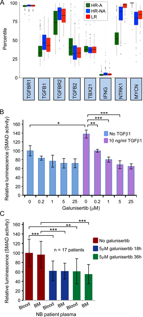
Experimental design: TGFB1 and TGFBR1 mRNA expression was determined for 249 primary neuroblastoma tumors by microarray analysis. The ability of galunisertib to inhibit SMAD activity induced by neuroblastoma patient blood and bone marrow plasmas in neuroblastoma cells was tested. The impact of galunisertib on TGFβ1-induced inhibition of aNK cytotoxicity and ADCC in vitro and on anti-neuroblastoma activity in NOD-scid gamma (NSG) mice was determined.

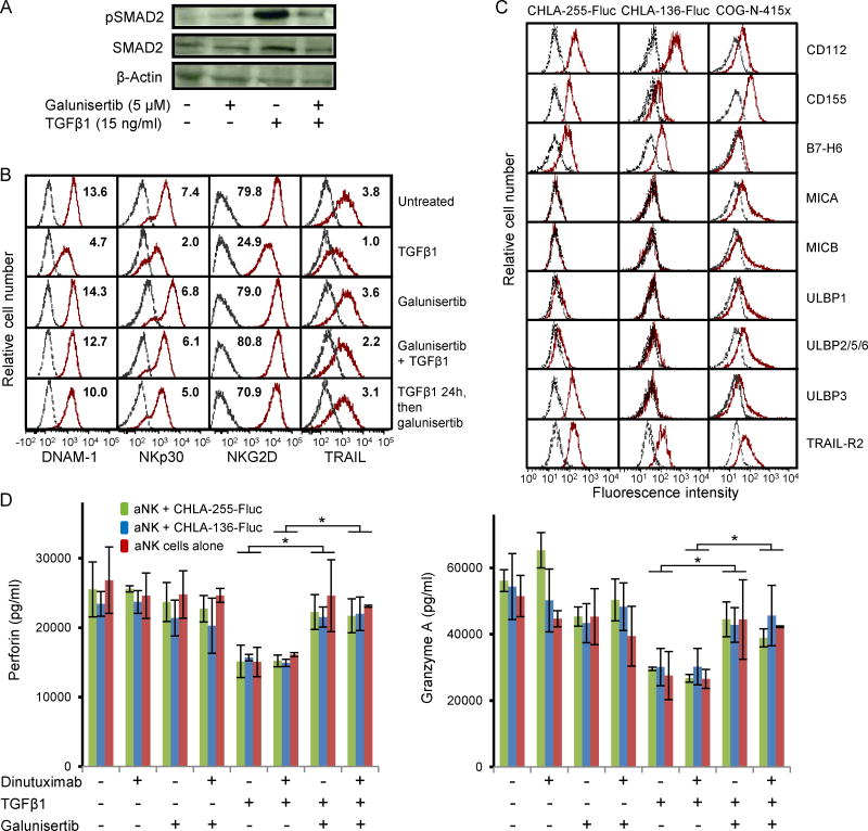
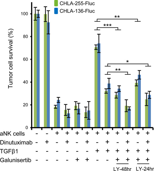
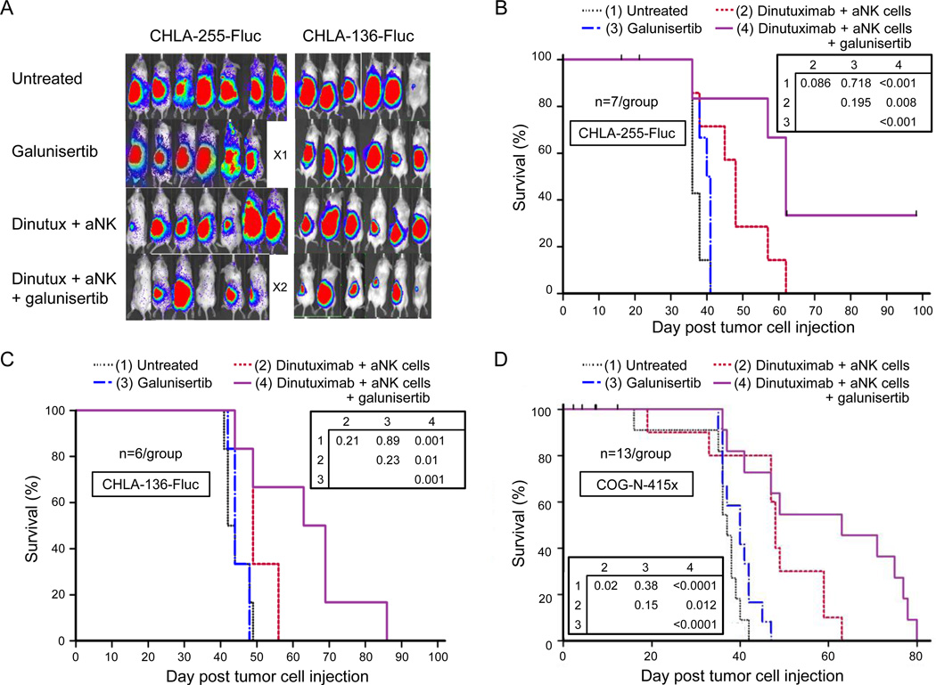
Results: Neuroblastomas express TGFB1 and TGFBR1 mRNA. Galunisertib suppressed SMAD activation in neuroblastoma cells induced by exogenous TGFβ1 or by patient blood and bone marrow plasma, and suppressed SMAD2 phosphorylation in human neuroblastoma cells growing in NSG mice. In NK cells treated in vitro with exogenous TGFβ1, galunisertib suppressed SMAD2 phosphorylation and restored the expression of DNAM-1, NKp30, and NKG2D cytotoxicity receptors and the TRAIL death ligand, the release of perforin and granzyme A, and the direct cytotoxicity and ADCC of aNK cells against neuroblastoma cells. Addition of galunisertib to adoptive cell therapy with aNK cells plus dinutuximab reduced tumor growth and increased survival of mice injected with two neuroblastoma cell lines or a patient-derived xenograft.

Conclusions: Galunisertib suppresses activation of SMAD2 in neuroblastomas and aNK cells, restores NK cytotoxic mechanisms, and increases the efficacy of dinutuximab with aNK cells against neuroblastoma tumors. Clin Cancer Res; 23(3); 804-13. ©2016 AACRSee related commentary by Zenarruzabeitia et al., p. 615.


Conflict of interest statement

Conflict of Interest: The authors declare no conflict of interest.

Figures

Figure 1
Figure 1
Expression of TGFBR1 and TGFB1 genes by human NB tumors and inhibition of SMAD activity in patient blood and bone marrow plasma by the TGFβR1 inhibitor galunisertib. A, expression levels of genes for 249 primary NB tumors were determined using microarray whole genome analysis (68 patients with high risk MYCN-amplified disease, 151 with high risk non-amplified disease, and 30 with low risk disease). High-risk tumors with MYCN amplification (HR-A) exhibited MYCN gene expression with a median percentile value at the 100th percentile. B, effect of galunisertib on SMAD activity in reporter cells induced by 10 ng/ml TGFβ1. Galunisertib was examined in serial 5-fold dilutions from 25 µM to 0.20 µM. *, p < 0.05; **, p < 0.01; ***, p < 0.001. C, effect of galunisertib on SMAD activation in reporter cells induced by blood or bone marrow plasma from 17 NB patients. Blood and bone marrow plasma were diluted ten-fold with IMDM and added into wells containing the reporter cell line CHLA255hRL-SmadFluc with or without 5 µM galunisertib for 18 or 36 hours. The mean induction of SMAD activity by the 17 blood plasmas was assigned a value of 100% (SD ± 28.9).
Figure 2
Figure 2
Effect of galunisertib on TGFβ1-induced SMAD2 phosphorylation, down-regulation of cytotoxicity receptors and TRAIL, and inhibition of release of perforin and granzyme A in aNK cells. A, aNK cells cultured with IL-2 (10 ng/ml) were treated with galunisertib alone (5 µM), TGFβ1 alone (15 ng/ml), or TGFβ1 and galunisertib for 18 hours. Whole lysates from aNK cells were subjected to pSMAD2, total SMAD2, and β-actin immunoblot assay. B, down-regulation by TGFβ1 of DNAM-1, NKp30, NKG2D, and TRAIL, and reversal by galunisertib. aNK cells were pretreated for 48 hours with TGFβ1 (10 ng/ml) alone, with galunisertib (5 µM) alone, or with TGFβ1 and galunisertib. An additional group received TGFβ1 alone for 24 hours, followed by addition of galunisertib for another 24 hours. Using 9-color flow cytometry, viable aNK cells were identified according to CD56 and CD16 expression and absence of CD3, CD14, and CD19 expression, and then their levels of cytotoxicity receptors and of TRAIL were determined. Stain Index values are given in each histogram overlay. The presented results are similar to those for aNK cells propagated from three additional human donors. Red lines, specific antibody; black lines, isotype control. C, expression of ligands for DNAM-1 (CD112 and CD155), NKp30 (B7-H6), and NKG2D (MICA, MICB, ULBP1, ULBP2/5/6, and ULBP3) and of TRAIL-R2 on NB cell lines and on PDX cells (COG-N-415x). D, suppression of perforin and granzyme A secretion from aNK cells by TGFβ1 is inhibited by galunisertib. After 6 hours of co-culturing aNK cells with NB cells, perforin and granzyme A were quantified in the supernatant of each treatment condition using a Luminex multiplexed microbead assay (*, p < 0.05).
Figure 3
Figure 3
Effect of TGFβ1 and galunisertib on human aNK cytotoxicity and ADCC against NB cells. A, aNK cells sustained with IL-2 (10 ng/ml) were pretreated 48 hours with TGFβ1 alone (10 ng/ml), galunisertib alone (5 µM), TGFβ1 and galunisertib, or TGFβ1 for 24 hours followed by addition of galunisertib for an additional 24 hours. aNK cells were then cultured with calcein-AM-labeled NB cell lines CHLA-255-Fluc or CHLA-136-Fluc at a 1:2 E:T ratio for 6 hours. aNK cell-mediated killing of NB cells was assayed by digital imaging microscopy. Galunisertib (5 µM) by itself had no cytotoxic effect (data not shown). *, p < 0.05; **, p < 0.01; ***, p < 0.001.
Figure 4
Figure 4
Effect of galunisertib on SMAD2 phosphorylation in human NB tumors growing in NSG mice. A, tumors derived from CHLA-255-Fluc cells. B, tumors derived from CHLA-136-Fluc cells. C, tumors from COG-N-415x cells. NB cells (1×10⁶ CHLA-255-Fluc or CHLA-136-Fluc cell lines or COG-N-415x PDX cells) were injected into the left kidney of each NSG mouse on day 0. Formulated galunisertib (75 mg/kg) was gavaged twice a day from days 3–10 and then five days per week from days 13–31. aNK cells (1×10⁷) plus dinutuximab (15 µg/mouse) were injected intravenously twice a week from day 3 along with IL-2 (2 µg) and IL-15 (4.9 µg/mouse) intraperitoneally. Paraffin-embedded tumor sections (5 µm) were prepared and immunostained with rabbit polyclonal anti-pSMAD2 as described in Materials and Methods. Tumors from the four treatment groups (untreated, aNK + dinutuximab, galunisertib alone, or aNK + dinutuximab + galunisertib) were harvested at 36 and 21 days after injection of CHLA-255-Fluc and CHLA-136-Fluc, respectively, and 28 days after injection of COG-N-415x PDX cells. Brown = phosho-SMAD2; blue = hematoxylin; galun = galunisertib; dinutux = dinutuximab.
Figure 5
Figure 5
Effect of galunisertib on growth of NB tumors and survival of NSG mice. Mice were treated as in figure 4. A, bioluminescence imaging on day 23 for mice bearing CHLA-255-Fluc or CHLA-136-Fluc cells. One mouse injected with CHLA-136-Fluc cells in the untreated group exhibited a weak signal but had a large tumor when sacrificed on day 49. Mouse X1 died on day 16 during anesthesia for imaging; mouse X2 died on day 21 from unknown reasons with no observable tumor. B, Kaplan Meier survival plot for mice injected with CHLA-255-Fluc cells. p values comparing treatment groups are shown in the inserted box. C, Kaplan Meier survival plot for mice injected with CHLA-136-Fluc cells. D, Kaplan Meier survival plot for mice injected with PDX COG-N-415x cells.

Comment in

Similar articles

Cited by

References

    1. Maris JM. Recent advances in neuroblastoma. N Engl J Med. 2010;362(23):2202–2211. Epub 2010/06/19. doi:362/23/2202 [pii] 10.1056/NEJMra0804577. PubMed PMID: 20558371. - PMC - PubMed
    1. Yu AL, Gilman AL, Ozkaynak MF, London WB, Kreissman SG, Chen HX, et al. Anti-GD2 antibody with GM-CSF, interleukin-2, and isotretinoin for neuroblastoma. N Engl J Med. 2010;363(14):1324–1334. Epub 2010/10/01. PubMed PMID: 20879881. - PMC - PubMed
    1. Wilson EB, El-Jawhari JJ, Neilson AL, Hall GD, Melcher AA, Meade JL, et al. Human tumour immune evasion via TGF-beta blocks NK cell activation but not survival allowing therapeutic restoration of anti-tumour activity. PLoS One. 2011;6(9):e22842. PubMed PMID: 21909397; PMCID: PMC3167809. - PMC - PubMed
    1. Massagué J, Blain SW, Lo RS. TGFbeta signaling in growth control, cancer, and heritable disorders. Cell. 2000;103(2):295–309. PubMed PMID: 11057902. - PubMed
    1. Padua D, Massagué J. Roles of TGFbeta in metastasis. Cell Res. 2009;19(1):89–102. PubMed PMID: 19050696. - PubMed

MeSH terms