Review
. 2016 Nov;101(11):1284-1294.
doi: 10.3324/haematol.2016.142463. Epub 2016 Oct 18.

New insights on hereditary erythrocyte membrane defects

Affiliations
Review

New insights on hereditary erythrocyte membrane defects

Immacolata Andolfo et al. Haematologica. 2016 Nov.

Abstract

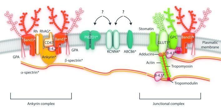
After the first proposed model of the red blood cell membrane skeleton 36 years ago, several additional proteins have been discovered during the intervening years, and their relationship with the pathogenesis of the related disorders have been somewhat defined. The knowledge of erythrocyte membrane structure is important because it represents the model for spectrin-based membrane skeletons in all cells and because defects in its structure underlie multiple hemolytic anemias. This review summarizes the main features of erythrocyte membrane disorders, dividing them into structural and altered permeability defects, focusing particularly on the most recent advances. New proteins involved in alterations of the red blood cell membrane permeability were recently described. The mechanoreceptor PIEZO1 is the largest ion channel identified to date, the fundamental regulator of erythrocyte volume homeostasis. Missense, gain-of-function mutations in the PIEZO1 gene have been identified in several families as causative of dehydrated hereditary stomatocytosis or xerocytosis. Similarly, the KCNN4 gene, codifying the so called Gardos channel, has been recently identified as a second causative gene of hereditary xerocytosis. Finally, ABCB6 missense mutations were identified in different pedigrees of familial pseudohyperkalemia. New genomic technologies have improved the quality and reduced the time of diagnosis of these diseases. Moreover, they are essential for the identification of the new causative genes. However, many questions remain to solve, and are currently objects of intensive studies.


Figures

Figure 1.
Figure 1.
Simplified cross-section of the erythrocyte membrane. The red blood cell membrane is composed of integral membrane proteins incorporated into a phospholipid bilayer. The network of cytoskeletal proteins is anchored to the membrane via several transmembrane proteins with a transport function: band 3, anion transporter; GLUT1, glucose and L-dehydroascorbic acid transporter; RhAG, gas transporter, in particular CO2; various cation pumps and transporters including, Na+-K+-ATPase, Ca++-ATPase, Na+-K+-2Cl and Na+-Cl, Na+-K+, K+-Cl co-transporters and Gardos channel. The most recently described proteins PIEZO1, KCNN4 and ABCB6, involved in the modulation of RBC membrane permeability, and their putative interactions are also shown. The relative positions of the proteins to each other within the various complexes are mostly unknown. The shapes of the major proteins are mostly imaginary. GPA, glycophorin A; Rh, Rhesus polypeptide; B-4.1, protein band 4.1; B-4.2, protein band 4.2; GPC, glycophorin C; RhAG, Rh-associated glycoprotein; RBC: red blood cells. *Proteins that are known to be affected by pathogenic mutations so far.
Figure 2.
Figure 2.
Flow diagram for the differential diagnosis of hemolytic anemias due to RBC membrane defects. The flow diagram shows the main steps for guiding the clinical suspicion toward the diagnosis of different subtypes of hereditary erythrocyte membrane disorders. First-, second-, and third-line investigations are also shown. The cut-off for the EMA binding test is still debated: currently, a test with a reduction of EMA binding > 21%, in comparison with controls, is defined positive, whereas a test with a reduction of EMA binding < 16% is considered negative. Values between 16–21% are not conclusive, although a cut-off of 11% has been proposed. Hb: hemoglobin; MCH: mean corpuscular hemoglobin; MCV: mean cellular volume; MCHC; mean corpuscular hemoglobin concentration; CBC: complete blood count; RBC: red blood cells; OHS: overhydrated hereditary stomatocytosis; DHS: dehydrated hereditary stomatocytosis; AD: autosomal dominant; AR: autosomal recessive; EMA: eosin-5-maleimide; SDS: Sodium dodecyl sulfate; NGS: next generation sequencing; RHAG: rhesus blood group-associated glycoprotein. PB: peripheral blood.
Figure 3.
Figure 3.
Examples of ektacytometric curves of different hereditary erythrocyte membrane disorders. The ektacytometer is a laser diffraction viscometer that measures RBCs (red blood cells) deformability at constant shear stress as a continuous function of suspending Osmolarity. Three principal features of the osmotic gradient ektacytometry profiles are: the Omin point (red asterisk), which corresponds to the osmolarity at which 50% of the red cells are lysed in the classical osmotic fragility test and represents the surface area to volume ratio; the maximal deformability index (DImax, black asterisk) value, which represents the maximal cellular deformability of the red cell population; and the O′ or hyper point (blue asterisk), which corresponds to the osmolarity at DImax/2, which reflects the hydration status of the red cells. Ektacytometric analysis of (A) HS, (B) OHS, (C) DHS1 and (D) DHS2 patients are shown. The dotted line is those relative to the control subjects. HS: hereditary spherocytosis; OHS: overhydrated hereditary stomatocytosis; DHS1: dehydrated hereditary stomatocytosis 1; DHS2: dehydrated hereditary stomatocytosis 2.

References

    1. Lux SE. Dissecting the red cell membrane skeleton. Nature. 1979;281(5731):426–429. - PubMed
    1. Mohandas N, Gallagher PG. Red cell membrane: past, present, and future. Blood. 2008;112(10):3939–3948. - PMC - PubMed
    1. Lux SE., 4th Anatomy of the red cell membrane skeleton: unanswered questions. Blood. 2016;127(2):187–199. - PubMed
    1. Pesciotta EN, Sriswasdi S, Tang HY, Mason PJ, Bessler M, Speicher DW. A label-free proteome analysis strategy for identifying quantitative changes in erythrocyte membranes induced by red cell disorders. J Proteomics. 2012;76 Spec No.:194–202. - PMC - PubMed
    1. Liu SC, Derick LH, Palek J. Visualization of the hexagonal lattice in the erythrocyte membrane skeleton. J Cell Biol. 1987;104(3):527–536. - PMC - PubMed

Publication types

MeSH terms

Supplementary concepts