Malignant Pleural Effusion and ascites Induce Epithelial-Mesenchymal Transition and Cancer Stem-like Cell Properties via the Vascular Endothelial Growth Factor (VEGF)/Phosphatidylinositol 3-Kinase (PI3K)/Akt/Mechanistic Target of Rapamycin (mTOR) Pathway
- PMID: 27756837
- PMCID: PMC5207183
- DOI: 10.1074/jbc.M116.753236
Malignant Pleural Effusion and ascites Induce Epithelial-Mesenchymal Transition and Cancer Stem-like Cell Properties via the Vascular Endothelial Growth Factor (VEGF)/Phosphatidylinositol 3-Kinase (PI3K)/Akt/Mechanistic Target of Rapamycin (mTOR) Pathway
Abstract
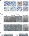
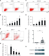
Malignant pleural effusion (PE) and ascites, common clinical manifestations in advanced cancer patients, are associated with a poor prognosis. However, the biological characteristics of malignant PE and ascites are not clarified. Here we report that malignant PE and ascites can induce a frequent epithelial-mesenchymal transition program and endow tumor cells with stem cell properties with high efficiency, which promotes tumor growth, chemoresistance, and immune evasion. We determine that this epithelial-mesenchymal transition process is mainly dependent on VEGF, one initiator of the PI3K/Akt/mechanistic target of rapamycin (mTOR) pathway. From the clinical observation, we define a therapeutic option with VEGF antibody for malignant PE and ascites. Taken together, our findings clarify a novel biological characteristic of malignant PE and ascites in cancer progression and provide a promising and available strategy for cancer patients with recurrent/refractory malignant PE and ascites.
Keywords: VEGF; ascites; cancer biology; cancer stem cells; epithelial-mesenchymal transition (EMT); malignant pleural effusion; tumor therapy.
© 2016 by The American Society for Biochemistry and Molecular Biology, Inc.
Figures


Similar articles
-
PI3K/AKT/mTOR and sonic hedgehog pathways cooperate together to inhibit human pancreatic cancer stem cell characteristics and tumor growth.Oncotarget. 2015 Oct 13;6(31):32039-60. doi: 10.18632/oncotarget.5055. Oncotarget. 2015. PMID: 26451606 Free PMC article.
-
Inhibition of PI3K/Akt/mTOR signaling pathway alleviates ovarian cancer chemoresistance through reversing epithelial-mesenchymal transition and decreasing cancer stem cell marker expression.BMC Cancer. 2019 Jun 24;19(1):618. doi: 10.1186/s12885-019-5824-9. BMC Cancer. 2019. PMID: 31234823 Free PMC article.
-
Activation of AKT signaling promotes epithelial-mesenchymal transition and tumor growth in colorectal cancer cells.Mol Carcinog. 2014 Feb;53 Suppl 1:E151-60. doi: 10.1002/mc.22076. Epub 2013 Sep 2. Mol Carcinog. 2014. PMID: 24000138
-
PI3K/Akt/mTOR signaling pathway in cancer stem cells.Pathol Res Pract. 2022 Sep;237:154010. doi: 10.1016/j.prp.2022.154010. Epub 2022 Jul 3. Pathol Res Pract. 2022. PMID: 35843034 Review.
-
Emergence of the phosphoinositide 3-kinase-Akt-mammalian target of rapamycin axis in transforming growth factor-β-induced epithelial-mesenchymal transition.Cells Tissues Organs. 2011;193(1-2):8-22. doi: 10.1159/000320172. Epub 2010 Nov 2. Cells Tissues Organs. 2011. PMID: 21041997 Free PMC article. Review.
Cited by
-
Apolipoprotein C-II induces EMT to promote gastric cancer peritoneal metastasis via PI3K/AKT/mTOR pathway.Clin Transl Med. 2021 Aug;11(8):e522. doi: 10.1002/ctm2.522. Clin Transl Med. 2021. PMID: 34459127 Free PMC article.
-
Oncological Frontiers in the Treatment of Malignant Pleural Mesothelioma.J Clin Med. 2021 May 25;10(11):2290. doi: 10.3390/jcm10112290. J Clin Med. 2021. PMID: 34070352 Free PMC article. Review.
-
Euphorbia factor L2 suppresses the generation of liver metastatic ascites in breast cancer via inhibiting NLRP3 inflammasome activation.Int J Mol Med. 2024 Jan;53(1):8. doi: 10.3892/ijmm.2023.5332. Epub 2023 Dec 8. Int J Mol Med. 2024. PMID: 38063231 Free PMC article.
-
Hexokinase 2 discerns a novel circulating tumor cell population associated with poor prognosis in lung cancer patients.Proc Natl Acad Sci U S A. 2021 Mar 16;118(11):e2012228118. doi: 10.1073/pnas.2012228118. Proc Natl Acad Sci U S A. 2021. PMID: 33836566 Free PMC article.
-
MicroRNAs Present in Malignant Pleural Fluid Increase the Migration of Normal Mesothelial Cells In Vitro and May Help Discriminate between Benign and Malignant Effusions.Int J Mol Sci. 2023 Sep 13;24(18):14022. doi: 10.3390/ijms241814022. Int J Mol Sci. 2023. PMID: 37762343 Free PMC article.
References
-
- Runyon B. A. (1994) Care of patients with ascites. N. Engl. J. Med. 330, 337–342 - PubMed
-
- Shen-Gunther J., and Mannel R. S. (2002) Ascites as a predictor of ovarian malignancy. Gynecol. Oncol. 87, 77–83 - PubMed
-
- Ryu J. S., Ryu H. J., Lee S. N., Memon A., Lee S. K., Nam H. S., Kim H. J., Lee K. H., Cho J. H., and Hwang S. S. (2014) Prognostic impact of minimal pleural effusion in non-small-cell lung cancer. J. Clin. Oncol. 32, 960–967 - PubMed
-
- Tan D. S., Agarwal R., and Kaye S. B. (2006) Mechanisms of transcoelomic metastasis in ovarian cancer. Lancet Oncol. 7, 925–934 - PubMed
-
- Kolomainen D. F., A'Hern R., Coxon F. Y., Fisher C., King D. M., Blake P. R., Barton D. P., Shepherd J. H., Kaye S. B., and Gore M. E. (2003) Can patients with relapsed, previously untreated, stage I epithelial ovarian cancer be successfully treated with salvage therapy? J. Clin. Oncol. 21, 3113–3118 - PubMed
MeSH terms
Substances
LinkOut - more resources
Full Text Sources
Other Literature Sources
Miscellaneous

