. 2016 Dec 2;291(49):25398-25410.
doi: 10.1074/jbc.M116.754655. Epub 2016 Oct 18.

Mechanism of Sirt1 NAD+-dependent Protein Deacetylase Inhibition by Cysteine S-Nitrosation

Affiliations

Mechanism of Sirt1 NAD+-dependent Protein Deacetylase Inhibition by Cysteine S-Nitrosation

Kelsey S Kalous et al. J Biol Chem. .

Abstract

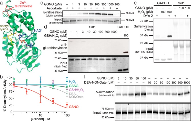
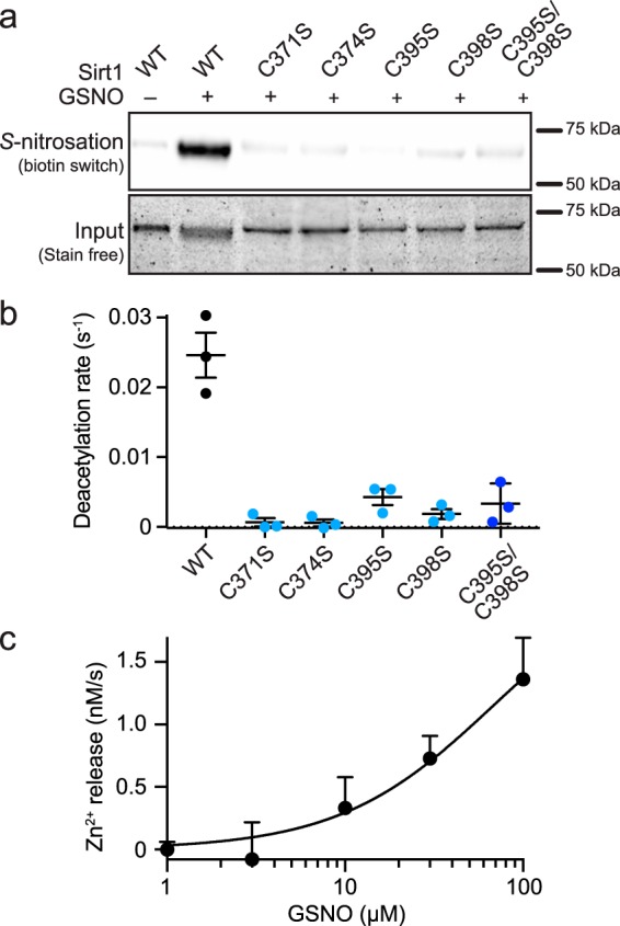
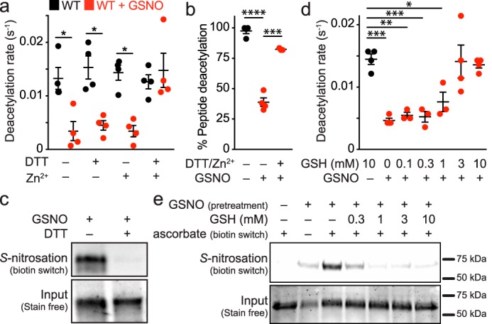
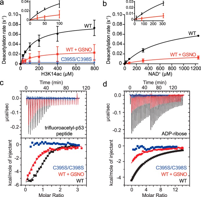
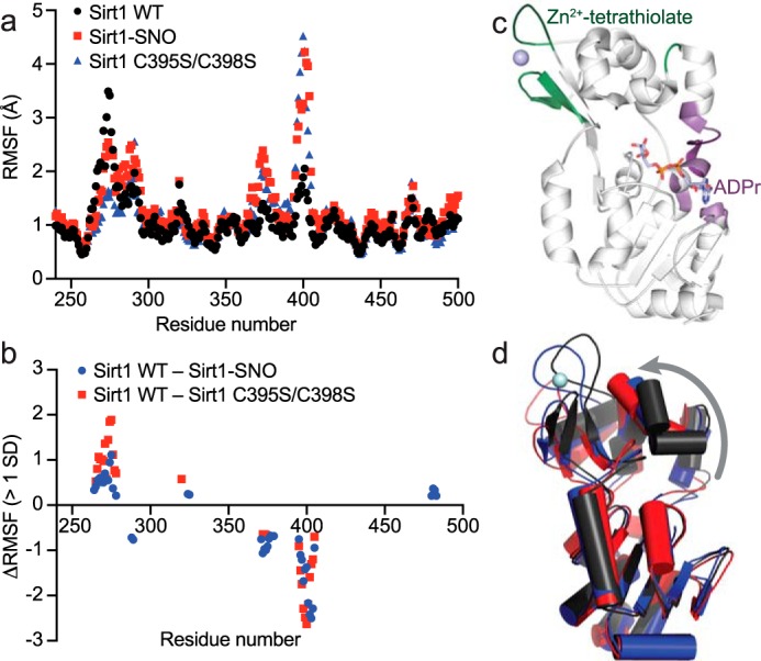
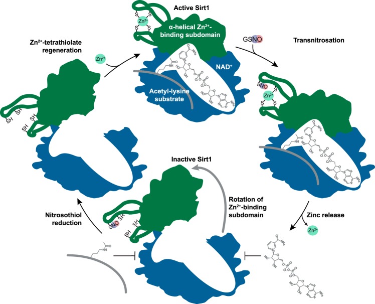
The sirtuin family of proteins catalyze the NAD+-dependent deacylation of acyl-lysine residues. Humans encode seven sirtuins (Sirt1-7), and recent studies have suggested that post-translational modification of Sirt1 by cysteine S-nitrosation correlates with increased acetylation of Sirt1 deacetylase substrates. However, the mechanism of Sirt1 inhibition by S-nitrosation was unknown. Here, we show that Sirt1 is transnitrosated and inhibited by the physiologically relevant nitrosothiol S-nitrosoglutathione. Steady-state kinetic analyses and binding assays were consistent with Sirt1 S-nitrosation inhibiting binding of both the NAD+ and acetyl-lysine substrates. Sirt1 S-nitrosation correlated with Zn2+ release from the conserved sirtuin Zn2+-tetrathiolate and a loss of α-helical structure without overall thermal destabilization of the enzyme. Molecular dynamics simulations suggested that Zn2+ loss due to Sirt1 S-nitrosation results in repositioning of the tetrathiolate subdomain away from the rest of the catalytic domain, thereby disrupting the NAD+ and acetyl-lysine-binding sites. Sirt1 S-nitrosation was reversed upon exposure to the thiol-based reducing agents, including physiologically relevant concentrations of the cellular reducing agent glutathione. Reversal of S-nitrosation resulted in full restoration of Sirt1 activity only in the presence of Zn2+, consistent with S-nitrosation of the Zn2+-tetrathiolate as the primary source of Sirt1 inhibition upon S-nitrosoglutathione treatment.

Keywords: acetylation; allosteric regulation; enzyme inactivation; inhibition mechanism; nicotinamide adenine dinucleotide (NAD); nitric oxide; nitrosation; nitrosylation; sirtuin 1 (SIRT1); zinc.


Figures

FIGURE 1.
FIGURE 1.
Sirt1 is inhibited by S-nitrosation. a, crystal structure (PDB code 4ZZJ (17)) of the Sirt1 catalytic domain with carba-NAD+ and an acetyl-lysine peptide bound in the Rossmann-fold subdomain, located proximal to the conserved Zn2+-tetrathiolate. b, comparison of Sirt1 inhibition by cysteine oxidants. Sirt1 (0.5–1 μm) deacetylase activity was assessed via an enzyme-coupled assay following treatment with varying concentrations of GSNO (red squares), DEA-NONOate (gray diamonds), H2O2 (blue circles), H2O2/GSH (purple triangles), and GSSG (green triangles) for 1 h at 37 °C. Percentage of deacetylase activity in the absence of oxidants was plotted versus log10 of the oxidant concentration (n = 3 ± S.E.). Relative Sirt1 S-nitrosation (c), glutathionylation (d), and sulfenylation (e) levels were assessed for Sirt1 WT treated with the indicated concentrations of GSNO for 1 h at 37 °C (representative blots, n = 3 for biotin switch S-nitrosation detection, n = 4 for anti-glutathionylation, and n = 3 for DYn-2 sulfenylation detection). GAPDH treated with 100 μm H2O2 and 100 μm GSH was used as a positive control for glutathionylation. GAPDH treated with 100 μm H2O2 was used as a positive control for sulfenylation. f, relative Sirt1 S-nitrosation levels for Sirt1 WT treated with varying concentrations of GSNO and DEA-NONOate for 1 h at 37 °C were assessed via the biotin switch assay (representative blot, n = 3). Full blots and loading control gels are shown in supplemental Fig. S1.
FIGURE 2.
FIGURE 2.
Sirt1 S-nitrosation of the Zn2+-tetrathiolate results in Zn2+ release. a, relative Sirt1 S-nitrosation levels were assessed for Sirt1 WT and Sirt1 tetrathiolate mutants (C371S, C374S, C395S, C398S, and C395S/C398S) treated with 100 μm GSNO for 1 h at 37 °C. Full blot and loading control gel are shown in supplemental Fig. S2. b, Sirt1 tetrathiolate mutants do not display deacetylase activity significantly above background. Deacetylase activity of Sirt1 WT and tetrathiolate mutants (1 μm) was assessed via continuous enzyme-coupled assay (n = 3 ± S.E.). c, Sirt1 S-nitrosation results in Zn2+ release. Sirt1 (7.5 μm) was treated with varying concentrations of GSNO in the presence of Zincon (40 μm). Rates of Zn2+ release were measured by following changes in absorbance at 529 and 620 nm for 30 min at 37 °C. Data were plotted as rate of Zn2+ release versus log10 of the GSNO concentration (n = 3 ± S.E.).
FIGURE 3.
FIGURE 3.
Sirt1 deacetylase inhibition by S-nitrosation is reversible in the presence of reductants and Zn2+. a, Sirt1 (2 μm) was treated with buffer or GSNO (100 μm) for 1 h at 37 °C, followed by incubation with DTT (10 mm) and/or ZnCl2 (1 μm) for 5 min at 37 °C. Sirt1 activity (0.5 μm) was assessed via the enzyme-coupled assay. Data were plotted as the mean deacetylation rate for each condition, and significance was determined using a one-way ANOVA and Tukey post-test (n = 4 ± S.E.). b, Sirt1 (2.5 μm) was treated with buffer or GSNO (60 μm) for 30 min at 37 °C followed by incubation with DTT (5 mm) and ZnCl2 (1–10 μm) for 5 min at 37 °C. Sirt1 activity (1 μm) was assessed via HPLC as detailed under “Experimental Procedures.” Data were plotted as the mean percent peptide deacetylation for each condition, and significance was determined via Student's t test (n = 3 ± S.E.). c, Sirt1 S-nitrosation is reversed by DTT. Sirt1 was treated with GSNO (100 μm) for 1 h at 37 °C, followed by immediate treatment with DTT (10 mm). The relative level of S-nitrosation was assessed via the biotin switch assay (n = 3, representative blot). d, Sirt1 inhibition by S-nitrosation is recovered by GSH. Sirt1 (2 μm) was treated with buffer or GSNO (100 μm) for 1 h at 37 °C, followed by incubation with varying concentrations of GSH and ZnCl2 (1 μm) for 5 min at 37 °C. Sirt1 activity (0.5 μm) was assessed via the enzyme-coupled assay, and data were plotted as the mean deacetylation rate versus GSH concentration (n = 4 ± S.E.). e, Sirt1 S-nitrosation is reversed by GSH. Sirt1 was treated with GSNO (100 μm) for 1 h at 37 °C followed by immediate treatment with varying concentrations of GSH for 5 min at 37 °C. The relative level of S-nitrosation was assessed via the biotin switch assay. Full blot and loading control gel are shown in supplemental Fig. S3 (n = 3, representative blot). Values that are significantly different are indicated by bars and asterisks as follows: *, p < 0.05; **, p < 0.01; ***, p < 0.005; ****, p < 0.001.
FIGURE 4.
FIGURE 4.
Effect of Sirt1 S-nitrosation on substrate binding and catalysis. a, titrations of H3K14ac peptide substrate were performed under saturating NAD+ concentrations (2 mm) following treatment of Sirt1 WT or Sirt1 C395S/C398S (2 μm) with buffer or GSNO (100 μm) for 1 h at 37 °C. Sirt1 (0.5 μm) deacetylase activity was assessed via the enzyme-coupled assay. Deacetylation rates were plotted versus H3K14ac concentration and fit to the Michaelis-Menten equation to obtain steady-state kinetic parameters (WT n = 3 ± S.D.; C395S/C398S n = 2 ± S.D.). Inset displays quality of nonlinear fit at low substrate concentrations. b, titrations of NAD+ were performed under saturating concentrations of H3K14ac peptide (1.4 mm) following treatment of Sirt1 (2 μm) with buffer or GSNO (100 μm) for 1 h at 37 °C. Sirt1 (1 μm) deacetylase activity was assessed via the enzyme-coupled assay. Deacetylation rates were plotted versus H3K14ac concentration and fit to the Michaelis-Menten equation to obtain steady-state kinetic parameters (n ≥3 ± S.D.). Inset displays quality of nonlinear fit at low substrate concentrations. c, determining the effect of Sirt1 S-nitrosation on binding to the acetyl-lysine-binding site by ITC. Sirt1 WT or C395S/C398S was pretreated with buffer or GSNO (100 μm) for 1 h at 37 °C. p53tfa peptide (200 μm) was titrated into Sirt1. The area under each peak was integrated and plotted as kcal/mol of p53tfa versus the molar ratio of p53tfa/Sirt1 (representative data shown, n ≥2 for each condition). d, determining the effect of Sirt1 S-nitrosation on binding to the NAD+-binding site by ITC. Sirt1 WT or C395S/C398S was pretreated with buffer or GSNO (100 μm) for 1 h at 37 °C. ADPr (400 μm) was titrated into Sirt1. The area under each peak was integrated and plotted as kcal/mol of ADPr versus the molar ratio of ADPr/Sirt1 (representative data shown, n ≥2 for each condition).
FIGURE 5.
FIGURE 5.
Sirt1 S-nitrosation results in selective alteration of Sirt1 secondary structure without a decrease in thermal stability. a, circular dichroism analyses of Sirt1 WT or C395S/C398S (14 μm) treated with buffer or GSNO (100 μm) for 1 h at 37 °C. CD spectra of desalted Sirt1 (2 mg/ml or 3.5 μm) were obtained by scanning from 190 to 280 nm in 0.1-nm intervals. b, percentages of α-helix, β-sheet, turn, and random coil were calculated by analyzing CD spectra with DICHROWEB software. Data were plotted as the percentage of each secondary structure observed under each condition, and significance was determined by two-way ANOVA and Tukey post-test (n = 4 ± S.E.). c, disruption of the Sirt1 Zn2+-tetrathiolate by S-nitrosation or mutation increases thermal stability. Sirt1 WT or C395S/C398S (14 μm) was treated with buffer, GSNO (100 μm), or DEA-NONOate (100 μm) for 1 h at 37 °C (n = 3 for each condition). Melting curves for desalted Sirt1 (7.5 μm) were obtained by following SYPRO Orange fluorescence as a function of temperature using a temperature gradient of 20–95 °C. Values that are significantly different are indicated by bars and asterisks as follows: *, p < 0.05; **, p < 0.01; ***, p < 0.005; ****, p < 0.001.
FIGURE 6.
FIGURE 6.
Molecular dynamics simulations support an allosteric mechanism of Sirt1 inhibition by S-nitrosation. 100-ns molecular dynamics simulations of Sirt1 WT, Sirt1-SNO, and Sirt1 C395S/C398S were performed using the Schrödinger Maestro molecular dynamics modeling platform. a, RMSF of each residue was calculated using Schrödinger Maestro and plotted against residue number. b, RMSF values of Sirt1-SNO and Sirt1 C395S/C398S were subtracted from Sirt1 WT RMSF values. ΔRMSF values within 1 S.D. of the average difference in RMSF were excluded, revealing localized regions of significant dynamic change between wild-type and modified Sirt1. c, differences in Sirt1-SNO or Sirt1 C395S/C398S RMSF outside of 1 S.D. plotted in b were mapped to the crystal structure of Sirt1 with ADP-ribose bound (PDB code 4KXQ (33)). Residues colored green indicate predicted regions of increased dynamics in Sirt1-SNO and Sirt1 C395S/C398S. Residues colored purple indicate predicted regions of increased rigidity in Sirt1-SNO and Sirt1 C395S/C398S. d, aligned end point structures from the Sirt1 WT (black), Sirt1-SNO (blue), and Sirt1 C395S/C398S (red) molecular dynamics simulations. The gray arrow represents the observed tilting away of the Zn2+-tetrathiolate subdomain relative to the Rossmann-fold subdomain in Sirt1-SNO and Sirt1 C395S/C398S compared with Sirt1 WT.
FIGURE 7.
FIGURE 7.
Proposed mechanism of Sirt1 inhibition by S-nitrosation. S-Nitrosation of Sirt1 occurs at one or more cysteines within the Zn2+-tetrathiolate, resulting in loss of Zn2+ coordination and loss of α-helical secondary structure. Local conformational changes are allosterically translated to the Sirt1 substrate-binding sites where acetyl-lysine and NAD+ binding and deacetylation are inhibited through rotation of the α-helical Zn2+-binding subdomain (green) away from the substrate-binding domain (blue) as indicated by the gray arrow. S-Nitrosation is reversible in the presence of reductants (e.g. GSH), and deacetylase activity is recoverable upon restoration of the Zn2+-tetrathiolate in the presence of Zn2+ and reductants.

References

    1. Feldman J. L., Dittenhafer-Reed K. E., and Denu J. M. (2012) Sirtuin catalysis and regulation. J. Biol. Chem. 287, 42419–42427 - PMC - PubMed
    1. Smith B. C., Hallows W. C., and Denu J. M. (2008) Mechanisms and molecular probes of sirtuins. Chem. Biol. 15, 1002–1013 - PMC - PubMed
    1. Sebastián C., Satterstrom F. K., Haigis M. C., and Mostoslavsky R. (2012) From sirtuin biology to human diseases: an update. J. Biol. Chem. 287, 42444–42452 - PMC - PubMed
    1. Kornberg M. D., Sen N., Hara M. R., Juluri K. R., Nguyen J. V., Snowman A. M., Law L., Hester L. D., and Snyder S. H. (2010) GAPDH mediates nitrosylation of nuclear proteins. Nat. Cell Biol. 12, 1094–1100 - PMC - PubMed
    1. Shinozaki S., Chang K., Sakai M., Shimizu N., Yamada M., Tanaka T., Nakazawa H., Ichinose F., Yamada Y., Ishigami A., Ito H., Ouchi Y., Starr M. E., Saito H., Shimokado K., et al. (2014) Inflammatory stimuli induce inhibitory S-nitrosylation of the deacetylase SIRT1 to increase acetylation and activation of p53 and p65. Sci. Signal. 7, ra106. - PMC - PubMed

LinkOut - more resources