. 2016 Dec 2;291(49):25617-25628.
doi: 10.1074/jbc.M116.748046. Epub 2016 Oct 18.

Electrostatic and Structural Bases of Fe2+ Translocation through Ferritin Channels

Affiliations

Electrostatic and Structural Bases of Fe2+ Translocation through Ferritin Channels

Balasubramanian Chandramouli et al. J Biol Chem. .

Abstract

Ferritin molecular cages are marvelous 24-mer supramolecular architectures that enable massive iron storage (>2000 iron atoms) within their inner cavity. This cavity is connected to the outer environment by two channels at C3 and C4 symmetry axes of the assembly. Ferritins can also be exploited as carriers for in vivo imaging and therapeutic applications, owing to their capability to effectively protect synthetic non-endogenous agents within the cage cavity and deliver them to targeted tissue cells without stimulating adverse immune responses. Recently, X-ray crystal structures of Fe2+-loaded ferritins provided important information on the pathways followed by iron ions toward the ferritin cavity and the catalytic centers within the protein. However, the specific mechanisms enabling Fe2+ uptake through wild-type and mutant ferritin channels is largely unknown. To shed light on this question, we report extensive molecular dynamics simulations, site-directed mutagenesis, and kinetic measurements that characterize the transport properties and translocation mechanism of Fe2+ through the two ferritin channels, using the wild-type bullfrog Rana catesbeiana H' protein and some of its variants as case studies. We describe the structural features that determine Fe2+ translocation with atomistic detail, and we propose a putative mechanism for Fe2+ transport through the channel at the C3 symmetry axis, which is the only iron-permeable channel in vertebrate ferritins. Our findings have important implications for understanding how ion permeation occurs, and further how it may be controlled via purposely engineered channels for novel biomedical applications based on ferritin.

Keywords: ferritin; ferritin channels; ion channel; iron; iron uptake; molecular dynamics; multi-ion mechanism; structure; translocation.


Figures

FIGURE 1.
FIGURE 1.
Ferritin structural elements. A, X-ray crystal structure of frog H′ ferritin (PDB ID: 4MJY). One of the ferroxidase cavities, with irons bound to the dinuclear catalytic site, is circled. B and C, three-dimensional arrangement of the C3 (B) and C4 (C) channels. D and E, magnified views of C3 (D) and C4 (E) channels. Residues subjected to mutation are shown as colored balls and sticks, Fe2+ ions within the channel, as observed in the X-ray structure, are in orange, and water oxygen is in red.
FIGURE 2.
FIGURE 2.
C3 channel dimension analysis. Shown is a profile of the channel radius along the C3 channel (i.e. Z-coordinate). Black lines represent the minimum and maximum observed radius size, whereas the red line is the average. In panels A, B, and C, simulations with no Fe2+ ions in the channel. In panels A′ and B′, simulations with Fe2+ ions in the channel; the average Fe2+ ion positions are represented as colored dots. In panel A′, the green line is the radius size as observed in the X-ray structure, and in panels A′ and B′, the vertical dashed lines are Fe2+ ion positions as observed in the X-ray structure.
FIGURE 3.
FIGURE 3.
C3 channel hydration analysis. Shown is the density of water molecules along the C3 channel. The left panels depict the channel hydration in the absence of Fe2+. The right panels depict the channel hydration in the presence of Fe2+ ions. Individual ferritin subunits constituting the channel are colored differently. Fe2+ ions are shown as colored spheres.
FIGURE 4.
FIGURE 4.
Fe2+ ions and protein carboxylic groups localization. Shown is the spatial occupancy of Fe2+ ions and protein carboxylic groups along the C3 channel pathway in C3WT (A–A″) and C3SM (B, B′). A, Fe2+ ions at the external site (purple), internal site (blue), Cγ centroid of Asp-127 (green), and Cδ centroid of Glu-130 (magenta). Dist., distribution. A′, simulation initiated after moving Fe2+ ions at the internal site into the bulk. A″, simulation initiated with increased concentration of bulk Fe2+ ions. B, Fe2+ ions at internal site (blue) and Cγ centroid of Asp-127 (green). B′, simulation initiated after moving Fe2+ ion in the internal site into the bulk. The dotted lines represent the position of Fe2+ ion in the X-ray structure of the WT protein.
FIGURE 5.
FIGURE 5.
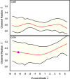
C4 channel dimension analysis. The channel radius profile is shown as a function of Z-coordinate. Data represent the minimum and maximum radius values (black lines) along with the time average (red line). The filled circle indicates the average position of the Fe2+ ion that entered the mutant C4 channel during the dynamics.
FIGURE 6.
FIGURE 6.
C4 channel hydration analysis. Shown is the grid density of water molecules along the C4 channel. The four individual subunits constituting the channel are colored differently. The purple sphere indicates the Fe2+ ion.
FIGURE 7.
FIGURE 7.
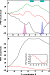
Free energy of Fe2+ translocation. A and B, free energy profiles for single Fe2+ ion translocation through C3 (A) and C4 (B) channels. A, data for C3WT, C3SM, and C3TM are shown in black, red, and green, respectively. The distribution shows the spatial occupancy of Fe2+ ions at external and internal sites in the C3WT model. B, data for C4WT and C4TM are shown in black and red. The inset plot depicts an enlarged view of the PMF profile for the C4TM system.
FIGURE 8.
FIGURE 8.
Iron uptake rate measurements at 2 Fe2+/subunit ratio. The C3 channel properties modulate activity in structurally intact ferritin cage variants. A and B, formation of DFP intermediate (A650 nm) (A) and DFO(H) (A350 nm) (B) products after the addition of 2 Fe2+/subunit to wild-type (blue), C3SM (red), and C3TM (green) variants. The lower panels show enlargements of the first 0.5 s to highlight the differences in the initial rates. Shown is a set of curves (mean ± S.D.) of a representative experiment from at least three experiments, each one performed in triplicate. Abs, absorbance.
FIGURE 9.
FIGURE 9.
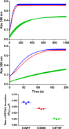
Iron uptake rate measurements at 20 Fe2+/subunit ratio. Top, reaction progress at a high iron:protein ratio (20 Fe2+/subunit; 480 Fe2+/cage) in wild-type (blue), C3SM (red), and C3TM (green) variants monitored as DFO(H) product (A350 nm) formation. The central panel shows an enlargement of the first 200 s. Upper and lower panels, a set of curves (mean ± S.D.) of a representative experiment of at least three (upper panels), each one performed in triplicate, and the corresponding scatter plots of the average initial rates (lower panel). *, significantly different from the corresponding value in wild type; p < 0.05. Abs, absorbance.
SCHEME 1.
SCHEME 1.
Fe2+ translocation mechanisms through the C3 channel. Shown are putative translocation mechanisms of Fe2+ through the wild-type C3 ferritin channel. In Pathway A, singly and doubly occupied channel states alternate with each other. Starting from the doubly occupied state, one Fe2+ ion is released into the cavity from the channel, dragged by an electrochemical gradient, and the remaining Fe2+ ion is shifted inward, leading to the singly occupied intermediate. Then, a new Fe2+ ion is introduced into the channel from the environment, thus restoring the initial state. In Pathway B, Fe2+ ions displace one another inwardly, as pushed by one Fe2+ entry. When tested in simulation, such a mechanism proved to be unfeasible, and no ion displacement occurred even if the entry ion was subject to an unrealistically large pulling force.

Similar articles

Cited by

References

    1. Arosio P., Carmona F., Gozzelino R., Maccarinelli F., and Poli M. (2015) The importance of eukaryotic ferritins in iron handling and cytoprotection. Biochem. J. 472, 1–15 - PubMed
    1. Theil E. C., Tosha T., and Behera R. K. (2016) Solving biology's iron chemistry problem with ferritin protein nanocages. Acc. Chem. Res. 49, 784–791 - PubMed
    1. Bou-Abdallah F. (2010) The iron redox and hydrolysis chemistry of the ferritins. Biochim. Biophys. Acta 1800, 719–731 - PubMed
    1. Honarmand Ebrahimi K., Hagedoorn P.-L., and Hagen W. R. (2015) Unity in the biochemistry of the iron-storage proteins ferritin and bacterioferritin. Chem. Rev. 115, 295–326 - PubMed
    1. Takahashi T., and Kuyucak S. (2003) Functional properties of threefold and fourfold channels in ferritin deduced from electrostatic calculations. Biophys. J. 84, 2256–2263 - PMC - PubMed

Associated data