. 2016 Dec 2;291(49):25553-25566.
doi: 10.1074/jbc.M116.751453. Epub 2016 Oct 18.

Oxidized Guanine Base Lesions Function in 8-Oxoguanine DNA Glycosylase-1-mediated Epigenetic Regulation of Nuclear Factor κB-driven Gene Expression

Affiliations

Oxidized Guanine Base Lesions Function in 8-Oxoguanine DNA Glycosylase-1-mediated Epigenetic Regulation of Nuclear Factor κB-driven Gene Expression

Lang Pan et al. J Biol Chem. .

Abstract

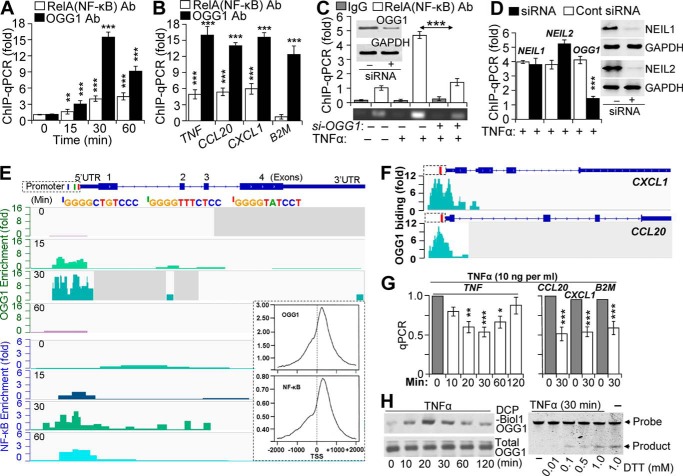
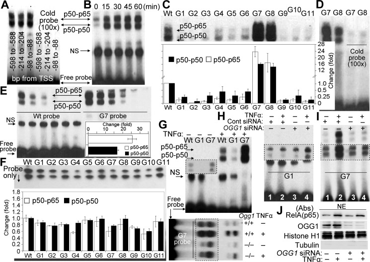
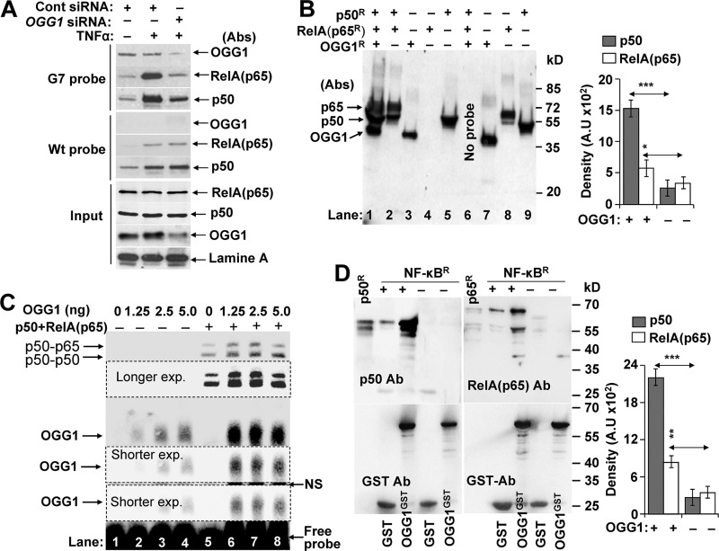
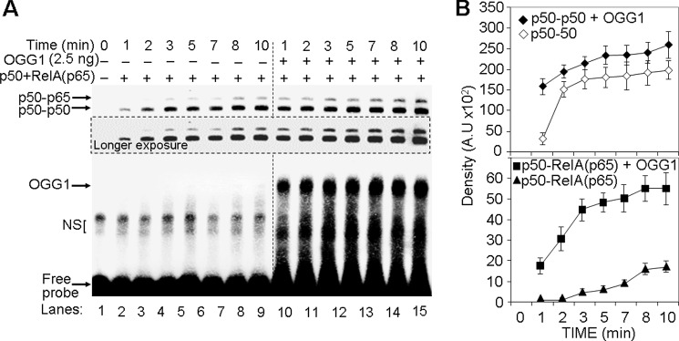
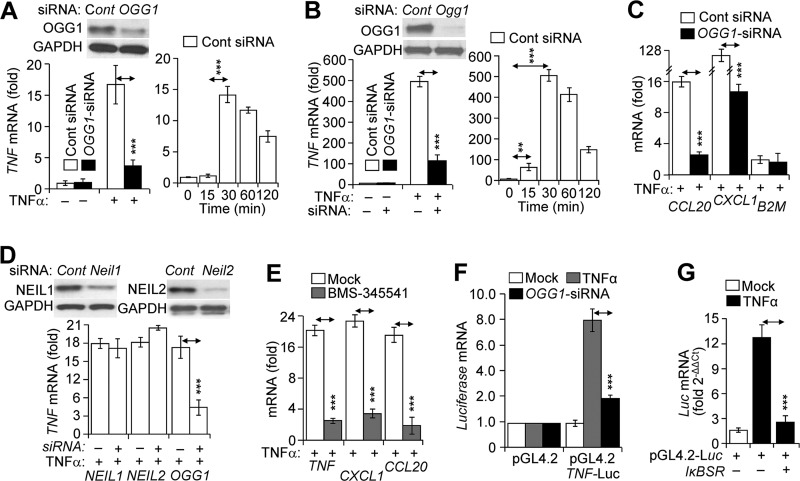
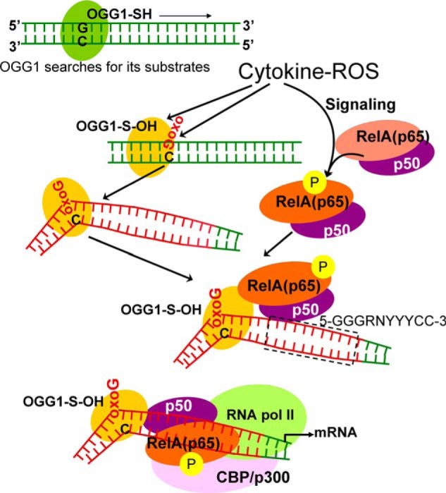
A large percentage of redox-responsive gene promoters contain evolutionarily conserved guanine-rich clusters; guanines are the bases most susceptible to oxidative modification(s). Consequently, 7,8-dihydro-8-oxoguanine (8-oxoG) is one of the most abundant base lesions in promoters and is primarily repaired via the 8-oxoguanine DNA glycosylase-1 (OOG1)-initiated base excision repair pathway. In view of a prompt cellular response to oxidative challenge, we hypothesized that the 8-oxoG lesion and the cognate repair protein OGG1 are utilized in transcriptional gene activation. Here, we document TNFα-induced enrichment of both 8-oxoG and OGG1 in promoters of pro-inflammatory genes, which precedes interaction of NF-κB with its DNA-binding motif. OGG1 bound to 8-oxoG upstream from the NF-κB motif increased its DNA occupancy by promoting an on-rate of both homodimeric and heterodimeric forms of NF-κB. OGG1 depletion decreased both NF-κB binding and gene expression, whereas Nei-like glycosylase-1 and -2 had a marginal effect. These results are the first to document a novel paradigm wherein the DNA repair protein OGG1 bound to its substrate is coupled to DNA occupancy of NF-κB and functions in epigenetic regulation of gene expression.

Keywords: 8-oxoguanine (8-oxoG); 8-oxoguanine glycosylase (OGG1); NF-κB transcription factor; promoter; reactive oxygen species (ROS).


Figures

FIGURE 1.
FIGURE 1.
Binding of NF-κB to promoters is OGG1-dependent. A, TNFα-induced enrichment of OGG1 and NF-κB on TNF promoter. Cells expressing FLAG-OGG1 were mock- or TNFα-exposed, and ChIP was performed at 0, 15, 30, and 60 min using anti-FLAG(OGG1) or anti-RelA(NF-κB) Ab. Uncross-linked DNA was subjected to PCR amplification as described under “Experimental Procedures” (n = 4). B, TNFα-induced enrichment of OGG1 and NF-κB on CCL20, CXCL1, and B2M promoters. Cells expressing FLAG-OGG1 were mock- or TNFα-exposed, and ChIP was performed at 30 min using anti-FLAG(OGG1) or anti-RelA(NF-κB) Ab. Uncross-linked DNA was subjected to PCR amplification of TNF, CCL20, CXCL1, and B2M promoters as described under “Experimental Procedures.” C, OGG1 expression-dependent enrichment of NF-κB on TNF promoter. OGG1 expression was down-regulated by siRNA (scrambled siRNA was used as a control, see under “Experimental Procedures”), and cells were TNFα-exposed. ChIP was performed at 30 min using anti-RelA(NF-κB) Ab (in controls, IgG). The levels of the TNF promoter in ChIP-ed DNA were determined by qPCR. The upper panel is a graphical depiction of changes in NF-κB binding to the TNF promoter in the presence and absence of OGG1 (n = 3). Lower panel, ethidium bromide-stained agarose gel from a representative experiment. Inset, cells were transfected with control and target-specific (OGG1) siRNA. At 48 h thereafter, cells were lysed to prepare protein extracts, and levels of OGG1 were determined by Western blotting. D, OGG1 depletion, but not that of NEIL1 or NEIL2, decreases NF-κB's association with promoters. NEIL1, NEIL2, or OGG1 expression was down-regulated by siRNA as described under “Experimental Procedures,” and cells were TNFα-exposed. ChIP was performed using Ab to RelA(NF-κB). The levels of TNF promoter in ChIP-ed DNA were determined by qPCR (30-min time point is shown). E, enrichment of OGG1 and NF-κB on the TNF promoter. FLAG-OGG1-expressing cells were TNFα-exposed and ChIP-ed at 0, 15, 30, and 60 min using Abs against RelA(NF-κB) or FLAG(OGG1). ChIP-ed DNA was subjected to sequencing, and sequence data were analyzed as described under “Experimental Procedures.” Images are directly taken from the Integrative Genomics Viewer. Enrichment levels (fold) of OGG1 and NF-κB for each time points are shown (left-hand side). Blue, green, and red bars are positions of NF-κB-binding motifs in promoter. Inset, distribution of OGG1 and NF-κB on TSS-adjacent sequences ±2000 bp at whole genome level. F, enrichment of OGG1 at the CXCL1 and CCL20 promoters. FLAG-OGG1-expressing cells were TNFα-exposed and ChIP-ed using Ab FLAG(OGG1), and ChIP-ed DNA was sequenced (see under “Experimental Procedures”). Images show enrichment peaks of OGG1 at 30 min after TNFα exposure of cells. Images are directly taken from the Integrative Genomics Viewer. Red bars within CXCL1 and CCL20 promoter regions are the locations of NF-κB-binding motifs. G, OGG1 digestion decreases qPCR product levels of TNF, CCL20, CXCL1, and B2M promoters. Cells were exposed to TNFα (20 ng/ml) for various lengths of time, and then the isolated DNAs were OGG1-digested and subjected to qPCR as under “Experimental Procedures” (n = 3). H, TNFα exposure of cells induces oxidative modifications of OGG1 and decreases its enzymatic activity. FLAG-OGG1-expressing cells were TNFα-exposed, and nuclear extracts were made in the presence or absence of DCP-Bio1 (a cysteine sulfenic acid-reacting agent). Levels of DCP-Bio1-tagged OGG1 were determined by streptavidin-coupled chemiluminescence. Total OGG1 levels were determined by immunoblotting using Ab to FLAG (OGG1). Lower panel: enzymatic activity of OGG1 is decreased in TNFα-exposed cells. NE was isolated at the 30-min post-exposure, and OGG1's excision activity was determined in the presence or absence of DTT in the reaction buffer, as described under “Experimental Procedures.” * = p < 0.05; ** = p < 0.01; *** = p < 0.001; Cells used are as follows: HEK293; CCL20, promoter of that chemokine (C-C motif) ligand 20 gene; CXCL1, promoter of chemokine (CXC motif) ligand 1 gene; B2M, promoter of β2-microglobulin gene; NEIL1, gene human Nei-like DNA glycosylase-1; NEIL2, gene human Nei-like DNA glycosylase 2; OGG1, gene of human 8-oxoguanine DNA glycosylase.
FIGURE 2.
FIGURE 2.
NF-κB's DNA occupancy as a function of OGG1 expression. A, putative NF-κB motifs located in TNF promoter are functional. NEs isolated from OGG1-expressing TNFα (20 ng per ml)-exposed cells were incubated with synthetic DNA (probe) containing NF-κB-binding sites derived from TSS-adjacent region of TNF promoter (5′-GGGGTTTCTCC-3′, −598 bp to −588 bp; 5′-GGGGTATCC-3′, −214 bp to −204 bp; and 5′-GGGGCTGTCC-3′, −98 bp to −88 bp). EMSA was performed using 2 μg of NE per individual probes. A cold probe containing canonical NF-κB motif (5′-GGGATCATTCCC-3′ (26)) prevented binding of NF-κB. B, nuclear accumulation of NF-κB in OGG1-expressing cells as assessed by EMSA. Cells were TNFα-exposed, and NEs were prepared at the time points indicated in the figure. EMSA was carried out (see under “Experimental Procedures”) using a probe containing an NF-κB-binding motif (5′-GGGGTATCC-3′, −214 bp to −204 bp upstream from TSS in the TNF promoter). C, OGG1 present in crude NE modulates NF-κB's DNA occupancy in an 8-oxoG position manner. NEs from TNFα-exposed OGG1-expressing cells were isolated at 30 min, and EMSAs were performed using probes lacking (WT) or containing 8-oxoG (G1 to G11) in sense (5′-TG7GGG8AGT GTG5AG1G2G3G4TAT CCG6AT GCTTG-3′) and complementary strand (3′-ACCCCTCACACTCCCCATAG9G10AACTAC G11AAC-5). Inset, graphical depiction of NF-κB binding to individual probes (n = 3). Band intensities of p50 homodimers and p50-p65 heterodimers were determined by densitometry using ImageJ software (version 1.44). D, NF-κB's occupancy of its binding site within G7 and G8 probes is specific. Crude NEs from OGG1-expressing cells were incubated with cold probe containing the IκB motif, and then G7 or G8 was added, and EMSAs were performed. E, OGG1 increases in occupancy of NF-κB's on DNA containing 8-oxoG 11 bp upstream of its binding site. Crude NEs from TNFα-exposed, OGG1-expressing cells were diluted stepwise (2, 1, 0.5, and 0.25, 0.125, and 0.006 μg per assay) and added to WT (left side) and G7 (right side) probes for 5 min, and EMSA was performed. Inset, fold increases in occupancy of homo- and heterodimeric NF-κB on the G7 probe (n = 3). Band intensities were determined by densitometry using ImageJ software, and fold changes in binding were calculated. F, binding of NF-κB to WT and 8-oxoG-containing probes. Recombinant p50 (3.75 ng) and RelA(p65) (2.75 ng) proteins were annealed in binding buffer for 60 min, and then individual probes were added for 5 min, and mixtures were subjected to EMSA. Inset, graphical depictions of changes in band intensities of p50-p50 homodimers and p50-p65 heterodimers. Band intensities were determined using ImageJ software (version 1.44), and fold changes were calculated (n = 3). G, occupancy of NF-κB on DNA, containing 8-oxoG within and adjacent to its motif. Upper panel, crude NEs (2 μg per assay) from OGG1-expressing, mock-, or TNFα-exposed cells were added to probes lacking (WT) or containing 8-oxoG within (G1) or 11 bp upstream of NF-κB's binding sequence (G7). Inset, longer exposure. Lower panel, NF-κB's DNA occupancy on G7 probe in crude NEs from Ogg1+/+ and Ogg1−/− cells. H, NF-κB's occupancy of DNA-containing 8-oxoG within its binding motif is re-established in NE from OGG1-depleted cells. OGG1 siRNA and control siRNA-transfected cells were TNFα-exposed (30 min), and NEs were prepared to perform EMSAs using the G1 probe. I, occupancy of NF-κB on 8-oxoG-containing DNA is diminished in NEs isolated from OGG1-depleted cells. OGG1 and control siRNA-transfected cells were TNFα-exposed for 30 min, NEs were prepared to perform EMSAs using the 8-oxoG-containing G7 probe (8-oxoG is 11 bp distance from its binding site). Lane 1, control; lane 2, TNFα-exposed OGG1-expressing; lane 3, mock-exposed OGG1-depleted; lane 4, TNFα-exposed, OGG1-depleted. J, OGG1 has insignificant impact on TNFα-induced nuclear translocation of RelA(NF-κB). Cells were TNFα-exposed for 30 min, and NEs were prepared. Proteins were fractionated by SDS-PAGE and immunoblotted using Abs to RelA(NF-κB) or OGG1. Quality control of NEs: 5 μg per lane, NEs were SDS-PAGE-fractionated and immunoblotted using Abs to tubulin and histone H1. A, B, and D–J, NEs were prepared from HEK293 cells; NS, nonspecific; TNF, human gene encoding for TNFα; WT, is a 31-bp DNA probe without 8-oxoG; G1 to G11 are 31-bp DNA probes containing 8-oxoG.
FIGURE 3.
FIGURE 3.
OGG1 facilitates NF-κB binding to its motif. A, OGG1 and NF-κB's occupancy of 8-oxoG-containing DNA in NE. NE (50 μg per sample) from ±TNFα-exposed, OGG1-expressing, and depleted cells were incubated with G7 (upper panel) or WT probe (middle panel) for 5 min, and protein DNA complexes were pulled down using magnetic streptavidin beads. The washed pellets were subjected to SDS-PAGE and immunoblotting using Abs to OGG1, p50, or RelA(p65). Lower panel (input): NE (3 μg per sample) was subjected SDS-PAGE and immunoblotted by using Ab to OGG1 and lamin A. B, OGG1 increases DNA occupancy of NF-κB. Annealed RelA(p65)R and p50R and OGG1R were mixed with the 8-oxoG-containing G7 probe conjugated to magnetic streptavidin beads. Five min thereafter, streptavidin bead-G7-associated proteins were collected, washed, and subjected to SDS-PAGE and immunoblotted using Abs to OGG1, p50, and RelA(p65). Lane 1, RelA(p65)R + p50R+OGG1; lane 2, p65R+p50R; lane 3, OGG1R alone; lane 4, RelA(p65)R alone; lane 5, p50R alone. Lanes 7–9 are OGG1R, RelA(p65)R, and p50R protein loaded as molecular size markers, respectively. Right panel, fold changes in p50 and p65(RelA) binding to G7 probe ± OGG1 (n = 3). Band intensities were determined by densitometry using ImageJ software (version 1.44). C, mutually positive interactions between NF-κB and OGG1 in binding to 8-oxoG containing DNA. Lanes 1–4, OGG1's binding to 8-oxoG-containing G7 probe. Increasing amounts of OGG1 were mixed with G7 probes for 5 min and subjected to EMSA. Lanes 5–8, OGG1 increases NF-κB's DNA occupancy. Annealed p50-RelA(p65) was added to the probe simultaneously with increasing concentrations of OGG1 for 5 min, and EMSAs were carried out. Results from a representative experiment (n = 3) are shown. D, physical interactions between OGG1 and NF-κB. p50R (15 ng) and p65R (20 ng) was preincubated for annealing, and GST or GST-tagged OGG1 (OGG1GST) was added in binding buffer at 37 °C for 30 min. OGG1GST·NF-κB complexes were pulled down using glutathione-Sepharose; GST served as a control (see under “Experimental Procedures”). NF-κB subunit proteins as well as OGG1GST were detected by immunoblotting using Ab to p50, RelA(p65) (upper panels) or GST (lower panels). p50R or Rel(p65)Ralone served as molecular size markers. Results of a representative experiment out of three are shown. Band intensities were determined by densitometry using ImageJ software (version 1.44), and fold changes were calculated (n = 3). * = p < 0.05; ** = p < 0.01; *** = p < 0.001; cells used are as follows: HEK293; OGG1R, recombinant human 8-oxoguanine DNA glycosylase-1; OGG1GST, glutathione S-transferase-tagged OGG1; NFBR, recombinant p50R and RelA(p65)R.
FIGURE 4.
FIGURE 4.
OGG1 accelerates association of NF-κB with its DNA motif. A, kinetics of binding of homo- and heterodimeric NF-κB to DNA ± OGG1. Annealed NF-κB subunits were mixed with OGG1, and 8-oxoG-containing DNA (G7 probe) was added. Aliquots were taken at the times indicated, and the levels of NF-κB·DNA complexes were determined by EMSA. B, graphical depiction of NF-κB's binding kinetic to DNA. Band intensities of NF-κB' homo- and heterodimers were determined by using ImageJ software (version 1.44) (n = 3).
FIGURE 5.
FIGURE 5.
OGG1-dependent gene expression in TNFα-exposed cells. A and B, OGG1-dependent gene expression from TNF/Tnf promoters. Left panels, parallel cultures of cells were transfected with control siRNA (□) or siRNA to OGG1 (■), and cells were exposed to TNFα (20 ng/ml) for 30 min. TNF mRNA levels were determined by RT-PCR. A and B, insets, expression of OGG1 at protein levels in cells after two cycles of transfections of cells with siRNA (Western blotting analysis). A and B, right panels, HEK293 and MLE-12 cells were exposed to TNFα (20 ng/ml), and mRNA levels were determined by RT-PCR at the time points indicated (n = 3). C, OGG1-dependent expression of CCL20 and CXCL1 mRNA. Cells were transfected with control siRNA (□) or siRNA to OGG1 (■) and TNFα-exposed (20 ng/ml). In controls, expression from B2M was tested. mRNA levels were determined at 30 min by RT-PCR (n = 3). D, depletion of NEIL1 and NEIL2 has no significant impact on TNF mRNA levels. Parallel cultures of cells were sequentially transfected with control siRNA (Cont) or siRNA to NEIL1, NEIL2, or OGG1. Inset, Western blotting analysis of protein levels. TNFα (20 ng/ml) was added for 30 min, and RNAs were isolated. mRNA levels of TNF were determined by RT-PCR (n = 3). E, IκB kinase inhibitor BMS-345541 decreases gene expression. Cells were pretreated with BMS-345541 (10 μm) for 3 h, and TNFα-exposed (20 ng/ml). mRNA levels were determined at 30 min by RT-PCR (n = 4). F, decreases in luciferase mRNA expression driven by TNF promoter in OGG1-depleted cells. OGG1 was depleted by siRNA, and cells were transfected with pGL4.2-Luc vector containing a promoter region (−974 + 90 bp) of human TNF (see under “Experimental Procedures”). mRNA expression levels of firefly luciferase were assessed at 30 min after mock (□) or TNFα (■) exposure (n = 4). G, NF-κB-dependent expression from the TNF promoter. Cells were co-transfected with IκB super-repressor (IκBSR) and pGL4.2-Luc, and then mock-exposed (□) or TNFα-exposed (■, 20 ng/ml). mRNA levels of firefly luciferase were assessed by RT-PCR (n = 3). ** = p < 0.01; *** = p < 0.001. HEK293 cells were utilized for experiments described in C–G. OGG1, 8-oxoguanine DNA glycosylase-1; CCL20, mRNA encoded by chemokine (C-C motif) ligand 20 gene; CXCL1, mRNA encoded by chemokine (CXC motif) ligand 1 gene; B2M, mRNA encoded by β2-microglobulin gene; IκBSR, super-repressor of κ-light-chain-enhancer of activated B cells; pGL4.2, luciferase (Luc) reporter vector 4.2; NEIL1, gene human Nei-like DNA glycosylase-1; NEIL2, gene human Nei-like DNA glycosylase2; OGG1, gene of human 8-oxoguanine DNA glycosylase-1; Tnf-Luc, human TNF promoter-containing pGL4.2-Luc expressing vector.
FIGURE 6.
FIGURE 6.
Model is shown for OGG1-mediated “homing” of NF-κB. OGG1 is a prototypic base excision repair protein, which searches for its substrates oxoG (8-oxoG, FapyG) in DNA (48). ROS-induced damage to DNA and OGG1's oxidative modification at cysteine (OGG1-SH → OGG1-S-OH) are inevitable events. OGG1-S-OH has a compromised base excision activity, although it is capable of base extrusion from DNA helix and interaction with the opposite cytosine and structural DNA modifications adjacent to its DNA footprint (39). OGG1-driven architectural DNA modification is utilized by NF-κB subunits for DNA occupancy. OGG1, 8-oxoguanine DNA glycosylase-1; oxoG, 8-oxoguanine; CBP/p300, cAMP-response element-binding protein (transcriptional co-activator); RNA pol II, RNA polymerase II; RelA(p65), 65-kDa regulatory subunit of NF-κB; p50, 50-kDa protein A DNA-binding subunit of NF-κB; 5′-GGGRNYYYCC-3′, NF-κB binding motif; G, guanine; R, purine; Y, pyrimidine; N can be any nucleotide (26).

Similar articles

Cited by

References

    1. David S. S., O'Shea V. L., and Kundu S. (2007) Base-excision repair of oxidative DNA damage. Nature 447, 941–950 - PMC - PubMed
    1. Dizdaroglu M. (1992) Oxidative damage to DNA in mammalian chromatin. Mutat. Res. 275, 331–342 - PubMed
    1. Steenken S., and Jovanovic S. V. (1997) How easily oxidizable is DNA? One-electron reduction potentials of adenosine and guanosine radicals in aqueous solution. J. Am. Chem. Soc. 119, 617–618
    1. Mitra S., Hazra T. K., Roy R., Ikeda S., Biswas T., Lock J., Boldogh I., and Izumi T. (1997) Complexities of DNA base excision repair in mammalian cells. Mol. Cells 7, 305–312 - PubMed
    1. Lindahl T., and Barnes D. E. (2000) Repair of endogenous DNA damage. Cold Spring Harb. Symp. Quant. Biol. 65, 127–133 - PubMed