. 2016 Dec 13;7(50):82889-82901.
doi: 10.18632/oncotarget.12658.

Targeting the cancer-associated fibroblasts as a treatment in triple-negative breast cancer

Affiliations

Targeting the cancer-associated fibroblasts as a treatment in triple-negative breast cancer

Ken Takai et al. Oncotarget. .

Abstract

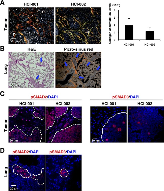
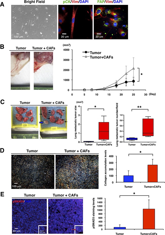
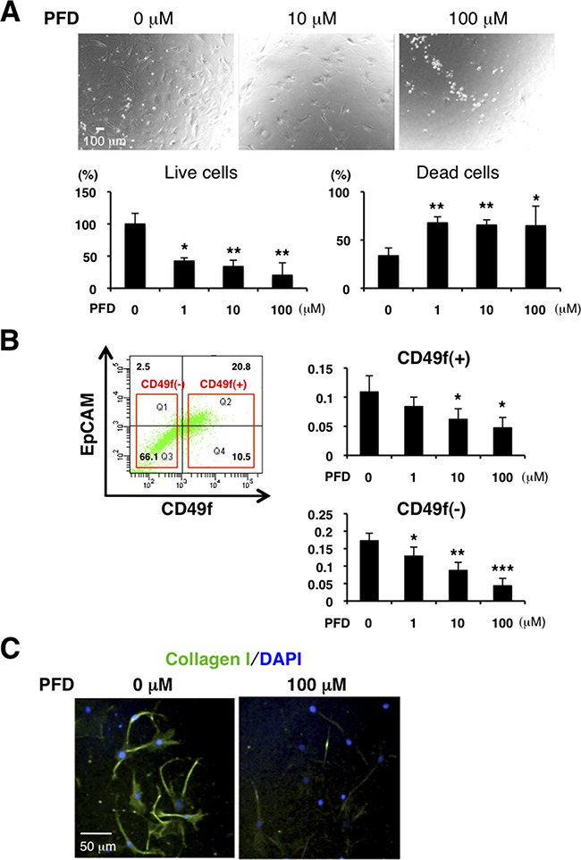
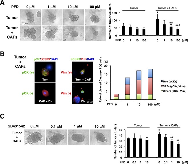
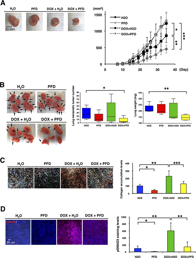
Increased collagen expression in tumors is associated with increased risk of metastasis, and triple-negative breast cancer (TNBC) has the highest propensity to develop distant metastases when there is evidence of central fibrosis. Transforming growth factor-β (TGF-β) ligands regulated by cancer-associated fibroblasts (CAFs) promote accumulation of fibrosis and cancer progression. In the present study, we have evaluated TNBC tumors with enhanced collagen to determine whether we can reduce metastasis by targeting the CAFs with Pirfenidone (PFD), an anti-fibrotic agent as well as a TGF-β antagonist. In patient-derived xenograft models, TNBC tumors exhibited accumulated collagen and activated TGF-β signaling, and developed lung metastasis. Next, primary CAFs were established from 4T1 TNBC homograft tumors, TNBC xenograft tumors and tumor specimens of breast cancer patients. CAFs promoted primary tumor growth with more fibrosis and TGF-β activation and lung metastasis in 4T1 mouse model. We then examined the effects of PFD in vitro and in vivo. We found that PFD had inhibitory effects on cell viability and collagen production of CAFs in 2D culture. Furthermore, CAFs enhanced tumor growth and PFD inhibited the tumor growth induced by CAFs by causing apoptosis in the 3D co-culture assay of 4T1 tumor cells and CAFs. In vivo, PFD alone inhibited tumor fibrosis and TGF-β signaling but did not inhibit tumor growth and lung metastasis. However, PFD inhibited tumor growth and lung metastasis synergistically in combination with doxorubicin. Thus, PFD has great potential for a novel clinically applicable TNBC therapy that targets tumor-stromal interaction.

Keywords: cancer-associated fibroblast; fibrosis; pirfenidone; transforming growth factor-β; triple-negative breast cancer.


Conflict of interest statement

CONFLICTS OF INTEREST

The authors declare no conflict of interest.

Figures

Figure 1
Figure 1. PDX models of TNBC exhibit enhanced collagen accumulation, activated TGF-β signaling and lung metastasis
A. TNBC xenograft tumors show enhanced collagen accumulation by picro-sirius red staining (left panel). Fibrillar collagen was quantified by picro-sirius red staining using ImageJ software. n=2-3 (right panel). B. Lung metastasis was detected in the TNBC xenograft model by H&E staining (left panel) and picro-sirius red staining (right panel). C. TNBC xenograft tumors were immunostained with anti-phospho-SMAD2 (red on left panel) and anti-phospho-SMAD3 (red on right panel) antibodies. DAPI (blue) stained nuclei. Phospho-SMAD2 was widely expressed in primary tumors and stroma. Phospho-SMAD3 was sporadically expressed in primary tumors, not in stroma. D. Lungs of the TNBC xenograft model were immunostained with anti-phospho-SMAD2 (red on left panel) and anti-phospho-SMAD3 (red on right panel) antibodies. DAPI (blue) stained nuclei. Phospho-SMAD2 and phospho-SMAD3 were expressed in the lung metastatic tumors, but not the stroma around the small tumors.
Figure 2
Figure 2. CAFs promote primary tumor growth and lung metastasis in TNBC mouse model
A. We cultured CAFs from fresh tumor specimens of breast cancer patients in a 3% O2 incubator (Bright Field, left panel). Immunofluorescence of cultured CAFs was conducted by using anti-vimentin (Vim, red), anti-pan-cytokeratin (pCK, green in middle panel), and anti-fibroblast activation protein (FAP, green in right panel) antibodies. DAPI (blue) stained nuclei. CAFs were stained with anti-vimentin and anti-FAP antibodies. B. We transplanted 4T1 cells (1x10⁴) without or with CAFs (2x10⁴) into mammary glands of BALB/c mice (n=5). Representative photographs of 4T1 primary tumor after transplantation with or without the CAFs are shown (left panel). CAFs promoted primary tumor growth. Tumor volume (mm3) was measured by “V=0.52xW2xL.” W=width (mm), L=length (mm). *p<0.02 (right panel). C. Representative photographs of lungs showed that CAFs promoted lung metastasis. Arrows indicate visible lung metastatic tumors and a broad arrow indicates a metastatic lymph node in the right brachium (left panel). Volume (mm3) of single metastatic tumor was measured by “V=0.52xW2xL.” W=width (mm), L=length (mm). *p<0.05 (middle panel). H&E staining showed that CAFs increased lung metastatic tumor number (*p<0.02) (right panel). n=5. D. Representative photographs of picro-sirius red staining showed that CAFs promoted primary tumor fibrosis (left panel). Collagen deposition marked by picro-sirius red staining was quantified by using ImageJ software. CAFs enhanced collagen accumulation in primary tumors. n=5, *p<0.01 (right panel). E. Primary tumors were immunostained with an anti-phospho-SMAD3 antibody (red). DAPI (blue) stained nuclei. Representative photographs showed that CAFs enhanced expression level of phospho-SMAD3 in primary tumors (left panel). Expression levels of phospho-SMAD3 in primary tumors were quantified by using ImageJ software. n=3, *p<0.02 (right panel).
Figure 3
Figure 3. PFD has inhibitory effects on cell viability and collagen production in CAFs
A. We cultured CAFs derived from tumor specimens of luminal-type breast cancer patients in a 3% O2 incubator and treated them with PFD in triplicate. Representative photographs are shown (upper panels). Total cells were stained with trypan blue on Day 4. Live cells decreased and dead cells increased with higher concentration of PFD (lower panels). *p<0.05, **p<0.01 compared to the control condition. B. FACS analysis was conducted from dissociated HCI-001 TNBC xenograft tumor cells by using anti-CD49f and anti-EpCAM antibodies and then CD49f+ cells (tumor cells) and CD49f cells (mainly stromal cells) were sorted. 4,000 CD49f+ cells or 10,000 CD49f cells were plated with each PFD concentration for culture and MTT assay was performed on day 15. Cell viability of CD49f+ and CD49f cells decreased with higher concentration of PFD. *p<0.05, **p<0.01, ***p<0.001 compared to the control conditions. C. We cultured CAFs derived from HCI-002 TNBC xenograft tumors. Immunofluorescence of the cultured CAFs used an anti-collagen I (green) antibody. DAPI (blue) stained nuclei. Collagen production was decreased by PFD.
Figure 4
Figure 4. Pirfenidone inhibits TNBC growth induced by CAFs
We cultured CAFs derived from TNBC xenograft tumors (HCI-001), and 3D co-culture assayed the CAFs and aggregated 4T1 cells (tumor cluster) in Matrigel. A. We examined the effects of PFD (triplicate). CAFs increased the tumor cluster size and PFD inhibited the size increased by CAFs (left panel). Also, CAFs increased the number of tumor clusters and PFD decreased the tumor cluster number increased by CAFs (right panel). *p<0.05 compared to tumor (0 μM). **p<0.05 or ***p<0.02 compared to 0 μM (Tumor + CAFs). B. We conducted immunofluorescence of the 3D Matrigel cultures by using anti-pan-cytokeratin (pCK, green, left panel), anti-vimentin (Vim, red, middle panel) and cleaved caspase-3 (cCSP3, red, left and green, middle panels, respectively) antibodies. DAPI (blue) stained nuclei. 4T1 tumor cells expressed both pan-cytokeratin and vimentin, and CAFs expressed vimentin. Therefore, pan-cytokeratin+ cells were 4T1 cells, and vimentin+ cells were either 4T1 cells or CAFs. Double-negative cells were regarded as other cell type. We counted the numbers of those cells (except 4T1 tumor clusters) with cleaved caspase-3 (representative photographs in left and middle panels) and quantified each apoptotic cells (right panel). We found that PFD induced apoptosis of 4T1 tumor cells and CAFs. C. We examined the effects of a TGF-β inhibitor (SB431542) in the 3D co-culture assay (triplicate). PFD decreased the tumor cluster size (left panel) and the tumor cluster number (right panel). *p<0.05 compared to tumor (0 μM). **p<0.01 compared to 0 μM (Tumor + CAFs).
Figure 5
Figure 5. Pirfenidone inhibits primary tumor growth and lung metastasis in combination with doxorubicin in TNBC mouse model
We transplanted 4T1 cells (1x10⁴) and 4T1-stimulated CAFs (2x10⁴) into mammary glands of BALB/c mice (n=5-6). PFD (50 mg/kg) or water was orally administered two times per day and doxorubicin (4 mg/kg) or PBS was injected from the mouse tail vain on day 0 and 19. A. Representative photographs of primary tumors after the treatments (Day 37) (left panel). Tumor volume (mm3) was measured by “V=0.52xW2xL.” W=width (mm), L=length (mm). PFD had no effect on primary tumor growth, but inhibited the tumor growth synergistically in combination with doxorubicin. *p<0.05, **p<0.01, ***p<0.001 (right panel). B. Representative photographs of lung after the treatments (Day 37) (left panel). Visible lung metastatic tumor numbers in five lobes were counted. PFD decreased the lung metastatic tumor numbers in combination with doxorubicin, but PFD monotherapy did not decrease the numbers. *p<0.02 (middle panel). Total lung weight was measured. PFD decreased tumor weight in combination with doxorubicin though PFD monotherapy did not decrease the weight. **p<0.002 (right panel). C. Representative photographs of primary tumors by picro-sirius red staining showed that doxorubicin enhanced and PFD inhibited collagen accumulation in primary tumors (left panel). Collagen deposition visualized by picro-sirius red staining was quantified by using ImageJ software. n=3, *p<0.01, **p<0.02, ***p<0.05 (right panel). D. Primary tumors were immunostained with an anti-phospho-SMAD3 antibody (red). DAPI (blue) stained nuclei (left panel). Expression levels of phospho-SMAD3 were quantified by using ImageJ software. Doxorubicin enhanced and PFD inhibited phospho-SMAD3 levels in primary tumors. n=4, *p<0.02, **p<0.01 (right panel).

Similar articles

Cited by

References

    1. Kalluri R, Zeisberg M. Fibroblasts in cancer. Nat Rev Cancer. 2006;6:392–401. - PubMed
    1. Sternlicht MD, Lochter A, Sympson CJ, Huey B, Rougier JP, Gray JW, Pinkel D, Bissell MJ, Werb Z. The stromal proteinase MMP3/stromelysin-1 promotes mammary carcinogenesis. Cell. 1999;98:137–146. - PMC - PubMed
    1. Radisky DC, Kenny PA, Bissell MJ. Fibrosis and cancer: do myofibroblasts come also from epithelial cells via EMT? J Cell Biochem. 2007;101:830–839. - PMC - PubMed
    1. Egeblad M, Rasch MG, Weaver VM. Dynamic interplay between the collagen scaffold and tumor evolution. Curr Opin Cell Biol. 2010;22:697–706. - PMC - PubMed
    1. Hasebe T. Tumor-stromal interactions in breast tumor progression--significance of histological heterogeneity of tumor-stromal fibroblasts. Expert Opin Ther Targets. 2013;17:449–460. - PubMed

MeSH terms