

Anti-FIRs (PUF60) auto-antibodies are detected in the sera of early-stage colon cancer patients
- PMID: 27756887
- PMCID: PMC5347708
- DOI: 10.18632/oncotarget.12696
Anti-FIRs (PUF60) auto-antibodies are detected in the sera of early-stage colon cancer patients
Abstract
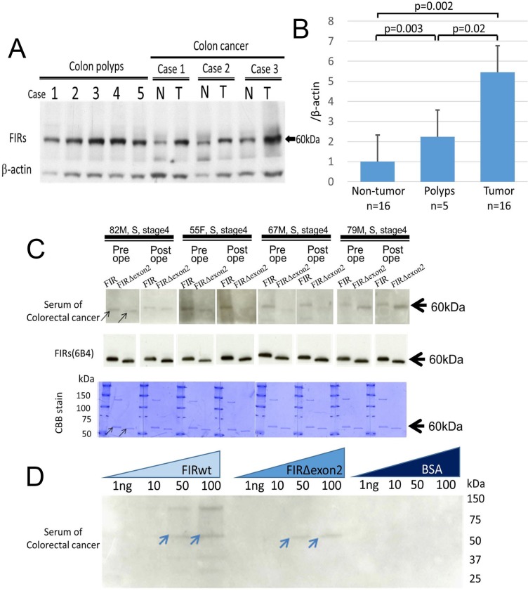
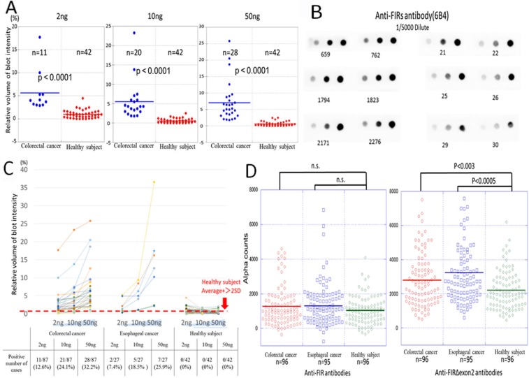
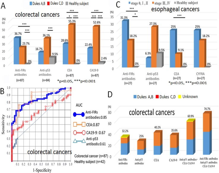
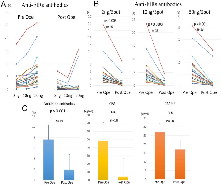
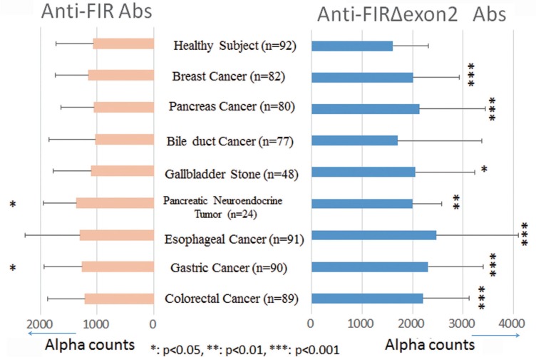
Anti-PUF60, poly(U)-binding-splicing factor, autoantibodies are reported to be detected in the sera of dermatomyositis and Sjogren's syndrome that occasionally associated with malignancies. PUF60 is identical with far-upstream element-binding protein-interacting repressor (FIR) that is a transcriptional repressor of c-myc gene. In colorectal cancers, a splicing variant of FIR that lacks exon2 (FIRΔexon2) is overexpressed as a dominant negative form of FIR. In this study, to reveal the presence and the significance of anti-FIRs (FIR/FIRΔexon2) antibodies in cancers were explored in the sera of colorectal and other cancer patients. Anti-FIRs antibodies were surely detected in the preoperative sera of 28 colorectal cancer patients (32.2% of positive rates), and the detection rate was significantly higher than that in healthy control sera (Mann-Whitney U test, p < 0.01). The level of anti-FIRs antibodies significantly decreased after the operation (p < 0.01). Anti-FIRs antibodies were detected in the sera of early-stage and/or recurrent colon cancer patients in which anti-p53 antibodies, CEA, and CA19-9 were not detected as well as in the sera of other cancer patients. Furthermore, the area under the curve of receiver operating characteristic for anti-FIRs antibodies was significantly larger (0.85) than that for anti-p53 antibodies or CA19-9. In conclusions, the combination of anti-FIRs antibodies with other clinically available tumor markers further improved the specificity and accuracy of cancer diagnosis.
Keywords: auto-antibodies; cancer biomarker; colorectal cancer; far-upstream element-binding protein-interacting repressor (FIR) = poly(U)-binding-splicing factor (PUF60).
Conflict of interest statement
The Authors do not have any conflicts of interest.
Figures
Similar articles
-
Anti-FIRΔexon2, a splicing variant form of PUF60, autoantibody is detected in the sera of esophageal squamous cell carcinoma.Cancer Sci. 2019 Jun;110(6):2004-2013. doi: 10.1111/cas.14024. Epub 2019 May 20. Cancer Sci. 2019. PMID: 30980774 Free PMC article.
-
PUF60: a prominent new target of the autoimmune response in dermatomyositis and Sjögren's syndrome.Ann Rheum Dis. 2016 Jun;75(6):1145-51. doi: 10.1136/annrheumdis-2015-207509. Epub 2015 Aug 7. Ann Rheum Dis. 2016. PMID: 26253095 Free PMC article.
-
Disturbed alternative splicing of FIR (PUF60) directed cyclin E overexpression in esophageal cancers.Oncotarget. 2018 May 1;9(33):22929-22944. doi: 10.18632/oncotarget.25149. eCollection 2018 May 1. Oncotarget. 2018. PMID: 29796163 Free PMC article.
-
[Alternative Splicing Detection as a Biomarker for Cancer Diagnosis: A Novel Progressive Mechanism of Acute Lymphoblastic Leukemia with Alternative Splicing as a Biomarker Candidate].Rinsho Byori. 2015 Sep;63(9):1091-102. Rinsho Byori. 2015. PMID: 26731899 Review. Japanese.
-
[Alternative Splicing Detection as Biomarker Candidates for Cancer Diagnosis and Treatment with Establishment of Clinical Biobank in Chiba University].Rinsho Byori. 2015 Mar;63(3):347-60. Rinsho Byori. 2015. PMID: 26524858 Review. Japanese.
Cited by
-
PUF60/AURKA Axis Contributes to Tumor Progression and Malignant Phenotypes in Bladder Cancer.Front Oncol. 2020 Oct 7;10:568015. doi: 10.3389/fonc.2020.568015. eCollection 2020. Front Oncol. 2020. PMID: 33117697 Free PMC article.
-
Serum anti-DIDO1, anti-CPSF2, and anti-FOXJ2 antibodies as predictive risk markers for acute ischemic stroke.BMC Med. 2021 Jun 9;19(1):131. doi: 10.1186/s12916-021-02001-9. BMC Med. 2021. PMID: 34103026 Free PMC article.
-
Identification of key gene modules and hub genes of human mantle cell lymphoma by coexpression network analysis.PeerJ. 2020 Mar 20;8:e8843. doi: 10.7717/peerj.8843. eCollection 2020. PeerJ. 2020. PMID: 32219041 Free PMC article.
-
Autoantibody Discovery, Assay Development and Adoption: Death Valley, the Sea of Survival and Beyond.Front Immunol. 2021 May 27;12:679613. doi: 10.3389/fimmu.2021.679613. eCollection 2021. Front Immunol. 2021. PMID: 34122443 Free PMC article. Review.
-
Integrated In Silico Analyses Identify PUF60 and SF3A3 as New Spliceosome-Related Breast Cancer RNA-Binding Proteins.Biology (Basel). 2022 Mar 22;11(4):481. doi: 10.3390/biology11040481. Biology (Basel). 2022. PMID: 35453681 Free PMC article.
References
-
- Matsushita K, Tomonaga T, Shimada H, Shioya A, Higashi M, Matsubara H, Harigaya K, Nomura F, Libutti D, Levens D, Ochiai T. An essential role of alternative splicing of c-myc suppressor fuse-binding protein-interacting repressor in carcinogenesis. Cancer Res. 2006;66:1409–1417. - PubMed
-
- Kajiwara T, Matsushita K, Itoga S, Tamura M, Tanaka N, Tomonaga T, Matsubara H, Shimada H, Habara Y, Matsuo M, Nomura F. Sap155-mediated c-myc suppressor far-upstream element-binding protein-interacting repressor splicing variants are activated in colon cancer tissues. Cancer Sci. 2013;104:149–156. - PMC - PubMed
-
- Malz M, Bovet M, Samarin J, Rabenhorst U, Sticht C, Bissinger M, Roessler S, Bermejo JL, Renner M, Calvisi DF, Singer S, Ganzinger M, Weber A, et al. Overexpression of far upstream element (fuse) binding protein (fbp)-interacting repressor (fir) supports growth of hepatocellular carcinoma. Hepatology. 2014;60:1241–1250. - PubMed
-
- Rahmutulla B, Matsushita K, Satoh M, Seimiya M, Tsuchida S, Kubo S, Shimada H, Ohtsuka M, Miyazaki M, Nomura F. Alternative splicing of fbp-interacting repressor coordinates c-myc, p27kip1/cycline and ku86/xrcc5 expression as a molecular sensor for bleomycin-induced dna damage pathway. Oncotarget. 2014;5:2404–2417. doi: 10.18632/oncotarget.1650. - DOI - PMC - PubMed
MeSH terms
Substances
LinkOut - more resources
Full Text Sources
Other Literature Sources
Molecular Biology Databases
Research Materials
Miscellaneous
