

Corneal endothelial changes after accelerated corneal collagen cross-linking in keratoconus and postLASIK ectasia
- PMID: 27757009
- PMCID: PMC5053387
- DOI: 10.2147/OPTH.S113412
Corneal endothelial changes after accelerated corneal collagen cross-linking in keratoconus and postLASIK ectasia
Abstract
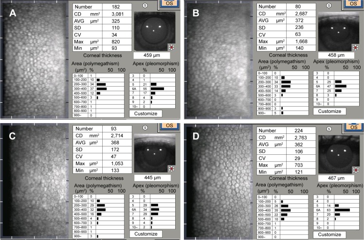
Purpose: The purpose of this study was to evaluate the effects of accelerated cross-linking (CXL) on corneal endothelium in keratoconus and postlaser-assisted in situ keratomileusis (LASIK) ectasia.
Design: This study is a prospective nonrandomized controlled study.
Setting: This study was conducted in Mansoura Ophthalmic Center (Mansoura University) and Al-Mostakbal Ophthalmic Center, Mansoura, Egypt.
Methods: In total, 40 eyes with progressive keratoconus and 10 eyes with postLASIK ectasia were subjected to an accelerated CXL (10 mW/cm2 for 9 minutes). Qualitative and quantitative analyses of the corneal endothelial cells were conducted before CXL and 3, 6, and 12 months after CXL by using a specular microscope (Tomy EM-3000).
Results: There was a significant reduction in endothelial cell count particularly at 3 and 6 months postCXL. In addition, the coefficient of variance was also statistically significantly higher at 3 and 6 months postoperatively than the preCXL value. There was a slight change in the percentage of hexagonal cells.
Conclusion: The use of accelerated CXL (10 mW/cm2 for 9 minutes) has a transient negative impact on endothelial cell density and/or endothelial morphology.
Keywords: accelerated cross-linking; corneal endothelium; keratoconus; postLASIK ectasia.
Conflict of interest statement
The author reports no conflicts of interest in this work.
Figures
Similar articles
-
Why Non-contact Tonometry Tests Cannot Evaluate the Effects of Corneal Collagen Cross-linking.J Refract Surg. 2017 Mar 1;33(3):184-192. doi: 10.3928/1081597X-20161206-02. J Refract Surg. 2017. PMID: 28264133
-
Prospective, randomized contralateral eye study of accelerated and conventional corneal cross-linking in pediatric keratoconus.Int Ophthalmol. 2019 May;39(5):971-979. doi: 10.1007/s10792-018-0898-y. Epub 2018 Mar 21. Int Ophthalmol. 2019. PMID: 29564806 Clinical Trial.
-
Conventional versus accelerated corneal collagen cross-linking in the treatment of keratoconus.Clin Exp Ophthalmol. 2016 Jan-Feb;44(1):8-14. doi: 10.1111/ceo.12571. Epub 2015 Jul 22. Clin Exp Ophthalmol. 2016. PMID: 26140309
-
Comparison of standard and accelerated corneal cross-linking for the treatment of keratoconus: a meta-analysis.Acta Ophthalmol. 2019 Feb;97(1):e22-e35. doi: 10.1111/aos.13814. Epub 2018 May 31. Acta Ophthalmol. 2019. PMID: 29855152 Review.
-
Combined laser in-situ keratomileusis and accelerated corneal cross-linking: an update.Curr Opin Ophthalmol. 2016 Jul;27(4):304-10. doi: 10.1097/ICU.0000000000000281. Curr Opin Ophthalmol. 2016. PMID: 27152484 Review.
Cited by
-
Tuck-in Lamellar keratoplasty with an lenticule obtained by small incision lenticule extraction for treatment of Post- LASIK Ectasia.Sci Rep. 2017 Dec 19;7(1):17806. doi: 10.1038/s41598-017-18201-4. Sci Rep. 2017. PMID: 29259313 Free PMC article.
-
Impact of Corneal Crosslinking on Endothelial and Biomechanical Parameters in Keratoconus.J Clin Med. 2025 Jun 25;14(13):4489. doi: 10.3390/jcm14134489. J Clin Med. 2025. PMID: 40648862 Free PMC article.
-
The effect of accelerated pulsed high-fluence corneal cross-linking on corneal endothelium; a prospective specular microscopy study.BMC Ophthalmol. 2023 Apr 18;23(1):163. doi: 10.1186/s12886-023-02912-6. BMC Ophthalmol. 2023. PMID: 37072730 Free PMC article.
-
Individual Risk Assessment and Prognostication of Outcomes After Corneal Cross-Linking.J Ophthalmol. 2025 Jul 1;2025:3678453. doi: 10.1155/joph/3678453. eCollection 2025. J Ophthalmol. 2025. PMID: 40630907 Free PMC article.
-
Ectasia After Corneal Refractive Surgery: A Systematic Review.Ophthalmol Ther. 2021 Dec;10(4):753-776. doi: 10.1007/s40123-021-00383-w. Epub 2021 Aug 20. Ophthalmol Ther. 2021. PMID: 34417707 Free PMC article. Review.
References
LinkOut - more resources
Full Text Sources
Other Literature Sources
