

Clinical safety and efficacy of implantation of octacalcium phosphate collagen composites in tooth extraction sockets and cyst holes
- PMID: 27757220
- PMCID: PMC5051665
- DOI: 10.1177/2041731416670770
Clinical safety and efficacy of implantation of octacalcium phosphate collagen composites in tooth extraction sockets and cyst holes
Abstract
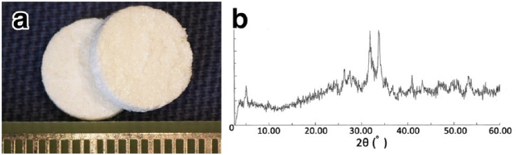
It was demonstrated that octacalcium phosphate collagen composite achieved notable bone regeneration in bone defects in preclinical studies. On the basis of the research results, an investigator-initiated exploratory clinical trial was conducted after approval from a local Institutional Review Board. This clinical study was performed as a single-arm non-randomized intervention study. Octacalcium phosphate collagen composite was implanted into a total of 10 cases of alveolar bone defects after tooth extractions and cystectomy. Safety assessment was performed in terms of the clinical course and several consecutive laboratory examinations, and sequential radiographs were used for efficacy assessment. All participants uneventfully completed the clinical trial without major problems in their general condition. Postoperative wound swelling was observed, as also commonly seen in tooth extraction or cystectomy. Although no serious liver dysfunction, renal dysfunction, electrolyte imbalance, or abnormal urinalysis results were recognized, the number of white blood cells and C-reactive protein level temporarily increased after the operation. An increase in radiopacity in the octacalcium phosphate collagen composite-implanted site was observed in all cases. Finally, the border between the original bone and the octacalcium phosphate collagen composite-implanted site became indistinguishable. These results suggest that octacalcium phosphate collagen composite could be utilized safely in clinical situations in the future.
Keywords: Bone regeneration; bone tissue engineering; calcium phosphate; collagen.
Conflict of interest statement
Declaration of conflicting interest: The authors (S.K. and O.S.) have obtained a patent for OCP/Col (#5046511) in Japan.
Figures


Similar articles
-
Clinical study of octacalcium phosphate and collagen composite in oral and maxillofacial surgery.J Tissue Eng. 2020 Jan 23;11:2041731419896449. doi: 10.1177/2041731419896449. eCollection 2020 Jan-Dec. J Tissue Eng. 2020. PMID: 32030119 Free PMC article.
-
Implantation of octacalcium phosphate collagen composites (OCP/Col) after extraction of canine deciduous teeth achieved undisturbed permanent tooth eruption.Arch Oral Biol. 2016 Dec;72:179-186. doi: 10.1016/j.archoralbio.2016.08.027. Epub 2016 Aug 26. Arch Oral Biol. 2016. PMID: 27598455
-
The regenerated bone quality by implantation of octacalcium phosphate collagen composites in a canine alveolar cleft model.Cleft Palate Craniofac J. 2014 Jul;51(4):420-30. doi: 10.1597/12-096. Epub 2013 Jan 31. Cleft Palate Craniofac J. 2014. PMID: 23369014
-
Octacalcium phosphate collagen composite facilitates bone regeneration of large mandibular bone defect in humans.J Tissue Eng Regen Med. 2017 May;11(5):1641-1647. doi: 10.1002/term.2110. Epub 2015 Nov 27. J Tissue Eng Regen Med. 2017. PMID: 26612731 Clinical Trial.
-
Octacalcium phosphate bone substitute materials: Comparison between properties of biomaterials and other calcium phosphate materials.Dent Mater J. 2020 Mar 31;39(2):187-199. doi: 10.4012/dmj.2020-001. Epub 2020 Mar 12. Dent Mater J. 2020. PMID: 32161239 Review.
Cited by
-
Influence of pre-freezing conditions of octacalcium phosphate and collagen composite for reproducible appositional bone formation.J Biomed Mater Res B Appl Biomater. 2020 Oct;108(7):2827-2834. doi: 10.1002/jbm.b.34613. Epub 2020 Apr 2. J Biomed Mater Res B Appl Biomater. 2020. PMID: 32239797 Free PMC article.
-
Comparative Analysis of Bone Regeneration According to Particle Type and Barrier Membrane for Octacalcium Phosphate Grafted into Rabbit Calvarial Defects.Bioengineering (Basel). 2024 Feb 24;11(3):215. doi: 10.3390/bioengineering11030215. Bioengineering (Basel). 2024. PMID: 38534489 Free PMC article.
-
Radiographic and Histomorphometric Evaluation of Sinus Floor Augmentation Using Biomimetic Octacalcium Phosphate Alloplasts: A Prospective Pilot Study.Materials (Basel). 2022 Jun 7;15(12):4061. doi: 10.3390/ma15124061. Materials (Basel). 2022. PMID: 35744118 Free PMC article.
-
Bone augmentation by octacalcium phosphate and collagen composite coated with poly-lactic acid cage.Clin Exp Dent Res. 2020 Aug;6(4):391-399. doi: 10.1002/cre2.287. Epub 2020 Mar 18. Clin Exp Dent Res. 2020. PMID: 32187863 Free PMC article.
-
Clinical study of octacalcium phosphate and collagen composite in oral and maxillofacial surgery.J Tissue Eng. 2020 Jan 23;11:2041731419896449. doi: 10.1177/2041731419896449. eCollection 2020 Jan-Dec. J Tissue Eng. 2020. PMID: 32030119 Free PMC article.
References
-
- Boyne PJ. Application of bone morphogenetic proteins in the treatment of clinical oral and maxillofacial osseous defects. J Bone Joint Surg Am 2001; 83-A: S146–S150. - PubMed
-
- Burg KJ, Porter S, Kellam JF. Biomaterial developments for bone tissue engineering. Biomaterials 2000; 21: 2347–2359. - PubMed
-
- Bucholz RW. Nonallograft osteoconductive bone graft substitutes. Clin Orthop Relat Res 2002; 395: 44–52. - PubMed
-
- LeGeros RZ. Properties of osteoconductive biomaterials: calcium phosphates. Clin Orthop Relat Res 2002; 395: 81–98. - PubMed
-
- Pourebrahim N, Hashemibeni B, Shahnaseri S, et al. A comparison of tissue-engineered bone from adipose-derived stem cell with autogenous bone repair in maxillary alveolar cleft model in dogs. Int J Oral Maxillofac Surg 2013; 42: 562–568. - PubMed
LinkOut - more resources
Full Text Sources
Other Literature Sources
Research Materials
