

Intra-tumoral heterogeneity in the expression of programmed-death (PD) ligands in isogeneic primary and metastatic lung cancer: Implications for immunotherapy
- PMID: 27757309
- PMCID: PMC5048760
- DOI: 10.1080/2162402X.2016.1213934
Intra-tumoral heterogeneity in the expression of programmed-death (PD) ligands in isogeneic primary and metastatic lung cancer: Implications for immunotherapy
Abstract
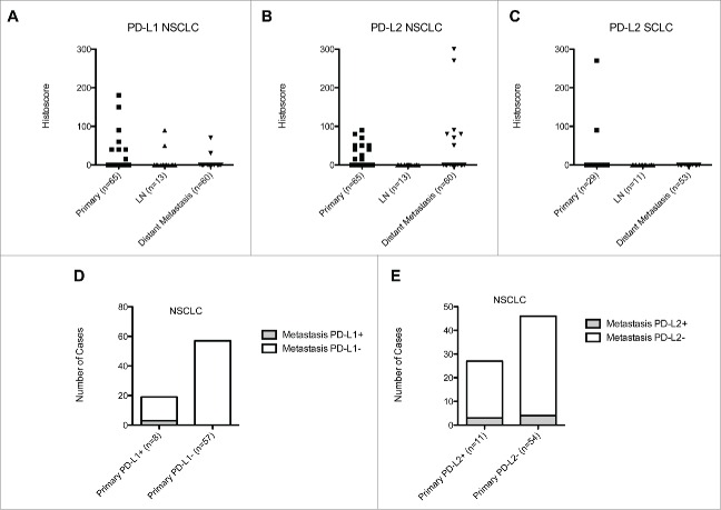
Purpose: There is inconclusive evidence to suggest the expression of programmed cell death (PD) ligand 1 (PD-L1) is a putative predictor of response to PD-1/PD-L1-targeted therapies in lung cancer. We evaluated the heterogeneity in the expression of PD-1 ligands in isogeneic primary and metastatic LC specimens. Experimental Design: From 12,580 post mortem cases, we identified 214 patients with untreated metastatic LC, of which 98 had adequately preserved tissues to construct a syngeneic primary LC/metastasis tissue microarray. Immunostaining for PD-L1 and 2 was evaluated in paired primary and metastatic lesions and correlated with clinicopathologic features. Results: We included 98 patients with non-small cell (NSCLC, n = 65, 66%), small cell histology (SCLC, n = 29, 30%) and four (4%) atypical carcinoids (AC). In total 8/65 (12%) primary PD-L1 positive NSCLC, had discordant matched metastases (14/17, 82%). PD-L1 negative primaries had universally concordant distant metastases. SCLCs were universally PD-L1 negative across primary and metastatic disease. PD-L2 positive NSCLC (n = 11/65, 17%) had high rate of discordant metastases (n = 24/27, 88%) and four cases (6%) had PD-L2 positive metastases with negative primaries. 2/29 SCLC (7%) and 1/4 AC (25%) were PD-L2 positive with discordance in all the sampled metastatic sites (n = 5). We found no correlation between the expression of PD ligands and clinicopathologic features of LC. Conclusions: Intra-tumoral heterogeneity in the expression of PD ligands is common in NSCLC, while PD-L1 is homogeneously undetectable in primary and metastatic SCLC. This holds implications in the clinical development of immune response biomarkers in LC.
Keywords: Heterogeneity; PD-1; PD-L1; PD-L2; lung cancer.
Figures


References
-
- Rizvi NA, Mazieres J, Planchard D, Stinchcombe TE, Dy GK, Antonia SJ, Horn L, Lena H, Minenza E, Mennecier B et al.. Activity and safety of nivolumab, an anti-PD-1 immune checkpoint inhibitor, for patients with advanced, refractory squamous non-small-cell lung cancer (CheckMate 063): a phase 2, single-arm trial. Lancet Oncol 2015; 16:257-65; PMID:25704439; http://dx.doi.org/ 10.1016/S1470-2045(15)70054-9 - DOI - PMC - PubMed
-
- Brahmer J, Reckamp KL, Baas P, Crino L, Eberhardt WE, Poddubskaya E, Antonia S, Pluzanski A, Vokes EE, Holgado E et al.. Nivolumab versus Docetaxel in Advanced Squamous-Cell Non-Small-Cell Lung Cancer. N Engl J Med 2015; 373:123-35; PMID:26028407; http://dx.doi.org/ 10.1056/NEJMoa1504627 - DOI - PMC - PubMed
-
- Anagnostou VK, Brahmer JR. Cancer immunotherapy: a future paradigm shift in the treatment of non-small cell lung cancer. Clin Cancer Res 2015; 21:976-84; PMID:25733707; http://dx.doi.org/ 10.1158/1078-0432.CCR-14-1187 - DOI - PubMed
-
- Brahmer JR, Drake CG, Wollner I, Powderly JD, Picus J, Sharfman WH, Stankevich E, Pons A, Salay TM, McMiller TL et al.. Phase I study of single-agent anti-programmed death-1 (MDX-1106) in refractory solid tumors: safety, clinical activity, pharmacodynamics, and immunologic correlates. J Clin Oncol 2010; 28:3167-75; PMID:20516446; http://dx.doi.org/ 10.1200/JCO.2009.26.7609 - DOI - PMC - PubMed
-
- Patel SP, Kurzrock R. PD-L1 Expression as a Predictive Biomarker in Cancer Immunotherapy. Mol Cancer Ther 2015; 14:847-56; PMID:25695955; http://dx.doi.org/ 10.1158/1535-7163.MCT-14-0983 - DOI - PubMed
Publication types
Grants and funding
LinkOut - more resources
Full Text Sources
Other Literature Sources
Research Materials