

Microcirculatory Response In Vivo on Local Intraarterial Infusion of Autogenic Adipose-derived Stem Cells or Stromal Vascular Fraction
- PMID: 27757364
- PMCID: PMC5055030
- DOI: 10.1097/GOX.0000000000001067
Microcirculatory Response In Vivo on Local Intraarterial Infusion of Autogenic Adipose-derived Stem Cells or Stromal Vascular Fraction
Abstract
Both adipose-derived stem cells (ASCs) and stromal vascular fraction (SVF) have been demonstrated to have regenerative properties with therapeutic potential for numerous diseases through local or topical applications. However, it is unclear whether ASC or SVF can be delivered systemically through an intra-arterial infusion. The purpose of this study was to examine the microcirculatory response in vivo on local intraarterial infusion of autogenic ASCs or SVF in a vascular pedicle isolated rat cremaster microcirculation model.
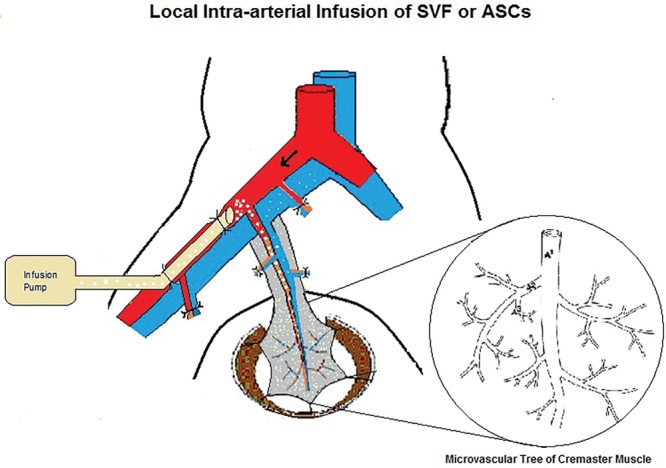
Materials and methods: Fat tissue was surgically harvested from the flanks of male Sprague-Dawley rats (n = 12) and processed for SVF isolation. Some SVF samples were cultured for 24 hours for ASC purification. The autogenic SVF (1 × 10⁵) cells (n = 6) or purified ASC (1 × 10⁵) cells (n = 6) cells were infused into the microcirculation of cremaster muscle at a speed of 0.05 mL/min through the cannulation of femoral artery. As this is a vascular pedicle isolated preparation, the infused SVF or ASC cells went nowhere but the cremaster muscle. The video image of the microcirculation was monitored in real time during infusion.
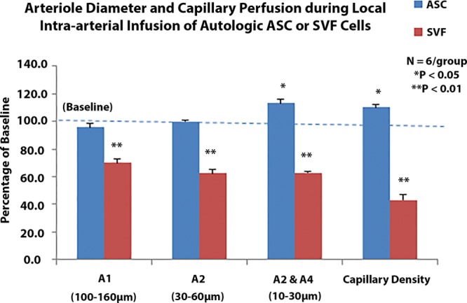
Results: Arteriole diameter was measured as A1 (100-160 µm), A2 (40-80 µm), and A3/A4 (10-30 µm). Capillary perfusion was quantified in 18 capillary fields of each muscle. There was a significant increase in the diameter of terminal arterioles (P = 0.049) and the capillary density (P = 0.02) after ASC intraarterial infusion. However, a significant cell aggregation, embolisms, and arterial obstruction were observed in the microcirculation in every case during SVF infusion.
Conclusions: Intraarterial infusion is an appropriate route for the delivery of autogenic ASCs, but not of SVF. SVF-induced microembolisms were the reason for narrowing or blocking the lumen of terminal arterioles, resulting in no flow in the corresponding capillaries.
Conflict of interest statement
The author has no financial interest to declare in relation to the content of this article. The Article Processing Charge was paid for by the author.
Figures


Similar articles
-
Autologous Stromal Vascular Fraction in the Intravenous Treatment of End-Stage Chronic Obstructive Pulmonary Disease: A Phase I Trial of Safety and Tolerability.J Clin Med Res. 2017 Aug;9(8):701-708. doi: 10.14740/jocmr3072w. Epub 2017 Jul 1. J Clin Med Res. 2017. PMID: 28725319 Free PMC article.
-
Acute microvascular action of vascular endothelial growth factor in skeletal muscle ischemia/reperfusion injury.Plast Reconstr Surg. 2005 Apr 15;115(5):1355-65. doi: 10.1097/01.prs.0000156980.38387.8d. Plast Reconstr Surg. 2005. PMID: 15809599
-
Comparative Clinical Outcomes After Intra-articular Injection With Adipose-Derived Cultured Stem Cells or Noncultured Stromal Vascular Fraction for the Treatment of Knee Osteoarthritis.Am J Sports Med. 2019 Sep;47(11):2577-2583. doi: 10.1177/0363546519864359. Epub 2019 Aug 2. Am J Sports Med. 2019. PMID: 31373830
-
Augmentation of Dermal Wound Healing by Adipose Tissue-Derived Stromal Cells (ASC).Bioengineering (Basel). 2018 Oct 26;5(4):91. doi: 10.3390/bioengineering5040091. Bioengineering (Basel). 2018. PMID: 30373121 Free PMC article. Review.
-
Stromal vascular fraction technologies and clinical applications.Expert Opin Biol Ther. 2019 Dec;19(12):1289-1305. doi: 10.1080/14712598.2019.1671970. Epub 2019 Sep 27. Expert Opin Biol Ther. 2019. PMID: 31544555 Review.
Cited by
-
Possibility of Injecting Adipose-Derived Stromal Vascular Fraction Cells to Accelerate Microcirculation in Ischemic Diabetic Feet: A Pilot Study.Int J Stem Cells. 2019 Mar 30;12(1):107-113. doi: 10.15283/ijsc18101. Int J Stem Cells. 2019. PMID: 30836733 Free PMC article.
-
The Effect of Mesenchymal Stem Cells, Adipose Tissue Derived Stem Cells, and Cellular Stromal Vascular Fraction on the Repair of Acute Anal Sphincter Injury in Rats.Bioengineering (Basel). 2022 Jul 15;9(7):318. doi: 10.3390/bioengineering9070318. Bioengineering (Basel). 2022. PMID: 35877369 Free PMC article.
References
-
- Uzbas F, May ID, Parisi AM, et al. Molecular physiognomies and applications of adipose-derived stem cells. Stem Cell Rev. 2015;11:298–308. - PubMed
-
- Nguyen A, Guo J, Banyard DA, et al. Stromal vascular fraction: a regenerative reality? Part 1: current concepts and review of the literature. J Plast Reconstr Aesth Surg. 2015;69:170–179. - PubMed
-
- Guo J, Nguyen A, Banyard DA, et al. Stromal vascular fraction: a regenerative reality? Part 2: mechanisms of regenerative action. J Plast Reconstr Aesth Surg. 2015;69:180–188. - PubMed
-
- Michalek J, Moster R, Lukac L, et al. Autologous adipose tissue-derived stromal vascular fraction cells application in patients with osteoarthritis. Cell Transplant. 2015 [Epub ahead of print] - PubMed
LinkOut - more resources
Full Text Sources
Other Literature Sources
Research Materials
Miscellaneous