. 2016 Oct 12;2(10):e1600760.
doi: 10.1126/sciadv.1600760. eCollection 2016 Oct.

Promiscuous targeting of bromodomains by bromosporine identifies BET proteins as master regulators of primary transcription response in leukemia

Affiliations

Promiscuous targeting of bromodomains by bromosporine identifies BET proteins as master regulators of primary transcription response in leukemia

Sarah Picaud et al. Sci Adv. .

Abstract

Bromodomains (BRDs) have emerged as compelling targets for cancer therapy. The development of selective and potent BET (bromo and extra-terminal) inhibitors and their significant activity in diverse tumor models have rapidly translated into clinical studies and have motivated drug development efforts targeting non-BET BRDs. However, the complex multidomain/subunit architecture of BRD protein complexes complicates predictions of the consequences of their pharmacological targeting. To address this issue, we developed a promiscuous BRD inhibitor [bromosporine (BSP)] that broadly targets BRDs (including BETs) with nanomolar affinity, creating a tool for the identification of cellular processes and diseases where BRDs have a regulatory function. As a proof of principle, we studied the effects of BSP on leukemic cell lines known to be sensitive to BET inhibition and found, as expected, strong antiproliferative activity. Comparison of the modulation of transcriptional profiles by BSP after a short exposure to the inhibitor resulted in a BET inhibitor signature but no significant additional changes in transcription that could account for inhibition of other BRDs. Thus, nonselective targeting of BRDs identified BETs, but not other BRDs, as master regulators of context-dependent primary transcription response.

Keywords: BET; Bromodomains; epigenetics; inhibition; leukemias.


Figures

Fig. 1
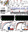
Fig. 1. BSP is a pan-BRD inhibitor in vitro.
(A) Triazolopyridazine scaffold of BSP. (B) 2FoFc map of BSP bound to BRD4(1) contoured at 2σ. (C) Complex of BSP with the BRD of BRD9. The compound adopts an acetyl-lysine mimetic pose within the BRD cavity, initiating interactions with the conserved asparagine (N100). The sulfonamide function initiates contacts with ZA-loop residues (G43), further stabilizing the interaction without displacing any of the structurally conserved water molecules (red spheres). (D) BSP binding with low micromolar to nanomolar affinity to most structural classes within the human BRD family. Dissociation constants (KD) measured in-solution using ITC are displayed on the human BRD tree as spheres (size and color as indicated in the inset). BRD structural classes are annotated with roman numerals. (E) Overlay of ITC measurements of typically strong BSP interactions with the BRDs of CECR2, BRD9, and TAF1L(2). Raw injection heats for the titrations of proteins into solutions of BSP are shown on the left panels. The right panel shows the normalized binding enthalpies corrected for the heat of protein dilution as a function of binding site saturation (symbols as indicated in the figure). Solid lines represent a nonlinear least-squares fit using a single-site binding model. All titrations were carried out in 50 mM Hepes buffer (pH 7.5; 25°C) and 150 mM NaCl at 15°C while stirring at 1000 rpm.
Fig. 2
Fig. 2. BSP engages its target BRDs in cells.
(A) BSP binds to the BRD acetyl-lysine cavity, allowing for further functionalization toward the front channel within the ZA loop (Ra vector annotated in orange) or the back of the pocket (Rb vector annotated in orange). The vectors are shown in the complex of BSP with BRD4(1). (B) Two variants of biotinylated BSP (BSP-a and BSP-b) were prepared to explore binding to human BRDs in cells by pull-down experiments. (C) Biotinylated BSP (BSP-a or BSP-b) immobilized on magnetic beads was used to pull down human CECR2 from Flp-In T-REx HEK293 cells stably expressing 3×FLAG CECR2. The protein captured from whole-cell lysate was identified using anti-FLAG. (D) Cell lysate from HEK293T cells was incubated with biotinylated BSP (BSP-a or BSP-b) immobilized on magnetic streptavidin beads in the presence or absence of 30 nmol of BSP for 2 hours at 4°C. After pull-down and tryptic digestion with trypsin, proteins were identified in a TripleTOF 5600 mass spectrometer. (Top) Normalized abundance of each BRD-containing protein in HEK293 cells (data from Proteomics DB; https://www.proteomicsdb.org/). (Bottom) Ratio of peptide to peptide abundance in the presence and absence of competing BSP, shown as a bar graph. BRD families are annotated with roman numerals. (E) FRAP evaluation of full-length GFP-tagged BRD4 dissociation from chromatin in U2OS cells. Nuclei of DMSO-treated (top) or BSP-treated (1 μM; bottom) cells. Target regions of photobleaching are indicated with a white circle. Scale bar, 10 μm. FL-BRD4, full-length BRD4; FL-BRD9, full-length BRD9. (F) Quantitative comparison of time to half-maximal fluorescence recovery for BRD4 FRAP studies using BSP (red bars) as a function of ligand concentration. (G) FRAP evaluation of full-length GFP-tagged BRD9 dissociation from chromatin in U2OS cells. Nuclei of DMSO-treated (top) or BSP-treated (1 μM; bottom) cells in the presence of 10 μM SAHA (added to increase the experimental window). Target regions of photobleaching are indicated with a white circle. Scale bars, 10 μm. (H) Quantitative comparison of time to half-maximal fluorescence recovery for BRD9 FRAP studies using BSP (red bars) as a function of ligand concentration. Data in (F) and (H) represent means ± SEM (n = 30) and are annotated with P values obtained from a two-tailed t test (*P < 0.05 and ***P < 0.001).
Fig. 3
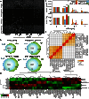
Fig. 3. BSP inhibits growth in leukemia cell lines.
(A) BSP inhibits clonogenic growth in leukemia cell lines. K562, KASUMI-1, MV4;11, and OCI-AML3 in methylcellulose were treated with vehicle (DMSO) or BSP (0.1, 0.5, or 1 μM) (n = 4). (B) Colony formation assay in K562, KASUMI-1, MV4;11, and OCI-AML3 cells using 0.1, 0.5, or 1.0 μM BSP (top) and the number of cells counted after treatment of cells with BSP for 6 to 10 days (n = 4) (bottom). CFU, colony-forming units; ns, not significant. (C) Similarity comparison of significantly expressed genes (P < 0.001 and fold change > 1.5) in the four cell lines after drug treatment. The heat map represents the intersect matrix for all pairwise comparisons (cell lines and treatments) using euclidean distances and complete linkage after transformation of the intersect counts into similarity Jaccard measures. (D) Venn diagrams showing overlap of the top statistically significant (Benjamini-Hochberg adjusted P < 0.001) genes (up- or down-regulated with a fold change of >1.5) differentially expressed by BSP or the pan-BET inhibitor JQ1 in four leukemia cell lines (K562, KASUMI-1, OCI-AML3, and MV4;11) after 8 hours of treatment with the inhibitor (0.5 μM) (top) and breakdown of the expression in terms of up- and down-regulated genes for each cell line (bottom). (E) Heat map of log fold changes in the expression of the top 50 statistically significant genes in the four cell lines tested, identified using Benjamini-Hochberg adjusted P < 0.001. Data in (B) represent means ± SEM (n = 4) and are annotated with P values obtained from a two-tailed t test (*P < 0.05, **P < 0.01, ***P < 0.001, and ****P < 0.0001).
Fig. 4
Fig. 4. Comparison of the effects of BET inhibition on cell cycle and transcription in leukemias.
(A and B) Cell cycle analysis of BSP (blue scale) and JQ1 (red scale) inhibition in resistant cells (A; K562) or sensitive cells (B; MV4;11) after 48 hours of treatment with inhibitors (amounts as indicated in the inset). The quantification given below each graph depicts the percent S-phase content measured under each condition, indicating a clear arrest in the sensitive line with minor effects on the resistant line. Data are means ± SEM (n = 3) and are annotated with P values obtained from analysis of variance (ANOVA) followed by Dunnett’s test (*P < 0.05 and **P < 0.01). (C) Heat map of fold changes (expressed in log2 scale as indicated in the inset) in the top 1000 significantly differentially expressed genes (Benjamini-Hochberg adjusted P < 0.001) in K562 cells (top) and MV4;11 cells (bottom) after 6 hours of treatment with selective BRD inhibitors or DMSO. The effects of JQ1 and BSP are very similar and much stronger than the effects of any other compounds targeting non-BET BRDs. (D) A published set of genes constituting a “JQ1 signature” is only attenuated by BSP and JQ1 in K562 cells (left) and MV4;11 cells (right) after 6 hours of treatment with a series of inhibitors (500 nM). The heat map depicts row-normalized values for gene expression, as indicated in the inset.
Scheme 1
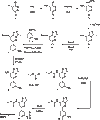
Scheme 1. Synthesis of BSP.

Similar articles

  • Interactome Rewiring Following Pharmacological Targeting of BET Bromodomains.
    Lambert JP, Picaud S, Fujisawa T, Hou H, Savitsky P, Uusküla-Reimand L, Gupta GD, Abdouni H, Lin ZY, Tucholska M, Knight JDR, Gonzalez-Badillo B, St-Denis N, Newman JA, Stucki M, Pelletier L, Bandeira N, Wilson MD, Filippakopoulos P, Gingras AC. Lambert JP, et al. Mol Cell. 2019 Feb 7;73(3):621-638.e17. doi: 10.1016/j.molcel.2018.11.006. Epub 2018 Dec 13. Mol Cell. 2019. PMID: 30554943 Free PMC article.
  • BET bromodomain inhibitors in leukemia.
    Basheer F, Huntly BJ. Basheer F, et al. Exp Hematol. 2015 Aug;43(8):718-31. doi: 10.1016/j.exphem.2015.06.004. Epub 2015 Jul 9. Exp Hematol. 2015. PMID: 26163798 Review.
  • PFI-1, a highly selective protein interaction inhibitor, targeting BET Bromodomains.
    Picaud S, Da Costa D, Thanasopoulou A, Filippakopoulos P, Fish PV, Philpott M, Fedorov O, Brennan P, Bunnage ME, Owen DR, Bradner JE, Taniere P, O'Sullivan B, Müller S, Schwaller J, Stankovic T, Knapp S. Picaud S, et al. Cancer Res. 2013 Jun 1;73(11):3336-46. doi: 10.1158/0008-5472.CAN-12-3292. Epub 2013 Apr 10. Cancer Res. 2013. PMID: 23576556 Free PMC article.
  • Inhibition of BET bromodomains as a therapeutic strategy for cancer drug discovery.
    Fu LL, Tian M, Li X, Li JJ, Huang J, Ouyang L, Zhang Y, Liu B. Fu LL, et al. Oncotarget. 2015 Mar 20;6(8):5501-16. doi: 10.18632/oncotarget.3551. Oncotarget. 2015. PMID: 25849938 Free PMC article. Review.
  • Selective inhibition of the BD2 bromodomain of BET proteins in prostate cancer.
    Faivre EJ, McDaniel KF, Albert DH, Mantena SR, Plotnik JP, Wilcox D, Zhang L, Bui MH, Sheppard GS, Wang L, Sehgal V, Lin X, Huang X, Lu X, Uziel T, Hessler P, Lam LT, Bellin RJ, Mehta G, Fidanze S, Pratt JK, Liu D, Hasvold LA, Sun C, Panchal SC, Nicolette JJ, Fossey SL, Park CH, Longenecker K, Bigelow L, Torrent M, Rosenberg SH, Kati WM, Shen Y. Faivre EJ, et al. Nature. 2020 Feb;578(7794):306-310. doi: 10.1038/s41586-020-1930-8. Epub 2020 Jan 22. Nature. 2020. PMID: 31969702

Cited by

References

    1. Filippakopoulos P., Picaud S., Mangos M., Keates T., Lambert J.-P., Barsyte-Lovejoy D., Felletar I., Volkmer R., Müller S., Pawson T., Gingras A.-C., Arrowsmith C. H., Knapp S., Histone recognition and large-scale structural analysis of the human bromodomain family. Cell 149, 214–231 (2012). - PMC - PubMed
    1. Filippakopoulos P., Knapp S., Targeting bromodomains: Epigenetic readers of lysine acetylation. Nat. Rev. Drug Discov. 13, 337–356 (2014). - PubMed
    1. Vidler L. R., Brown N., Knapp S., Hoelder S., Druggability analysis and structural classification of bromodomain acetyl-lysine binding sites. J. Med. Chem. 55, 7346–7359 (2012). - PMC - PubMed
    1. Belkina A. C., Denis G. V., BET domain co-regulators in obesity, inflammation and cancer. Nat. Rev. Cancer 12, 465–477 (2012). - PMC - PubMed
    1. Filippakopoulos P., Qi J., Picaud S., Shen Y., Smith W. B., Fedorov O., Morse E. M., Keates T., Hickman T. T., Felletar I., Philpott M., Munro S., McKeown M. R., Wang Y., Christie A. L., West N., Cameron M. J., Schwartz B., Heightman T. D., La Thangue N., French C. A., Wiest O., Kung A. L., Knapp S., Bradner J. E., Selective inhibition of BET bromodomains. Nature 468, 1067–1073 (2010). - PMC - PubMed

MeSH terms