

Spinal myxopapillary ependymoma with interval drop metastasis presenting as cauda equina syndrome: case report and review of literature
- PMID: 27757435
- PMCID: PMC5067269
- DOI: 10.21037/jss.2016.08.06
Spinal myxopapillary ependymoma with interval drop metastasis presenting as cauda equina syndrome: case report and review of literature
Abstract
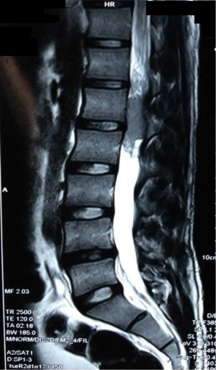
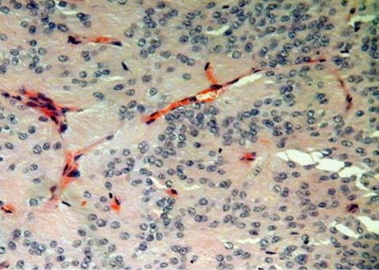
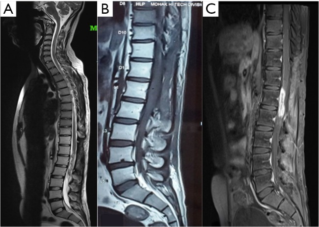
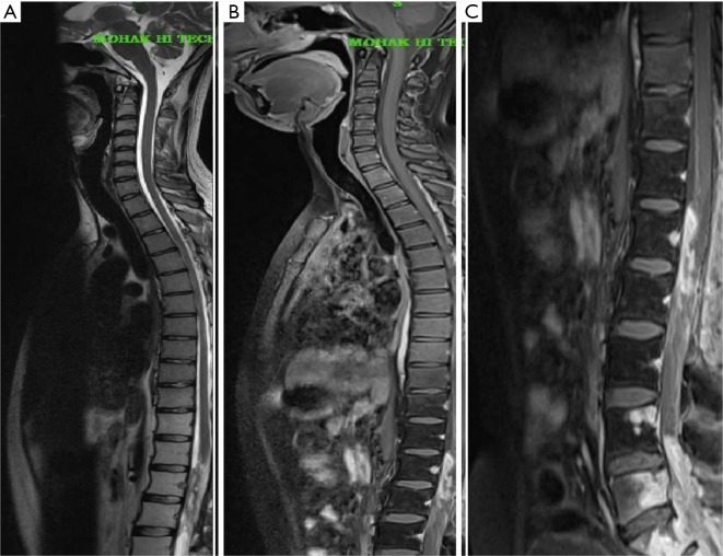
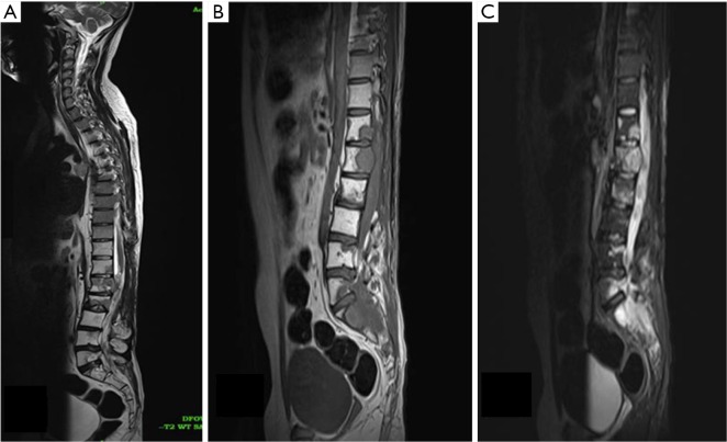
Myxopapillary ependymoma is a benign slow-growing tumour, arising predominantly in the region of the filum terminale. It has been designated histologically as grade I neoplasm according to the 2007 WHO classification. Despite this benign character dissemination and metastasis along the cerebrospinal axis and metastasis to distant sites have occasionally been reported. There have been previously reported cases of drop metastasis from MPE, however in three of these cases the drop metastasis was diagnosed with concurrent primary spinal MPE. There has been only one previously published case of interval drop metastasis in a case of operated spinal MPE in literature. We hereby present the second case of interval drop metastasis in a case of conus MPE, with history of having undergone a subtotal resection and post operative adjuvant radiotherapy prior to second surgery. The patient presented months after the primary surgery with symptoms of cauda equina syndrome attributable to the drop metastasis.
Keywords: Myxopapillary ependymoma; adjuvant radiotherapy; cauda equina syndrome; drop metastasis.
Conflict of interest statement
The authors have no conflicts of interest to declare.
Figures
References
-
- McLendon RE, Rosenblum MK, Schiffer D, et al. Myxopapillary ependymoma. In: Louis DN, Ohgaki H, Weistler OD, et al. editors. WHO classification of tumors of the central nervous system. Lyon: International Agency for Research on Cancer, 2007:72-3.
-
- Kernohan JW. Primary tumors of the spinal cord and intradural filum terminale. In: Penfield W. Cytology and cellular pathology of the nervous system. New York: Hoeber; 19323;3:993-1025.
Publication types
LinkOut - more resources
Full Text Sources
Other Literature Sources