. 2017 Jan;133(1):79-90.
doi: 10.1007/s00401-016-1633-2. Epub 2016 Oct 18.

Pathological TDP-43 changes in Betz cells differ from those in bulbar and spinal α-motoneurons in sporadic amyotrophic lateral sclerosis

Affiliations

Pathological TDP-43 changes in Betz cells differ from those in bulbar and spinal α-motoneurons in sporadic amyotrophic lateral sclerosis

Heiko Braak et al. Acta Neuropathol. 2017 Jan.

Abstract

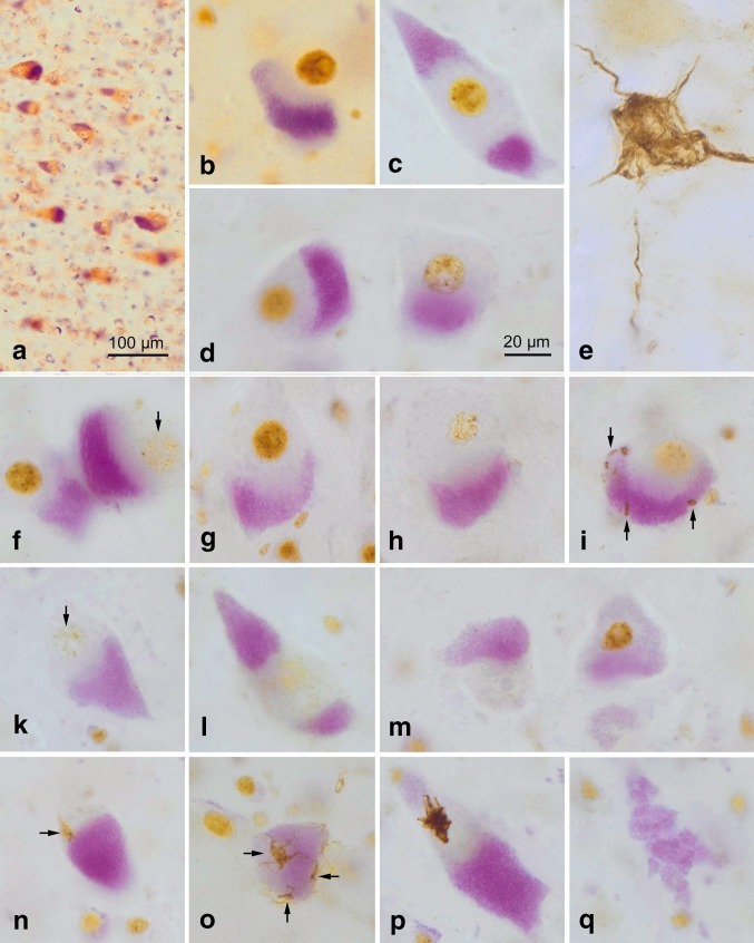
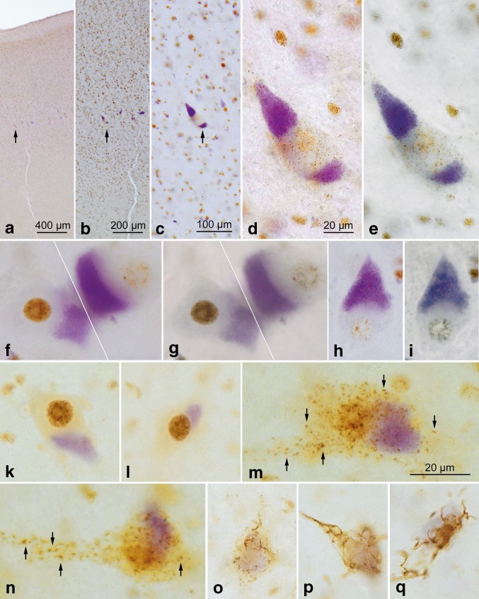
Two nerve cells types, Betz cells in layer Vb of the primary motor neocortex and α-motoneurons of the lower brainstem and spinal cord, become involved at the beginning of the pathological cascade underlying sporadic amyotrophic lateral sclerosis (sALS). In both neuronal types, the cell nuclei forfeit their normal (non-phosphorylated) expression of the 43-kDa transactive response DNA-binding protein (TDP-43). Here, we present initial evidence that in α-motoneurons the loss of normal nuclear TDP-43 expression is followed by the formation of phosphorylated TDP-43 aggregates (pTDP-43) within the cytoplasm, whereas in Betz cells, by contrast, the loss of normal nuclear TDP-43 expression remains mostly unaccompanied by the development of cytoplasmic aggregations. We discuss some implications of this phenomenon of nuclear clearing in the absence of cytoplasmic inclusions, namely, abnormal but soluble (and, thus, probably toxic) cytoplasmic TDP-43 could enter the axoplasm of Betz cells, and following its transmission to the corresponding α-motoneurons in the lower brainstem and spinal cord, possibly contribute in recipient neurons to the dysregulation of the normal nuclear protein. Because the cellular mechanisms that possibly inhibit the aggregation of TDP-43 in the cytoplasm of involved Betz cells are unknown, insight into such mechanisms could disclose a pathway by which the development of aggregates in this cell population could be accelerated, thereby opening an avenue for a causally based therapy.

Keywords: Amyotrophic lateral sclerosis; Betz cells; Motor neuron disease; Primary motor cortex; TAR DNA-binding protein; TDP-43; Therapeutics; Transsynaptic spreading; α-Motoneurons.


Conflict of interest statement

The authors have no current or potential conflicts of interest.

Figures

Fig. 1
Fig. 1
a Overview of the Betz giant pyramidal cells in layer Vb of the primary motor neocortex of a control individual (Table 1, case #17). Layer V is shown at right angles to the cortical surface (at left). The pigment-Nissl staining (violet-red) here and the pigment-staining (violet) in the remaining micrographs marks the location of the closely packed lipofuscin pigment granules within the cell somata. bd, fq Pigment-staining and TDP-43 immunohistochemistry in individuals with sALS. b, f (at left), and g Micrographs showing normal Betz cells with strong nuclear TDP-43 immunostaining. c, d A Betz cell with reduced nuclear TDP-43 immunostaining (d, at left) alongside of Betz cells in which TDP-43 immunostaining is nearly absent (d, at right; see also h, i) or absent (f, at right, k and l, at left; see also m, n, o). e A pTDP-43-immunopositive α-motoneuron filled with aggregates in the motor nucleus of the hypoglossal nerve (N. XII) (Table 1, case #13). In the other micrographs here, the Betz cells that become involved in sALS do not display this kind of lesional profile. hm These micrographs show the growing reduction and loss of intranuclear TDP-43 immunoreactivity in Betz cells; note also, however, that the cytoplasmic inclusions (aggregates) that do develop are slight. i A Betz cell containing discreet dot-like or granular cytoplasmic inclusions (arrows). An exception to this discreet cytoplasmic pathology is seen in p. m A Betz cell with a completely ‘empty’ (i.e., TDP-43 immunonegative) cell nucleus (at left) shown next to a Betz cell with normal nuclear TDP-43 immunostaining. In the cell at the left, only the nucleolus is still visible. n, o Subtle TDP inclusions (arrows) in Betz cells, including skein-like lesions in o. q Remnants of lipofuscin pigment granules mark the site of a dead Betz cell. Scale bar in d is valid for all micrographs except a. 100 µm polyethylene glycol (PEG) sections. Micrographs b, o (Table 1, case #1), c, d, f, kn, p, q (Table 1, case #3), gl (Table 1, case #2). A plan apochromat 40:1 lens was utilized to evaluate and photograph individual Betz cells (ad, fq)
Fig. 2
Fig. 2
Upper panels ai Betz cells as seen in sections stained for lipofuscin deposits (aldehyde fuchsin) combined with an immunoreaction for normal TDP-43 (chromogen: brown). Following an overview micrograph (a) and additional micrographs taken at higher magnifications (b and c), additional micrographs of individual Betz cells were taken using an apochromat 40:1 lens to show both an immunonegative cell nucleus and immunonegative cytoplasm of the cell body (d). Then, the coverslip was removed in xylene and the tissue section was transferred through a descending ethanol series to H2O before performing a second immunoreaction, this time using an antibody against phosphorylated TDP-43 (chromogen: blue). With the aid of the initial micrographs, the same cells of interest photographed previously were located in the same section again and studied in the double immunoreactions (e). Immunonegative cytoplasm of the cell body in immunoreactions for normal TDP-43 remained immunonegative after the second immunoreaction for phosphorylated TDP-43 was performed, i.e., no inclusion bodies or traces of aggregated material were found that only appeared in immunoreactions with phosphorylated TDP-43 but remained immunonegative in sections with normal TDP-43 immunostaining. f and g, h and i Additional pairs of cells were visualized in the manner described above. f, g Here, an involved Betz cell with an empty cell nucleus and empty cytoplasm is located directly adjacent to a normal Betz cell displaying a strongly immunopositive cell nucleus. Micrographs ai (Table 1, case #3). Lower panels, kq Bulbar α-motoneurons of the hypoglossal nerve. k and l Staining of lipofuscin deposits (aldehyde fuchsin) combined with an immunoreaction for normal TDP-43 (chromogen: brown) sometimes revealed normal α-motoneurons with strongly immunopositive cell nuclei. mn With the onset of nuclear clearing, i.e., when a weakly immunoreactive cell nucleus clearly was discernible, the formation of cytoplasmic dash-like TDP-43-immunopositive particles (arrows) began to develop in the cell soma, and these particles were widely distributed there. oq A possibly more advanced phase in the development of the TDP-43 lesions could be the coalescence of the dash-like particles into the fine (o) and coarser (p and q) forms of skein-like inclusion bodies. In contrast to the situation encountered in the Betz cells, α-motoneurons with immunonegative cell nuclei that were accompanied by an immunonegative cytoplasm did not occur. Scale bar in d is valid for el and oq. 100 µm polyethylene glycol (PEG) sections. Micrographs kn (Table 1, case #2), o (Table 1, case #1), p, q (Table 1, case #13). A plan apochromat 40:1 lens was utilized to evaluate and photograph individual Betz cells (ai)
Fig. 3
Fig. 3
Schematic diagram summarizing the findings in giant Betz cells (a, ce, g, h) and in α-motoneurons (b, f) in the cases of sporadic amyotrophic lateral sclerosis studied. We hypothesize that the pathology may be transferred by means of still soluble but toxic or pathogenic axoplasmic TDP-43 directly (monosynaptically) from involved Betz cells to α-motoneurons. This postulated route is marked by means of the red arrow in the synapse between e and f. Deposits of lipofuscin granules serve as a marker of the cortical cellular type and are represented here by violet shading. a, b Betz cells in controls as well as in non-involved Betz cells and in non-involved α-motoneurons displayed normal, strongly immunoreactive intranuclear TDP-43 staining (here, in brown). The long axon of the Betz cell projects to, and synapses directly on, the corresponding α-motoneuron in the lower brainstem or in the ventral horn of the spinal cord. c, d Involved Betz cells of the cases examined displayed increasingly weak (c) and severe reduction (d) of TDP-43 intranuclear immunostaining. e The putative end-point of this development may be reached when the cell nucleus is completely ‘empty’, i.e., TDP-43 immunonegative. Theoretically, the protein could be present (although no longer immunoreactive) in the somatodendritic and/or axonal cytoplasm in a soluble state (here, in pink), where, for an unknown time interval, it does not convert into insoluble aggregates. f At the same time the above-described abnormalities became visible within the Betz cells (depicted in ce), somatic cytoplasmatic pTDP-43 inclusions were found in α-motoneurons (here, as red blasts). It should be emphasized that the aggregates were only found in the somatic and axonal cytoplasm. g Adjacent to the Betz cells that have TDP-43 immunonegative nuclei but lack pTDP-43 cytoplasmic inclusions, one sometimes encountered Betz cells containing discreet (dot-like, granular) aggregates (here, in red), which could indicate that these cells do not completely forfeit the potential to develop inclusion bodies. h Betz cells containing large aggregates were seldom. The dashed lines in a and e are intended to indicate that the involved axons are much longer than depicted schematically here

Similar articles

Cited by

References

    1. Al-Chalabi A, Jones A, Troakes C, King A, Al-Sarraj S, van den Berg LH. The genetics and neuropathology of amyotrophic lateral sclerosis. Acta Neuropathol. 2012;124:339–352. doi: 10.1007/s00401-012-1022-4. - DOI - PubMed
    1. Appel SH. Unifying hypothesis for the cause of amyotrophic lateral sclerosis, Parkinsonism and Alzheimer disease. Ann Neurol. 1981;10:499–505. doi: 10.1002/ana.410100602. - DOI - PubMed
    1. Arai T, Hasegawa M, Akiyama H, Ikeda K, Nonaka T, Mori H, et al. TDP-43 is a component of ubiquitin-positive tau-negative inclusions in frontotemporal lobar degeneration and amyotrophic lateral sclerosis. Biochem Biophys Res Commun. 2006;351:602–611. doi: 10.1016/j.bbrc.2006.10.093. - DOI - PubMed
    1. Ayala YM, Zago P, D’Ambrogio D, Xu YF, Buratti E, et al. Structural determinants of the cellular localization and shuttling of TDP-43. J Cell Sci. 2008;121:3778–3785. doi: 10.1242/jcs.038950. - DOI - PubMed
    1. Betz W. Anatomischer Nachweis zweier Gehirncentra. Centralblatt für die medizinischen Wissenschaften. 1874;12(578–580):595–599.

MeSH terms

Supplementary concepts