Characterization of a recombinant Newcastle disease virus expressing the glycoprotein of bovine ephemeral fever virus
- PMID: 27757685
- PMCID: PMC5306239
- DOI: 10.1007/s00705-016-3078-2
Characterization of a recombinant Newcastle disease virus expressing the glycoprotein of bovine ephemeral fever virus
Abstract
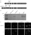
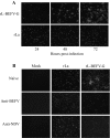
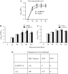
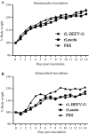
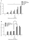
Bovine ephemeral fever (BEF) is caused by the arthropod-borne bovine ephemeral fever virus (BEFV), which is a member of the family Rhabdoviridae and the genus Ephemerovirus. BEFV causes an acute febrile infection in cattle and water buffalo. In this study, a recombinant Newcastle disease virus (NDV) expressing the glycoprotein (G) of BEFV (rL-BEFV-G) was constructed, and its biological characteristics in vitro and in vivo, pathogenicity, and immune response in mice and cattle were evaluated. BEFV G enabled NDV to spread from cell to cell. rL-BEFV-G remained nonvirulent in poultry and mice compared with vector LaSota virus. rL-BEFV-G triggered a high titer of neutralizing antibodies against BEFV in mice and cattle. These results suggest that rL-BEFV-G might be a suitable candidate vaccine against BEF.
Conflict of interest statement
The authors have declared that no competing interests exist.
Figures
Similar articles
-
Vaccinia virus-expressed bovine ephemeral fever virus G but not G(NS) glycoprotein induces neutralizing antibodies and protects against experimental infection.J Gen Virol. 1996 Apr;77 ( Pt 4):631-40. doi: 10.1099/0022-1317-77-4-631. J Gen Virol. 1996. PMID: 8627251
-
Bovine ephemeral fever in Australia and the world.Curr Top Microbiol Immunol. 2005;292:57-80. doi: 10.1007/3-540-27485-5_4. Curr Top Microbiol Immunol. 2005. PMID: 15981468 Review.
-
Genetically modified rabies virus vector-based bovine ephemeral fever virus vaccine induces protective immune responses against BEFV and RABV in mice.Transbound Emerg Dis. 2021 May;68(3):1353-1362. doi: 10.1111/tbed.13796. Epub 2020 Sep 5. Transbound Emerg Dis. 2021. PMID: 32805767
-
Immunogenicity of a plasmid DNA vaccine encoding G1 epitope of bovine ephemeral fever virus G glycoprotein in mice.Onderstepoort J Vet Res. 2018 Aug 28;85(1):e1-e6. doi: 10.4102/ojvr.v85i1.1617. Onderstepoort J Vet Res. 2018. PMID: 30198280 Free PMC article.
-
Epidemiology and control of bovine ephemeral fever.Vet Res. 2015 Oct 28;46:124. doi: 10.1186/s13567-015-0262-4. Vet Res. 2015. PMID: 26511615 Free PMC article. Review.
Cited by
-
Development and Scalable Production of Newcastle Disease Virus-Vectored Vaccines for Human and Veterinary Use.Viruses. 2022 May 6;14(5):975. doi: 10.3390/v14050975. Viruses. 2022. PMID: 35632717 Free PMC article. Review.
-
The Application of Newcastle Disease Virus (NDV): Vaccine Vectors and Tumor Therapy.Viruses. 2024 May 30;16(6):886. doi: 10.3390/v16060886. Viruses. 2024. PMID: 38932177 Free PMC article. Review.
-
Negative-Strand RNA Virus-Vectored Vaccines.Methods Mol Biol. 2024;2786:51-87. doi: 10.1007/978-1-0716-3770-8_3. Methods Mol Biol. 2024. PMID: 38814390 Review.
-
A SYBR green I-based quantitative RT-PCR assay for bovine ephemeral fever virus and its utility for evaluating viral kinetics in cattle.J Vet Diagn Invest. 2020 Jan;32(1):44-50. doi: 10.1177/1040638719895460. Epub 2019 Dec 17. J Vet Diagn Invest. 2020. PMID: 31845623 Free PMC article.
-
Intranasal Administration of Recombinant Newcastle Disease Virus Expressing SARS-CoV-2 Spike Protein Protects hACE2 TG Mice against Lethal SARS-CoV-2 Infection.Vaccines (Basel). 2024 Aug 16;12(8):921. doi: 10.3390/vaccines12080921. Vaccines (Basel). 2024. PMID: 39204044 Free PMC article.
References
-
- Alexander D (1997) Newcastle disease and other avian paramyxovirus infections. In: Diseases of poultry, vol 50014. Iowa State University Press, Ames, pp 541–569
-
- Bevan L. Ephemeral fever or three day sickness of cattle. Vet J. 1912;68:458–461.
-
- Bukreyev A, Collins PL. Newcastle disease virus as a vaccine vector for humans. Curr Opinion Mol Ther. 2008;10:46–55. - PubMed
MeSH terms
Substances
LinkOut - more resources
Full Text Sources
Other Literature Sources

