. 2017 Feb;13(1):127-138.
doi: 10.1007/s12015-016-9694-0.

Magnetic Resonance Imaging of Ferumoxytol-Labeled Human Mesenchymal Stem Cells in the Mouse Brain

Affiliations

Magnetic Resonance Imaging of Ferumoxytol-Labeled Human Mesenchymal Stem Cells in the Mouse Brain

Na Kyung Lee et al. Stem Cell Rev Rep. 2017 Feb.

Abstract

The success of stem cell therapy is highly dependent on accurate delivery of stem cells to the target site of interest. Possible ways to track the distribution of MSCs in vivo include the use of reporter genes or nanoparticles. The U.S. Food and Drug Administration (FDA) has approved ferumoxytol (Feraheme® [USA], Rienso® [UK]) as a treatment for iron deficiency anemia. Ferumoxytol is an ultrasmall superparamagnetic iron oxide nanoparticle (USPIO) that has recently been used to track the fate of transplanted cells using magnetic resonance imaging (MRI). The major objectives of this study were to demonstrate the feasibility of labeling hUCB-MSCs with ferumoxytol and to observe, through MRI, the engraftment of ferumoxytol-labeled human umbilical cord blood-derived mesenchymal stem cells (hUCB-MSCs) delivered via stereotactic injection into the hippocampi of a transgenic mouse model of familial Alzheimer's disease (5XFAD). Ferumoxytol had no toxic effects on the viability or stemness of hUCB-MSCs when assessed in vitro. Through MRI, hypointense signals were discernible at the site where ferumoxytol-labeled human MSCs were injected. Iron-positive areas were also observed in the engrafted hippocampi. The results from this study support the use of nanoparticle labeling to monitor transplanted MSCs in real time as a follow-up for AD stem cell therapy in the clinical field.

Keywords: Ferumoxytol; Magnetic resonance imaging; Mesenchymal stem cells; Monitoring; Stem cell therapy.


Conflict of interest statement

The authors declare that they have no conflict of interest.

Figures

Fig. 1
Fig. 1
Labeling hUCB-MSCs with Ferumoxytol. a Compared to unlabeled cells where no iron-positive blue areas were evident, blue staining could be observed in the cytoplasm of ferumoxytol–labeled hUCB-MSCs. Scale bar =50 μm. b Co-localization of iron- (black solid arrow) and ferumoxytol-labeled hUCB-MSCs (circular, pink) in a re-suspended state. Scale bar =100 μm. c Brown staining could be detected from the pellet of ferumoxytol-labeled hUCB-MSCs. Grid =1 mm. d Cell viability of hUCB-MSCs treated with various concentrations of protamine sulfate/ferumoxytol assessed using the Alamar Blue assay (average of 4 independent experiments, *P ≤ 0.05 compared to the unlabeled control sample)
Fig. 2
Fig. 2
Differentiation Potential of Ferumoxytol-Labeled hUCB-MSCs. Adipogenic (Oil Red O staining), osteogenic (alkaline phosphatase), and chondrogenic (Safranin O staining) differentiation were observed from both (a) unlabeled and (b) ferumoxytol-labeled hUCB-MSCs. Oil Red O: Scale bar =50 μm; Alkaline phosphatase and Safranin O = 100 μm
Fig. 3
Fig. 3
Differences in Hypointensities Generated from Sham-Operated and Ferumoxytol-labeled hUCB-MSC Injected Mice. a A linear hypointense signal from T2-weighted and T2* GRE images revealed the puncture site and needle track. b In contrast, diffusion of hypointense signals was discernible in the right hippocampus of mice that received injections of ferumoxytol-labeled hUCB-MSCs. Prussian blue stains revealed iron positive cells in the right hippocampus. Scale bar: 80 μm
Fig. 4
Fig. 4
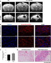
Post 1 Day MR Images Corroborated with IHC Staining. a A total of 2 × 105 unlabeled or ferumoxytol labeled hUCB-MSCs were injected into the left and right hippocampus, respectively, of 5XFAD mice. According to the T2-weighted and T2* MR images acquired post 1 day, compared to the left hemisphere, hypointense signals could be observed from the right hippocampus where ferumoxytol-labeled hUCB-MSCs were administered. Left (L) = unlabeled hUCB-MSCs, and right (R) = ferumoxytol-labeled hUCB-MSCs. b Human MSCs were observed in both the left and right hippocampi using the anti-mitochondria antibody. Scale bar: 20 μm. c Quantification of the number of cells positive for anti-mitochondria staining over the total number of Hoechst-positive cells (4 sections per hippocampus for each mouse). d Iron deposits were detected by Prussian blue staining in the right hippocampus. Scale bar: 90 μm
Fig. 5
Fig. 5
Absence of MSCs in the Vascular Regions of the Mouse Brain. No hUCB-MSCs (anti-mitochondria antibody) were discernible in the (a) dorsal 3rd ventricle or in either the (b) left or (c) right lateral ventricle. Scale bar: dorsal 3rd ventricle: 50 μm, lateral ventricles: 100 μm
Fig. 6
Fig. 6
Inflammatory Cells are not Observed at the Sites of hUCB-MSC Engraftment. a Iba-1 and GFAP expression was not observed at the locations where hUCB-MSCs were transplanted in either the left or right hippocampus. The white dotted line indicates the site of cell engraftment. Scale bar: 100 μm. b An adjacent slide was stained using the anti-mitochondria antibody to confirm the location of the cells enclosed by the white dotted line. (C) Quantification of number of cells positive for Iba-1 (left) or GFAP (right) over total number of Hoechst-positive cells (4 sections per hippocampus for each mouse)
Fig. 7
Fig. 7
Hypointense Signals Consistently Visualized at Extended Time Points. MR images acquired up to (a) 7 and (b) 14 days post transplantation. Similar to early time points, hypointense signals were observed in the right hippocampus where the ferumoxtyol-labeled hUCB-MSCs were transplanted. A gradual decrease in the size of the signal intensity was apparent at post 7 days while striking differences were not noted between post 7 and 14 days

Similar articles

Cited by

References

    1. Lindvall O, Kokaia Z. Stem cells in human neurodegenerative disorders--time for clinical translation? The Journal of Clinical Investigation. 2010;120(1):29–40. doi: 10.1172/JCI40543. - DOI - PMC - PubMed
    1. Holvoet B, De Waele L, Quattrocelli M, et al. Increased Understanding of Stem Cell Behavior in Neurodegenerative and Neuromuscular Disorders by Use of Noninvasive Cell Imaging. Stem Cells International. 2016;2016:6235687. doi: 10.1155/2016/6235687. - DOI - PMC - PubMed
    1. Tanna T, Sachan V. Mesenchymal stem cells: potential in treatment of neurodegenerative diseases. Current Stem Cell Research & Therapy. 2014;9(6):513–521. doi: 10.2174/1574888X09666140923101110. - DOI - PubMed
    1. Bali P, Lahiri DK, Banik A, Nehru B, Anand A. (2016). Potential for Stem Cells Therapy in Alzheimer’s Disease: Do Neurotrophic Factors Play Critical Role? Curr Alzheimer Res. - PMC - PubMed
    1. Tong LM, Fong H, Huang Y. Stem cell therapy for Alzheimer’s disease and related disorders: current status and future perspectives. Experimental & Molecular Medicine. 2015;47:e151. doi: 10.1038/emm.2014.124. - DOI - PMC - PubMed

Substances

LinkOut - more resources