

Comparison of the image-derived radioactivity and blood-sample radioactivity for estimating the clinical indicators of the efficacy of boron neutron capture therapy (BNCT): 4-borono-2-18F-fluoro-phenylalanine (FBPA) PET study
- PMID: 27757932
- PMCID: PMC5069228
- DOI: 10.1186/s13550-016-0230-7
Comparison of the image-derived radioactivity and blood-sample radioactivity for estimating the clinical indicators of the efficacy of boron neutron capture therapy (BNCT): 4-borono-2-18F-fluoro-phenylalanine (FBPA) PET study
Abstract
Background: In boron neutron capture therapy (BNCT), positron emission tomography (PET) with 4-borono-2-18F-fluoro-phenylalanine (FBPA) is the only method to estimate an accumulation of 10B to target tumor and surrounding normal tissue after administering 10B carrier of L-paraboronophenylalanine and to search the indication of BNCT for individual patient. Absolute concentration of 10B in tumor has been estimated by multiplying 10B concentration in blood during BNCT by tumor to blood radioactivity (T/B) ratio derived from FBPA PET. However, the method to measure blood radioactivity either by blood sampling or image data has not been standardized. We compared image-derived blood radioactivity of FBPA with blood sampling data and studied appropriate timing and location for measuring image-derived blood counts.
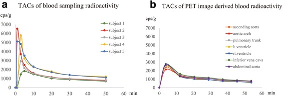
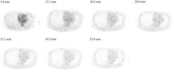
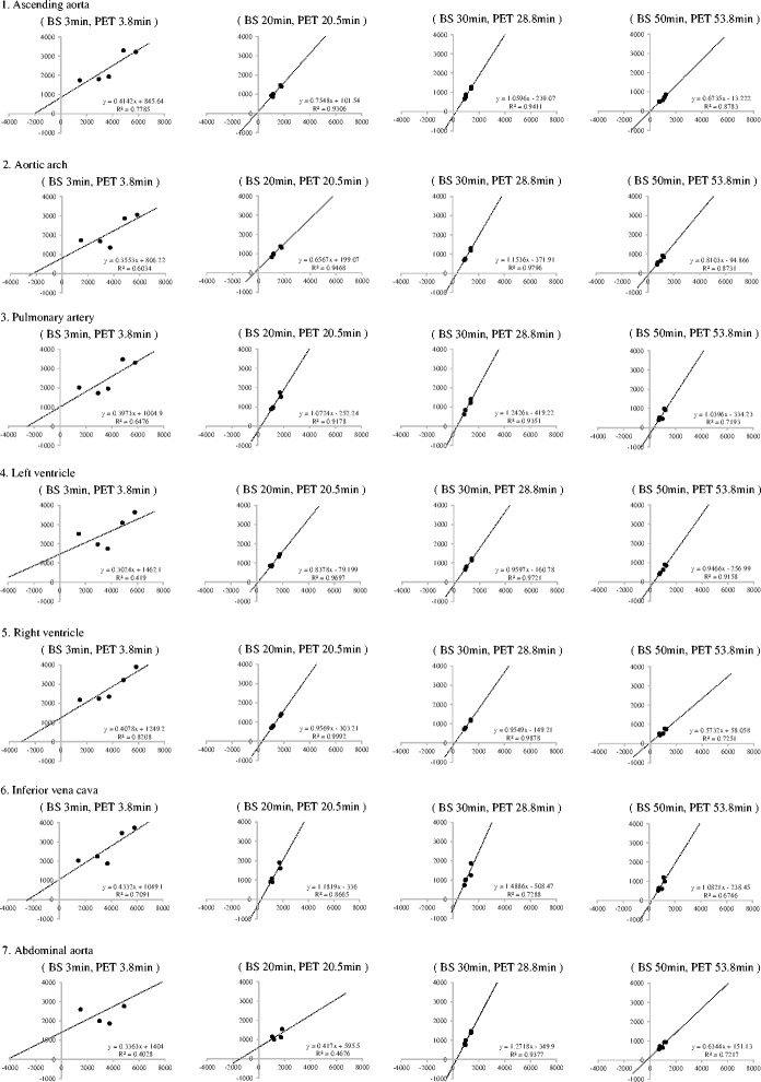
Methods: We obtained 7 repeated whole-body PET scans in five healthy subjects. Arterialized venous blood samples were obtained from the antecubital vein, heated in a heating blanket. Time-activity curves (TACs) of image-derived blood radioactivity were obtained using volumes of interest (VOIs) over ascending aorta, aortic arch, pulmonary artery, left and right ventricles, inferior vena cava, and abdominal aorta. Image-derived blood radioactivity was compared with those measured by blood sampling data in each location.
Results: Both the TACs of blood sampling radioactivity in each subject, and the TACs of image-derived blood radioactivity showed a peak within 5 min after the tracer injection, and promptly decreased soon thereafter. Linear relationship was found between blood sampling radioactivity and image-derived blood radioactivity in all the VOIs at any timing of data sampling (p < 0.001). Image-derived radioactivity measured in the left and right ventricles 30 min after injection showed high correlation with blood radioactivity. Image-derived blood radioactivity was lower than blood sampling radioactivity data by 20 %. Reduction of blood radioactivity of FBPA in left ventricle after 30 min of FBPA injection was minimal.
Conclusion: We conclude that the image-derived T/B ratio can be reliably used by setting the VOI on the left ventricle at 30 min after FBPA administration and correcting for underestimation due to partial volume effect and reduction of FBPA blood radioactivity.
Keywords: BNCT; FBPA; PET; T/B ratio.
Figures
References
-
- Barth RF, Soloway AH, Fairchild RG. Boron neutron capture therapy of cancer. Cancer Res. 1990;50:1061–1070. - PubMed
LinkOut - more resources
Full Text Sources
Other Literature Sources
