. 2016 Nov 25;291(48):25032-25049.
doi: 10.1074/jbc.M115.693473. Epub 2016 Oct 7.

A Novel Fucose-binding Lectin from Photorhabdus luminescens (PLL) with an Unusual Heptabladed β-Propeller Tetrameric Structure

Affiliations

A Novel Fucose-binding Lectin from Photorhabdus luminescens (PLL) with an Unusual Heptabladed β-Propeller Tetrameric Structure

Atul Kumar et al. J Biol Chem. .

Abstract

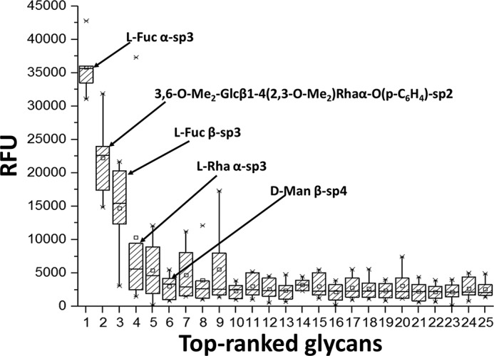
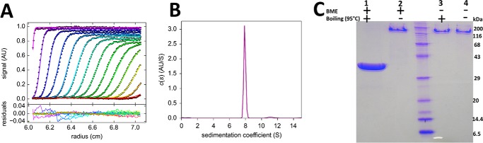
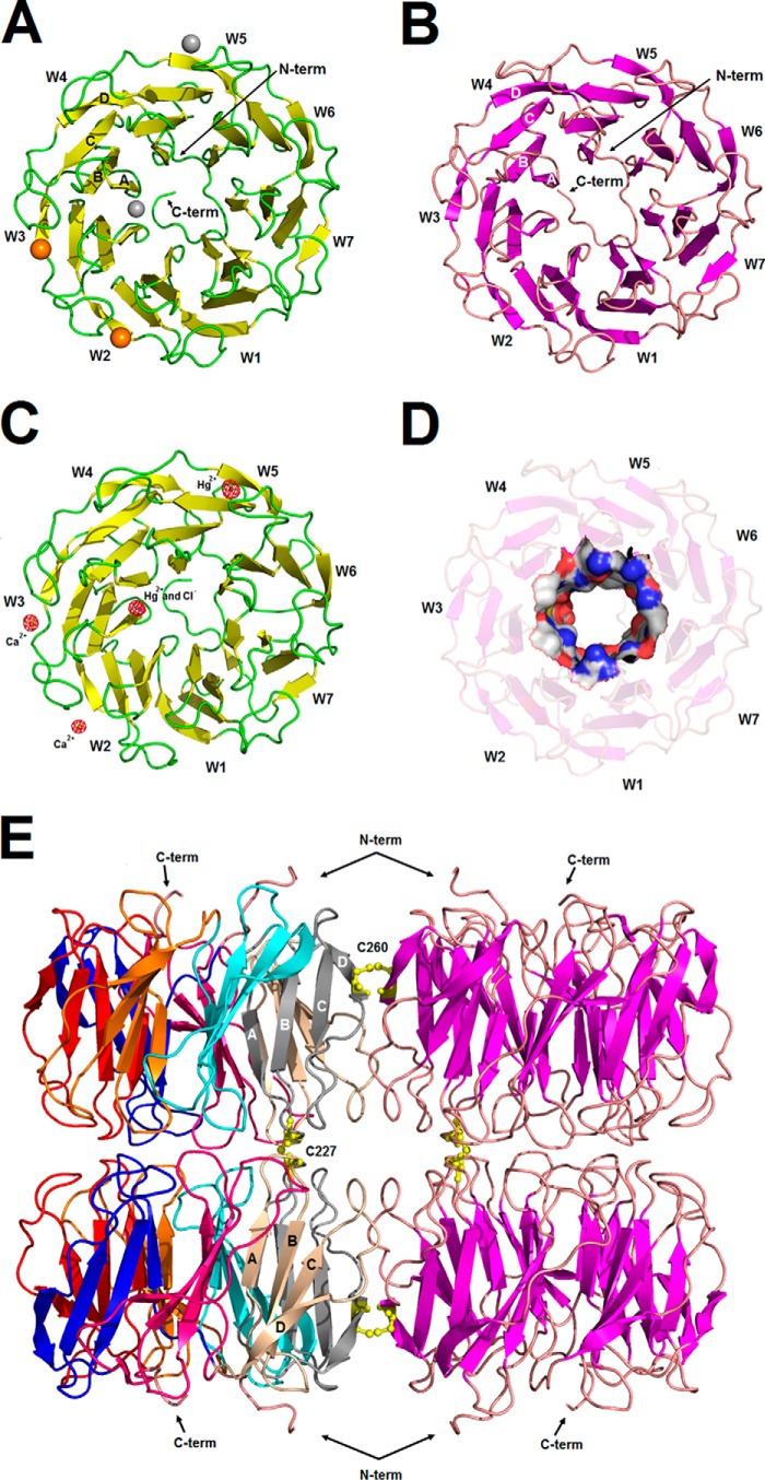
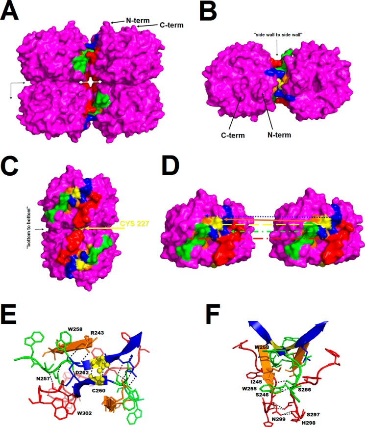
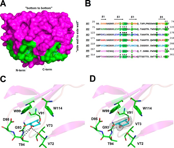
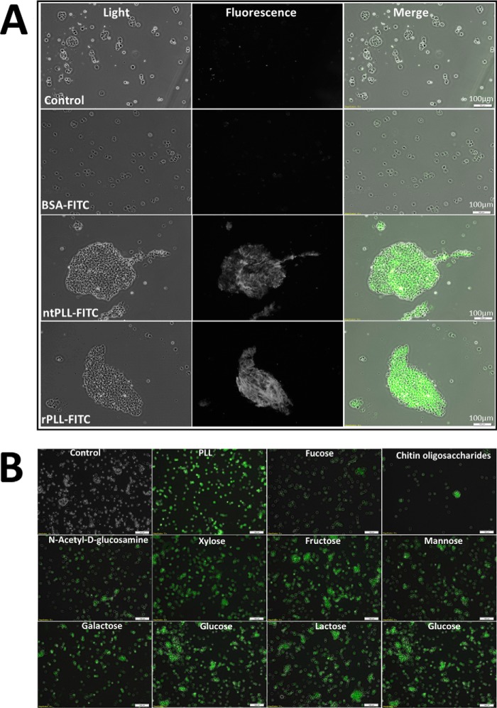
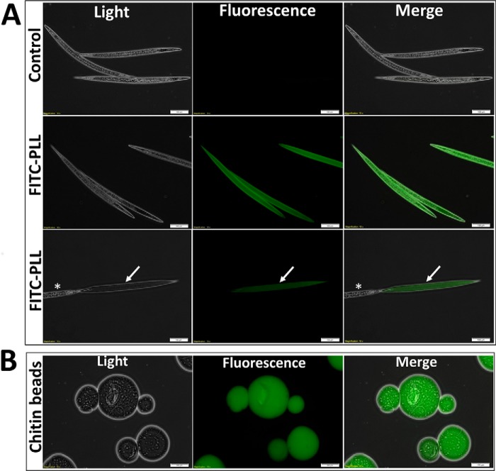
Photorhabdus luminescens is known for its symbiosis with the entomopathogenic nematode Heterorhabditis bacteriophora and its pathogenicity toward insect larvae. A hypothetical protein from P. luminescens was identified, purified from the native source, and characterized as an l-fucose-binding lectin, named P. luminescens lectin (PLL). Glycan array and biochemical characterization data revealed PLL to be specific toward l-fucose and the disaccharide glycan 3,6-O-Me2-Glcβ1-4(2,3-O-Me2)Rhaα-O-(p-C6H4)-OCH2CH2NH2 PLL was discovered to be a homotetramer with an intersubunit disulfide bridge. The crystal structures of native and recombinant PLL revealed a seven-bladed β-propeller fold creating seven putative fucose-binding sites per monomer. The crystal structure of the recombinant PLL·l-fucose complex confirmed that at least three sites were fucose-binding. Moreover, the crystal structures indicated that some of the other sites are masked either by the tetrameric nature of the lectin or by incorporation of the C terminus of the lectin into one of these sites. PLL exhibited an ability to bind to insect hemocytes and the cuticular surface of a nematode, H. bacteriophora.

Keywords: Galleria mellonella; Photorhabdus luminescens; bacterial pathogenesis; crystal structure; hemocytes from insect larvae; host/pathogen interaction; lectin; structural biology.


Figures

FIGURE 1.
FIGURE 1.
Purification and identification of native PLL from P. luminescens and cloning, expression, sequence variations, and purification of rPLL. A, SDS-PAGE (12%) gel stained with Coomassie Brilliant Blue R250 showing purification of ntPLL from P. luminescens using l-fucose-Sepharose column. Lanes 1–4, unbound fractions; lane 5, purified ntPLL; lane M, molecular weight marker. B, comparison of amino acid sequences of hypothetical protein from P. luminescens TTO1 (NCBI accession number WP_011145107.1) and PLL from P. luminescens subsp. kayaii; tryptic peptides identified by MS/MS analysis are in blue, and differences in amino acids are underlined. Five peptides exhibited 99% identity with the hypothetical protein Plu0732 from P. luminescens subsp. laumondii TT01 (NCBI accession number WP_011145107.1). The peptide identified by N-terminal sequencing is in red. C, amplification of pll coding sequence from total genomic DNA of P. luminescens; agarose gel showing amplified pll band of 1.1 kbp. D, SDS-PAGE showing purification of rPLL. Crude extract was loaded into a nickel-nitrilotriacetic acid column; washed with 0.02 m phosphate buffer (pH 7.5), 0.002 m trehalose; and eluted with 0.02 m phosphate buffer (pH 7.5), 0.002 m trehalose containing 0.35 m imidazole. Purified rPLL was found to be ∼41 kDa, as expected from the deduced amino acid sequence.
FIGURE 2.
FIGURE 2.
SPR sensorgrams displaying the binding of ntPLL onto sugar-immobilized chips. A, interaction of ntPLL with immobilized l-fucoside (black, top), mannan (gray, middle), and d-mannoside (light gray, bottom). B, ntPLL binding to l-fucoside channel in the presence of competing saccharides (l-fucose, methyl-α-l-fucoside, d-mannose, and d-galactose). Concentration of saccharides was 0–20 mm. The protein concentration was checked by measuring A280 using a theoretical molar extinction coefficient of 145,980 m−1 cm−1. C, hemagglutination of ntPLL with erythrocytes of blood group A. The concentrations of the protein were 19.8–0.019 μm. The last well was a negative control (n.c.; erythrocytes and PBS). The blue squares show the lowest ntPLL concentration at which the agglutination of erythrocytes was visible. D, hemagglutination inhibition assay. 0.02 ml of ntPLL (12 μm) and 0.02 ml of saccharide (geometry dilution from 33 mm) were used, and the minimal inhibitory concentrations of the tested saccharides are shown by blue squares. RU, resonance units.
FIGURE 3.
FIGURE 3.
Titration microcalorimetry of rPLL with l-fucose (A), methyl-α-l-fucoside (B) and chitin oligosaccharides (C) at 25 °C. A, titration curve of rPLL (0.1 mm) with l-fucose (60 mm); B, titration curve of rPLL (0.1 mm) with methyl-α-l-fucoside (60 mm). Twenty injections of 2.0 μl of sugars were added every 240 s to rPLL-containing cell. The protein and sugars were prepared in 0.02 m phosphate buffer, 0.05 m NaCl (pH 7.5). C, ITC measurement with chitin oligosaccharides. Twenty injections of 2.0 μl of rPLL (0.3 mm) were added every 240 s to sugar solution (30 mg ml−1) containing cells. The protein and sugar was prepared in 0.02 m phosphate buffer, 0.002 m trehalose (pH 7.5). The plots in the bottom panels show the total heat released as a function of total injectant concentration for the titration shown in the top panels. The solid lines represent the best least-squares fit to experimental data using a one-site model.
FIGURE 4.
FIGURE 4.
Box and whisker plot for glycan array screening with rPLL (200 μg ml−1). The top 25 saccharides are presented here; the five saccharides giving the highest average signals are depicted. Linker formulas are as follows: sp2, -O-CH2CH2NH2; sp3, -O-(CH2)3NH2; sp4, -NHCOCH2NH2. The bottom and top of the box are the first and third quartiles; the band inside the box is the second quartile (the median); the ends of the whiskers represent minimum and maximum values of the data. Asterisks represent marginal values, and small squares inside the boxes represent the mean. Complete glycan array results and the raw data are given in supplemental Tables S1 and Table S2, respectively. RFU, relative fluorescence units.
FIGURE 5.
FIGURE 5.
Analytical ultracentrifugation studies for the sedimentation velocity measurement of rPLL and confirmation of intersubunit disulfide bonds by SDS-PAGE. The AUC experiment was carried out at 42,000 rpm at 20 °C, and the scans were recorded every 6 min; every third scan is shown. A, sedimentation profiles and fitted curves of rPLL (0.22 mg ml−1) were obtained from continuous c(s) analysis using Sedfit (A, top); the residual plot (A, bottom) shows the differences between the experimental data and fitted curves. B, continuous size distribution of sedimenting species demonstrating a sedimentation coefficient of 7.91 S. (S20,w = 8.09 as calculated using Sednterp). The figures were created in GUSSI 1.0.8e. C, SDS-PAGE gel (14%) loaded with four rPLL (20 μg) samples treated with heating at 95 °C for 5 min, with and without BME (1.0%). Lanes contain reduced (lane 1) and intact rPLL (lanes 2–4).
FIGURE 6.
FIGURE 6.
A, crystal structure of ntPLL monomer shown in a secondary structure representation (β-strands (yellow), loops (green), Hg2+ ions (gray spheres), and Ca2+ ions (orange spheres)). B, crystal structure of rPLL monomer shown in secondary structure representation (β-strands (violet) and loops (pink)). C, secondary structure representation of ntPLL with shown Hg2+ (gray spheres), Cl (violet sphere), and Ca2+ ions (orange spheres) defined by an anomalous map at 8.0σ. D, the surface representation of the rPLL tunnel showing the charge distribution (negative potential (red), positive potential (blue), and neutral potential (gray)). E, quaternary structure of rPLL (color code as for B, disulfide bridges in yellow). The blades for two monomers are color-coded for each repeat (W1 (blue), W2 (red), W3 (orange), W4 (cyan), W5 (gray), W6 (beige), and W7 (pink)). The W4 motif (A and B) and the W5 motif (E) are defined by a twisted four-stranded antiparallel β-sheet (labeled as A, B, C, and D in panel E).
FIGURE 7.
FIGURE 7.
A, surface representation of rPLL (violet), color-coded according to the side wall-to-side wall association: β-strands D (blue) and C (orange; not visible in A) of repeat W5, loops connecting β-strands C and D of repeats W5 (green) and W6 (red), and cysteines forming disulfide bridges (yellow). The arrows indicate two pseudo-2-fold axes generating the tetramer. B, side wall-to-side wall association mode (surface representation) shown along a 2-fold pseudo-axis. C, bottom-to-bottom association mode (surface representation) shown along a 2-fold pseudo-axis with highlighted disulfide bridge (Cys-227; yellow). D, side wall-to-side wall association (surface representation) shown with two separated monomers of rPLL. The color-coded arrows show the surface interaction partners. E, ribbon and stick representation (disulfide bridge (Cys-260) shown in yellow spheres) of side wall-to-side wall association perpendicular to the 2-fold pseudo-axis. The polar interactions are shown as black dashed lines. Labeling is not shown for symmetrically related residues. F, ribbon and stick representation of side wall-to-side wall association along the 2-fold pseudo-axis. The polar interactions are shown as black dashed lines. Labeling is not shown for symmetrically related residues.
FIGURE 8.
FIGURE 8.
A, surface representation (violet) of rPLL. The fucose-binding site is shown in green with bound l-fucose as cyan sticks. B, sequence alignment of seven repeats (W1–W7) of rPLL lectin with main secondary structure elements indicated (residues color-coded to each β-blade, respectively). The amino acids of seven putative fucose-binding sites are highlighted with a green background. The corresponding binding pockets occupied by l-fucose in the rPLL structure are marked with the star (repeats W1 and W2), sphere (repeats W2 and W3), and triangle (repeats W6 and W7) symbols. C, stick representation of PLL fucose-binding site II (dashed square in A) (residues from repeats W1 and W2 in green) with l-fucose (cyan sticks) showing hydrogen bonds highlighted with dashed lines. D, omit map (2mFoDFc) at 1.0σ defining the l-fucose in binding site II.
FIGURE 9.
FIGURE 9.
Fluorescence microscopy of G. mellonella hemocytes. A, light (left), fluorescence (center), and merge (right) images of hemocytes treated with ntPLL-FITC, rPLL-FITC, and controls. Hemocytes were incubated with ntPLL-FITC and rPLL-FITC (experiment; 0.1 mg ml−1), buffer or BSA (controls, 0.1 mg ml−1) for 20 min at 17 °C and used for microscopy after three washes with PBS. B, inhibition of rPLL binding on larval hemocytes using 10 different sugars. rPLL-FITC was incubated with sugars for 30 min, followed by incubation with hemocytes for 30 min at 17 °C in a humid chamber, and used for microscopy after three washes with PBS.
FIGURE 10.
FIGURE 10.
Light (left), fluorescence (center), and merge (right) images of fluorescence microscopy of nematodes, H. bacteriophora, and chitin resin. A, nematodes treated with rPLL-FITC (0.1 mg ml−1). Nematodes were incubated with rPLL-FITC (experiment) for 30 min at 17 °C and used for microscopy after three washes with PBS. An arrow marks a recently shed cuticle of nematode, and an asterisk indicates the nematode itself. B, binding of rPLL-FITC with chitin resins. Chitin resin was incubated with rPLL-FITC for 20 min at room temperature and used for microscopy after washing the resin three times with PB/Treh buffer.

Similar articles

Cited by

References

    1. Joyce S. A., Watson R. J., and Clarke D. J. (2006) The regulation of pathogenicity and mutualism in Photorhabdus. Curr. Opin. Microbiol. 9, 127–132 - PubMed
    1. Forst S., Dowds B., Boemare N., and Stackebrandt E. (1997) Xenorhabdus and Photorhabdus spp.: bugs that kill bugs. Annu. Rev. Microbiol. 51, 47–72 - PubMed
    1. Forst S., and Nealson K. (1996) Molecular biology of the symbiotic pathogenic bacteria Xenorhabdus spp., and Photorhabdus spp. Microbiol. Rev. 60, 21–43 - PMC - PubMed
    1. Duchaud E., Rusniok C., Frangeul L., Buchrieser C., Givaudan A., Taourit S., Bocs S., Boursaux-Eude C., Chandler M., Charles J. F., Dassa E., Derose R., Derzelle S., Freyssinet G., Gaudriault S., et al. (2003) The genome sequence of the entomopathogenic bacterium Photorhabdus luminescens. Nat. Biotechnol. 21, 1307–13 - PubMed
    1. Williamson V. M., and Kaya H. K. (2003) Sequence of a symbiont. Nat. Biotechnol. 21, 1294–1295 - PubMed

MeSH terms