. 2016 Nov 25;291(48):24892-24899.
doi: 10.1074/jbc.M116.734673. Epub 2016 Oct 7.

Core Binding Factor β Protects HIV, Type 1 Accessory Protein Viral Infectivity Factor from MDM2-mediated Degradation

Affiliations

Core Binding Factor β Protects HIV, Type 1 Accessory Protein Viral Infectivity Factor from MDM2-mediated Degradation

Yusuke Matsui et al. J Biol Chem. .

Abstract

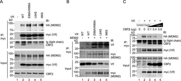
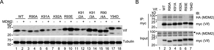
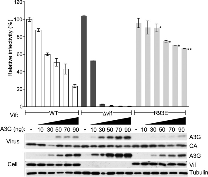
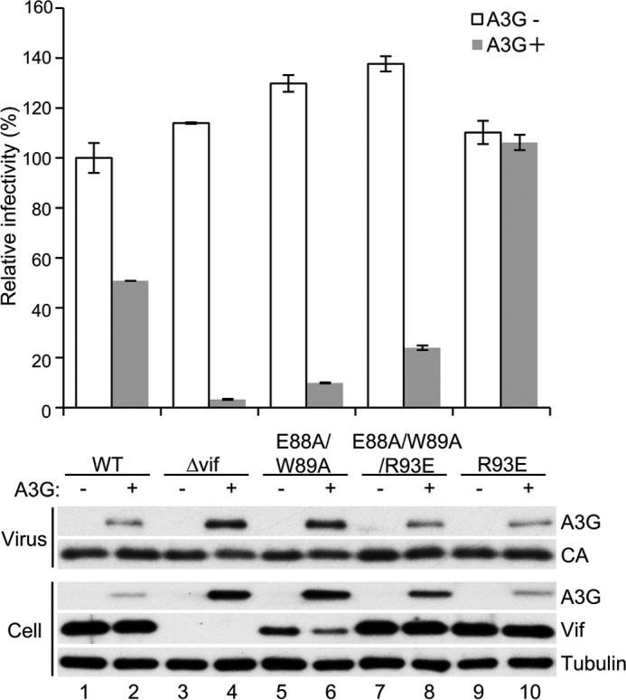
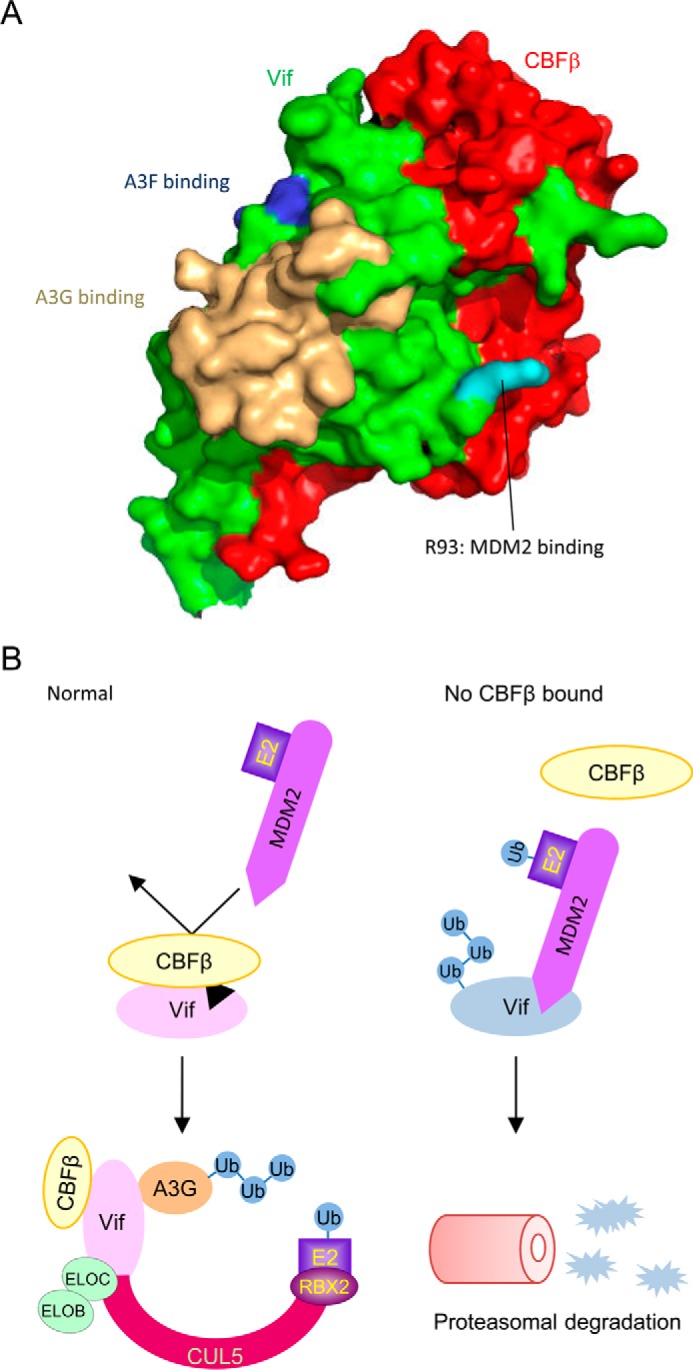
HIV, type 1 overcomes host restriction factor apolipoprotein B mRNA-editing enzyme catalytic polypeptide-like 3 (APOBEC3) proteins by organizing an E3 ubiquitin ligase complex together with viral infectivity factor (Vif) and a host transcription cofactor core binding factor β (CBFβ). CBFβ is essential for Vif to counteract APOBEC3 by enabling the recruitment of cullin 5 to the complex and increasing the steady-state level of Vif protein; however, the mechanisms by which CBFβ up-regulates Vif protein remains unclear. Because we have reported previously that mouse double minute 2 homolog (MDM2) is an E3 ligase for Vif, we hypothesized that CBFβ might protect Vif from MDM2-mediated degradation. Co-immunoprecipitation analyses showed that Vif mutants that do not bind to CBFβ preferentially interact with MDM2 and that overexpression of CBFβ disrupts the interaction between MDM2 and Vif. Knockdown of CBFβ reduced the steady-state level of Vif in MDM2-proficient cells but not in MDM2-null cells. Cycloheximide chase analyses revealed that Vif E88A/W89A, which does not interact with CBFβ, degraded faster than wild-type Vif in MDM2-proficient cells but not in MDM2-null cells, suggesting that Vif stabilization by CBFβ is mainly caused by impairing MDM2-mediated degradation. We identified Vif R93E as a Vif variant that does not bind to MDM2, and the virus with this substitution mutation was more resistant to APOBEC3G than the parental virus. Combinatory substitution of Vif residues required for CBFβ binding and MDM2 binding showed full recovery of Vif steady-state levels, supporting our hypothesis. Our data provide new insights into the mechanism of Vif augmentation by CBFβ.

Keywords: HIV; host-pathogen interaction; mouse double minute 2 homolog (MDM2); proteasome; protein-protein interaction; ubiquitin; viral protein.


Figures

FIGURE 1.
FIGURE 1.
CBFβ interferes with the interaction between Vif and MDM2. A, co-immunoprecipitation of MDM2 with Vif wild-type and variants that do not interact with CBFβ. 293T cells transiently expressing myc-tagged Vif wild-type or the variant and HA-tagged MDM2 were lysed and immunoprecipitated by anti-myc serum, and samples were analyzed by immunoblotting (IB) with anti-HA, anti-myc, and anti-CBFβ sera. B, reciprocal co-immunoprecipitation of Vif wild-type and variants with MDM2. Lysates of 293T cells with overexpression of untagged Vif wild-type or the variant and myc-tagged MDM2 were immunoprecipitated by anti-myc serum, and samples were analyzed by immunoblotting with anti-Vif and anti-myc sera. C, co-immunoprecipitation of MDM2 with Vif wild-type in the presence of CBFβ overexpression. Lysates of 293T cells transiently expressing myc-tagged Vif, HA-tagged MDM2, and various amounts of CBFβ were immunoprecipitated by anti-myc serum and analyzed for co-precipitation of MDM2 by immunoblotting.
FIGURE 2.
FIGURE 2.
Protection of Vif from MDM2-mediated degradation is responsible for Vif augmentation by CBFβ. A, p53−/− and p53−/−; MDM2−/− MEF cells were stably transfected with Vif wild-type or E88A/W89A, and clones that expressed comparable amount of Vif protein were picked according to immunoblotting. B, siRNA against murine Cbfb or control siRNA was transfected into MDM2-proficient and MDM2-null cells stably expressing Vif, and Vif levels were analyzed by immunoblotting. C, cycloheximide (CHX) chase analyses of Vif degradation. After cycloheximide treatment for the indicated time, lysates of p53−/− and p53−/−; MDM2−/− MEF cells stably expressing Vif wild-type or E88A/W89A were analyzed by immunoblotting with anti-Vif serum. Tubulin was also analyzed as a loading control. D, the amounts of Vif in C were quantified by densitometry, and those without cycloheximide treatment were normalized to 100%.
FIGURE 3.
FIGURE 3.
Vif R93 is required for MDM2 binding. A, degradation of Vif variants by MDM2. 293T cells were co-transfected with expression vectors for MDM2 and Vif wild-type or variant, and protein levels of Vif were analyzed by immunoblotting with anti-Vif serum. B, co-immunoprecipitation of MDM2 with Vif variants. 293T cells transiently expressing HA-tagged MDM2 and myc-tagged Vif wild-type or variant were lysed and immunoprecipitated by anti-myc serum. Samples were analyzed by immunoblotting (IB) with anti-HA and anti-myc sera.
FIGURE 4.
FIGURE 4.
Enhanced counteraction of Vif R93E against the restriction by APOBEC3G. Single-cycle infection experiments were performed by using VSV-G pseudotyped luciferase reporter viruses produced in 293T cells by co-transfection of pNL43/ΔEnv-Luc with the indicated Vif phenotype and pVSV-G in the presence or absence of co-transfection of pcDNA3/HA-A3G. Virus-containing supernatant was added to fresh 293T cells, and the luciferase activity of the cell lysates was measured by adding substrate and using a luminometer. The value of the virus without vif mutation in the absence of APOBEC3G was normalized to 100%, and mean ± S.E. of three independent experiments is shown (top panel). *, p < 0.05; **, p < 0.01 (statistically significant differences of infectivity between wild-type and R93E-harboring viruses). The levels of APOBEC3G in cells and virions and Vif in cells were also analyzed by immunoblotting (bottom panel). Virus p24 capsid protein (CA) and cellular tubulin were used as control.
FIGURE 5.
FIGURE 5.
The disruption of MDM2 binding restores the protein levels of the Vif variant that does not bind to CBFβ, but it does not fully restore the function to counteract APOBEC3G. Single-cycle infection experiments were performed by using VSV-G pseudotyped luciferase reporter viruses produced in 293T cells by co-transfection of pNL43/ΔEnv-Luc with the indicated vif phenotype and pVSV-G in the presence or absence of co-transfection of pcDNA3/HA-A3G. Virus-containing supernatant was added to fresh 293T cells, and the luciferase activity of the cell lysates was measured by adding substrate and using a luminometer. The value of the virus without vif mutation in the absence of APOBEC3G was normalized to 100%, and mean ± S.E. of three independent experiments is shown (top panel). The levels of APOBEC3G in cells and virions and Vif in cells were also analyzed by immunoblotting (bottom panel). Capsid (CA) and tubulin were used as control.
FIGURE 6.
FIGURE 6.
CBFβ protects Vif from MDM2. A, surface model of the Vif-CBFβ heterodimer based on the reported structure of the Vif-CBFβ-ELOB-ELOC-CUL5 complex (PDB code 4N9F). Vif and CBFβ are shown in green and red, respectively. Vif R93 is highlighted in cyan, and APOBEC3G and APOBEC3F binding sites are marked in wheat and blue, respectively. B, proposed model in which CBFβ stabilizes Vif protein. Under normal conditions, CBFβ interacts with Vif extensively just near the MDM2 binding site and sequesters MDM2 from Vif. The Vif variant that is defective in CBFβ binding is preferentially captured by MDM2 and rapidly cleared by the proteasome pathway. Ub, ubiquitin.

References

    1. Gabuzda D. H., Lawrence K., Langhoff E., Terwilliger E., Dorfman T., Haseltine W. A., and Sodroski J. (1992) Role of vif in replication of human immunodeficiency virus type 1 in CD4+ T lymphocytes. J. Virol. 66, 6489–6495 - PMC - PubMed
    1. Gabuzda D. H., Li H., Lawrence K., Vasir B. S., Crawford K., and Langhoff E. (1994) Essential role of vif in establishing productive HIV-1 infection in peripheral blood T lymphocytes and monocyte/macrophages. J. Acquir. Immune Defic. Syndr. 7, 908–915 - PubMed
    1. Sheehy A. M., Gaddis N. C., Choi J. D., and Malim M. H. (2002) Isolation of a human gene that inhibits HIV-1 infection and is suppressed by the viral Vif protein. Nature 418, 646–650 - PubMed
    1. Madani N., and Kabat D. (1998) An endogenous inhibitor of human immunodeficiency virus in human lymphocytes is overcome by the viral Vif protein. J. Virol. 72, 10251–10255 - PMC - PubMed
    1. Simon J. H., Gaddis N. C., Fouchier R. A., and Malim M. H. (1998) Evidence for a newly discovered cellular anti-HIV-1 phenotype. Nat. Med. 4, 1397–1400 - PubMed

MeSH terms

Substances

Associated data

LinkOut - more resources