

The Type B Flagellin of Hypervirulent Clostridium difficile Is Modified with Novel Sulfonated Peptidylamido-glycans
- PMID: 27758867
- PMCID: PMC5207245
- DOI: 10.1074/jbc.M116.749481
The Type B Flagellin of Hypervirulent Clostridium difficile Is Modified with Novel Sulfonated Peptidylamido-glycans
Abstract
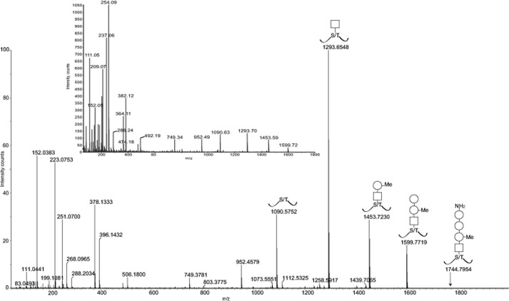
Glycosylation of flagellins is a well recognized property of many bacterial species. In this study, we describe the structural characterization of novel flagellar glycans from a number of hypervirulent strains of C. difficile We used mass spectrometry (nano-LC-MS and MS/MS analysis) to identify a number of putative glycopeptides that carried a variety of glycoform substitutions, each of which was linked through an initial N-acetylhexosamine residue to Ser or Thr. Detailed analysis of a LLDGSSTEIR glycopeptide released by tryptic digestion, which carried two variant structures, revealed that the glycopeptide contained, in addition to carbohydrate moieties, a novel structural entity. A variety of electrospray-MS strategies using Q-TOF technology were used to define this entity, including positive and negative ion collisionally activated decomposition MS/MS, which produced unique fragmentation patterns, and high resolution accurate mass measurement to allow derivation of atomic compositions, leading to the suggestion of a taurine-containing peptidylamido-glycan structure. Finally, NMR analysis of flagellin glycopeptides provided complementary information. The glycan portion of the modification was assigned as α-Fuc3N-(1→3)-α-Rha-(1→2)-α-Rha3OMe-(1→3)-β-GlcNAc-(1→)Ser, and the novel capping moiety was shown to be comprised of taurine, alanine, and glycine. This is the first report of a novel O-linked sulfonated peptidylamido-glycan moiety decorating a flagellin protein.
Keywords: Clostridium difficile; Gram-positive bacteria; bacteria; flagellin; glycosylation; mass spectrometry (MS); modification; nuclear magnetic resonance (NMR); sulfonated.
© 2016 by The American Society for Biochemistry and Molecular Biology, Inc.
Figures


References
-
- He M., Miyajima F., Roberts P., Ellison L., Pickard D. J., Martin M. J., Connor T. R., Harris S. R., Fairley D., Bamford K. B., D'Arc S., Brazier J., Brown D., Coia J. E., Douce G., et al. (2013) Emergence and global spread of epidemic healthcare-associated Clostridium difficile. Nat. Genet. 45, 109–113 - PMC - PubMed
-
- Cairns M. D., Stabler R. A., Shetty N., and Wren B. W. (2012) The continually evolving Clostridium difficile species. Future Microbiol. 7, 945–957 - PubMed
-
- Eidhin D. N., Ryan A. W., Doyle R. M., Walsh J. B., and Kelleher D. (2006) Sequence and phylogenetic analysis of the gene for surface layer protein, slpA, from 14 PCR ribotypes of Clostridium difficile. J. Med. Microbiol. 55, 69–83 - PubMed
-
- Hennequin C., Janoir C., Barc M. C., Collignon A., and Karjalainen T. (2003) Identification and characterization of a fibronectin-binding protein from Clostridium difficile. Microbiology 149, 2779–2787 - PubMed
MeSH terms
Substances
Grants and funding
- MR/K000551/1/MRC_/Medical Research Council/United Kingdom
- BB/D521849/1/BB_/Biotechnology and Biological Sciences Research Council/United Kingdom
- BB/F008309/1/BB_/Biotechnology and Biological Sciences Research Council/United Kingdom
- G1000214/MRC_/Medical Research Council/United Kingdom
- 102979/Z/13/Z/WT_/Wellcome Trust/United Kingdom
LinkOut - more resources
Full Text Sources
Other Literature Sources
