. 2016 Dec 15;128(24):2785-2796.
doi: 10.1182/blood-2016-03-705962. Epub 2016 Oct 6.

Alveolar macrophage development in mice requires L-plastin for cellular localization in alveoli

Affiliations

Alveolar macrophage development in mice requires L-plastin for cellular localization in alveoli

Elizabeth M Todd et al. Blood. .

Abstract

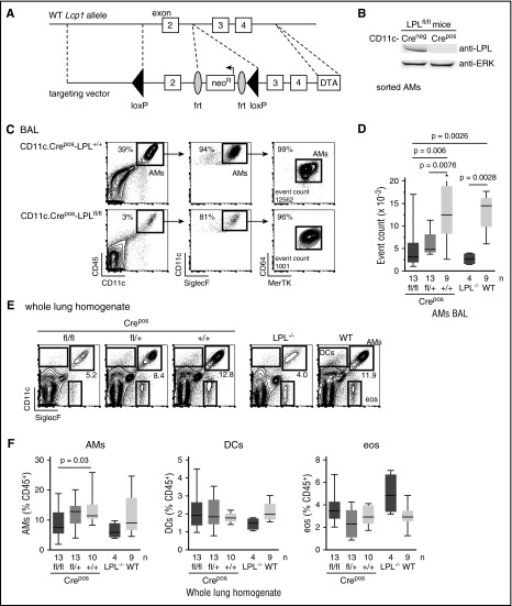
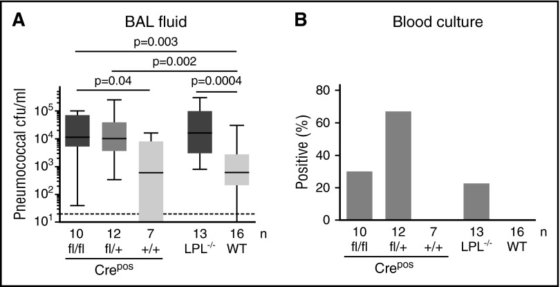
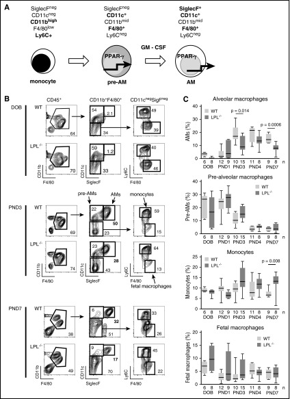
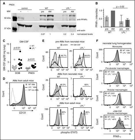
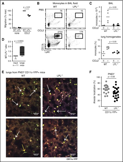
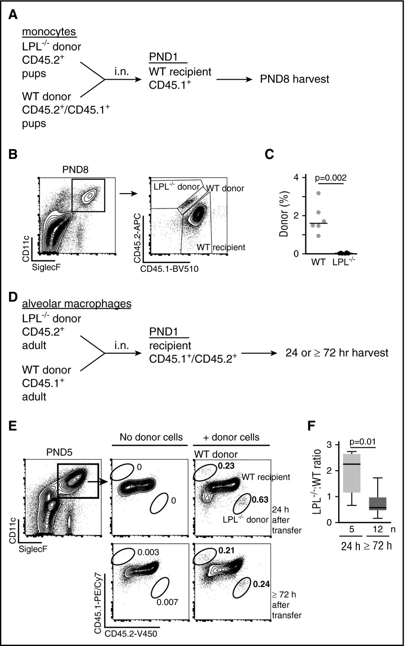
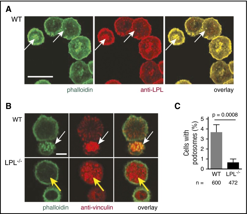
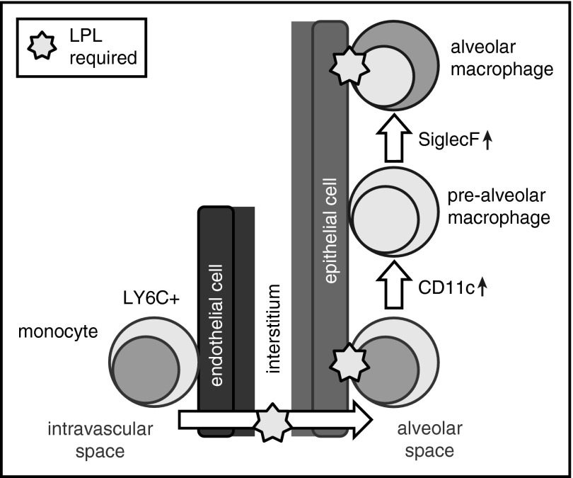
Alveolar macrophages are lung-resident sentinel cells that develop perinatally and protect against pulmonary infection. Molecular mechanisms controlling alveolar macrophage generation have not been fully defined. Here, we show that the actin-bundling protein L-plastin (LPL) is required for the perinatal development of alveolar macrophages. Mice expressing a conditional allele of LPL (CD11c.Crepos-LPLfl/fl) exhibited significant reductions in alveolar macrophages and failed to effectively clear pulmonary pneumococcal infection, showing that immunodeficiency results from reduced alveolar macrophage numbers. We next identified the phase of alveolar macrophage development requiring LPL. In mice, fetal monocytes arrive in the lungs during a late fetal stage, maturing to alveolar macrophages through a prealveolar macrophage intermediate. LPL was required for the transition from prealveolar macrophages to mature alveolar macrophages. The transition from prealveolar macrophage to alveolar macrophage requires the upregulation of the transcription factor peroxisome proliferator-activated receptor-γ (PPAR-γ), which is induced by exposure to granulocyte-macrophage colony-stimulating factor (GM-CSF). Despite abundant lung GM-CSF and intact GM-CSF receptor signaling, PPAR-γ was not sufficiently upregulated in developing alveolar macrophages in LPL-/- pups, suggesting that precursor cells were not correctly localized to the alveoli, where GM-CSF is produced. We found that LPL supports 2 actin-based processes essential for correct localization of alveolar macrophage precursors: (1) transmigration into the alveoli, and (2) engraftment in the alveoli. We thus identify a molecular pathway governing neonatal alveolar macrophage development and show that genetic disruption of alveolar macrophage development results in immunodeficiency.


Figures

Figure 1.
Figure 1.
Reduced alveolar macrophages in CD11c.Crepos -LPLfl/fl and CD11.Cre+ -LPLfl/+ mice. (A) Vector design for generation of conditional allele of LPL, encoded by gene Lcp1. A conditional allele was generated by flanking exon 2 of the gene encoding LPL, Lcp1, with loxP sequences. Exon 2 includes the ATG start site and was targeted in the generation of the LPL−/− mice, in which expression of LPL is deleted in all cells. (B) Confirmation of deletion of LPL from CD11c+ alveolar macrophages sorted from adult CD11c.Crepos-LPLfl/fl mice, with alveolar macrophages derived from CD11c.Creneg-LPLfl/fl mice shown as control. (C) Representative flow cytometry of CD45+ singlets from the BAL fluid from mice of indicated genotypes. Alveolar macrophages identified as CD11c+SiglecF+ cells, confirmed as macrophages by the expression of the pan-macrophage markers CD64 and MerTK. Percentage of alveolar macrophages shown in upper right corners of flow plots and event count of cells identified as alveolar macrophages given in last panels. (D) Quantification of number of alveolar macrophages in recovered BAL fluid. Data from 4 independent experiments; n given below graphs. *One outlier value of 89 070 not shown on graph but included in statistical analysis. Exclusion of this outlier does not alter the statistical significance of differences between indicated groups. (E) Representative flow cytometry of whole lung homogenates from mice of the indicated genotypes. (F) Percentage of CD45+ cells that were alveolar macrophages (AMs), dendritic cells (DCs), or eosinophils (eos) obtained from whole lung homogenates from mice of indicated genotypes.
Figure 2.
Figure 2.
Impaired pneumococcal clearance and increased pneumococcal dissemination in CD11c.Crepos -LPLfl/fl and CD11c.Crepos -LPLfl/+ mice. (A) Mice of indicated genotypes were challenged via intratracheal injection of 5 × 10⁴ colony-forming units (cfu) of S pneumoniae serotype 3. After 24 hours, bacterial colony-forming units in harvested BAL fluid were determined by serial dilution. Number of mice shown along the x-axis; data combined from at least 3 independent experiments. (B) Percentage of mice with positive blood cultures following pulmonary pneumococcal infection.
Figure 3.
Figure 3.
Transition from prealveolar macrophage intermediate to mature alveolar macrophage requires LPL. (A) During embryogenesis, fetal macrophages (not shown) derived from yolk-sac precursors are present in lung tissue. Around embryonic day 16, fetal monocytes migrate from the fetal liver to seed the lungs, presumably via the bloodstream., During the final days of fetal development, these monocytes downregulate Ly6C and upregulate CD11c, leading to their designation as prealveolar macrophages. Following birth, prealveolar macrophages upregulate SiglecF and are then identified as mature alveolar macrophages, which appear in the alveolar space around PND1. (B) Representative flow cytometry demonstrating gating scheme to identify fetal macrophages, fetal monocytes, prealveolar macrophages, and mature alveolar macrophages. Whole lung homogenates from WT or LPL−/− neonatal mice at indicated ages are shown. Percentages of each gate are given within each flow plot. Flow cytometric gates for each experiment were established using samples obtained from contemporaneous adult WT control animals (supplemental Figure 3A). (C) Quantification of percentage of CD45+ cells that were mature alveolar macrophages, prealveolar macrophages, fetal monocytes, or fetal macrophages from whole lung homogenates isolated from WT or LPL−/− pups. Age indicated as DOB, PND. Data for each age combined from at least 2 independent experiments; n of each sample given on the x-axis.
Figure 4.
Figure 4.
Developing alveolar macrophages in LPL−/− pups do not upregulate PPAR-γ despite abundant GM-CSF. (A) Representative immunoblot of PPAR-γ from prealveolar macrophages and alveolar macrophages sorted from whole lung homogenates of WT and LPL−/− neonatal (PND3) pups. Both PPAR-γ1 and PPAR-γ2 were detected in prealveolar macrophages and alveolar macrophages, although only PPAR-γ1 is found in the activated peritoneal macrophages used as a positive control. Positive control contained lysate from thioglycollate-elicited peritoneal macrophages, and the negative control contained lysate from thioglycollate-elicited peritoneal macrophages from PPAR-γ-deficient animals. Immunoblot of actin used as loading control. Density of PPAR-γ band normalized first to actin, then to WT alveolar macrophage sample, set to “1.” (B) PPAR-γ levels (normalized) from 4 independent experiments. Bar shows median of 4 values with interquartile range. (C) GM-CSF concentrations, measured by enzyme-linked immunosorbent assay, from whole lung homogenates from WT (gray symbols) or LPL−/− (black symbols) neonatal pups. Data normalized to lung weight obtained prior to lysis. Each symbol represents data from 1 animal, line at median. Data from 3 independent experiments combined. (D) Flow cytometric analysis of CD131 on alveolar macrophages from BAL fluid from adult WT (filled gray histogram) and LPL−/− (solid line) mice. Cells that do not express CD131 shown as negative control (dotted gray line). Representative of at least 3 independent experiments. (E) Flow cytometric analysis of phospho-STAT5 in cells incubated for 15 minutes with (solid line) or without (filled gray histogram) GM-CSF. Prealveolar macrophages and alveolar macrophages from whole lung homogenates from WT and LPL−/− neonatal pups (PND1-2) and in alveolar macrophages from BAL fluid of adult WT and LPL−/− mice, defined by flow cytometric analysis as in Figure 3. Few fully mature alveolar macrophages were present in neonatal WT and LPL−/− pups. Percentage of cells positive for phospho-STAT5 given in each histogram. Representative of 2 independent experiments. (F) Intracellular flow cytometric analysis of PPAR-γ expression in cells from whole lung homogenates of WT and LPL−/− neonatal pups (PND1-3), with cell types defined as in supplemental Figure 3C. Median fluorescence intensity of each histogram is given. In lowest panel, cells were incubated overnight in vitro with GM-CSF (20 ng/mL); percentage and median fluorescence intensity of cells with upregulated PPAR- γ are given. Representative of 2 independent experiments. MW, molecular weight.
Figure 5.
Figure 5.
Monocyte trafficking into alveoli requires LPL. (A) Monocytes isolated from the bone marrow of WT (light gray circles) and LPL−/− (black circles) mice were allowed to migrate across Transwell inserts for 90 minutes. The percentage of migrated cells was normalized to the maximum migration of WT monocytes stimulated with CCL2 (10 ng/mL) within each experiment. Averages from duplicate samples from 3 independent experiments shown; P < .01 by Student t test. (B) Representative flow cytometry of BAL fluid harvested 24 hours after intratracheal CCL2 challenge of adult WT and LPL−/− mice. Neutrophils and alveolar macrophages excluded using Ly6G and SiglecF, respectively. Monocytes identified as CD45+CD11b+Ly6C+ cells. Total number of cells in BAL fluid shown in the upper right corner; the entire BAL sample was acquired to enumerate cells. (C) Quantification of total number of monocytes recovered from BAL fluid and percentages of monocytes in whole lung homogenates from adult WT (gray circles) or LPL−/− (black circles) mice challenged with intratracheal injection of CCL2 (or PBS control). Each symbol represents data from 1 animal; data from 2 independent experiments. (D) Ratio of WT to LPL−/− monocytes isolated from adult animals, mixed and cotransferred via retro-orbital injection into WT neonatal mice and recovered 1 day following CCL2 intranasal challenge. Input ratio is used as the control. Data combined from 14 recipient mice in 3 independent experiments. (E) Examples of images acquired via 2PM of cleared lungs from PND7 CD11c.YFP+-WT or CD11c.YFP+-LPL−/− pups. PND7 mice were the smallest pups that could be consistently thoroughly perfused. CD11c+ prealveolar macrophages or alveolar macrophages were easily distinguished as round, YFP+ (green/yellow) cells (white arrows) from the thin and flat dendritic cells (yellow arrows). CD11c+ cells were also readily distinguished from many smaller, intensely autofluorescent bodies of unclear etiology located entirely within the lung parenchyma, which were found in lungs of both CD11c.YFP+-WT and CD11c.YFP+-LPL−/− pups and may be artifact from the clearing process. Brightness and contrast were adjusted using ImageJ for display in print. Images representative of randomly selected fields from lungs of 9 CD11c.YFP+-WT and 9 CD11c.YFP+-LPL−/− pups from 2 independent experiments. Scale bar represents 50 μM. (F) Percentage of round CD11c+ cells localized entirely within alveoli as determined by a blinded observer who scored Z-stacks from randomly selected fields from CD11c.YFP+-WT (gray circles) and CD11c.YFP+-LPL−/− (black circles) pups. Data combined from 2 independent experiments.
Figure 6.
Figure 6.
LPL is required for engraftment into the alveolar space. (A) Schematic of competitive transfer experiment. Fetal monocytes were sorted from the lungs of congenically marked WT (CD45.1+/CD45.2+) and LPL−/− (CD45.2+) neonatal pups. Precursors were mixed in equal proportions and transferred by intranasal (i.n.) administration into congenically marked WT (CD45.1+) neonatal pup recipients. Neonatal pups were employed as donors and recipients so as to evaluate engraftment and alveolar macrophage generation during the physiologically relevant developmental window. After 1 week, lungs were harvested and analyzed for alveolar macrophages derived from donor monocytes. (B) Representative flow cytometry from 1 recipient pup, demonstrating gating for mature alveolar macrophages and determination of origin. Flow cytometry plots showing controls for gating of CD45.1, CD45.2, and CD45.1/CD45.2 populations shown in supplemental Figure 2. (C) Percentage of alveolar macrophages derived from either WT (gray circles) or LPL−/− (black circles) donor pups. Each symbol represents data from 1 recipient animal from 2 independent experiments. (D) Schematic of competitive transfer experiment in which alveolar macrophages were isolated from BAL fluid of congenically marked adult WT and LPL−/− mice, mixed and coinjected intranasally into marked recipient PND1 pups. After 24 or ≥72 hours, lungs were harvested and analyzed for alveolar macrophages derived from donor mice. (E) Representative flow cytometric analysis of a recipient pup to evaluate presence of donor alveolar macrophages. As neonatal development of alveolar macrophages was actively ongoing in recipients, the proportion of transferred alveolar macrophages was low. (F) Ratios of LPL−/−-derived:WT-derived alveolar macrophages recovered from recipient pups at the indicated times after adoptive transfer. We delivered more alveolar macrophages from LPL−/− mice to ensure detection of LPL−/−-derived cells after 24 hours.
Figure 7.
Figure 7.
LPL is essential for alveolar macrophage podosome formation. (A) Alveolar macrophages from adult WT mice were applied to glass coverslips and fixed. F-actin was illuminated by staining with phalloidin-AlexaFluor 488 (green), and LPL was labeled with anti-LPL mAb followed by DyLight594 (red). LPL colocalizes with F-actin in podosomes (white arrows). Scale bar shows 20 μM. (B) Confocal image analysis demonstrating podosome formation in alveolar macrophages from adult WT animals (white arrow) with a podosome defined as an actin dot (green) surrounded by anti-vinculin staining (red). Podosomes did not form well, if at all, in alveolar macrophages from adult LPL−/− mice (yellow arrow). Scale bar shows 5 μM. (C) Percentage of alveolar macrophages with podosomes from adult WT (gray bar) or LPL−/− (black bar) mice. Data from 3 independent experiments combined; the standard errors of the mean of the proportions are shown and were calculated using the formula standard error = √(p*(1 − p)/n), where p represents proportion and n represents the number of samples; P value was determined using Fisher’s exact test.
Figure 8.
Figure 8.
LPL is required for the generation of alveolar macrophages and supports transmigration of cells into alveoli and retention within the alveolar microenvironment. Our data suggest a model in which LPL is required for transmigration of alveolar macrophage precursors into the alveoli during development. Our data also show that LPL is required for engraftment of monocytes and/or mature alveolar macrophages into alveoli.

Comment in

Similar articles

Cited by

References

    1. Martí-Lliteras P, Regueiro V, Morey P, et al. . Nontypeable Haemophilus influenzae clearance by alveolar macrophages is impaired by exposure to cigarette smoke. Infect Immun. 2009;77(10):4232-4242. - PMC - PubMed
    1. Steinwede K, Tempelhof O, Bolte K, et al. . Local delivery of GM-CSF protects mice from lethal pneumococcal pneumonia. J Immunol. 2011;187(10):5346-5356. - PMC - PubMed
    1. Schneider C, Nobs SP, Heer AK, et al. . Alveolar macrophages are essential for protection from respiratory failure and associated morbidity following influenza virus infection. PLoS Pathog. 2014;10(4):e1004053. - PMC - PubMed
    1. Kopf M, Schneider C, Nobs SP. The development and function of lung-resident macrophages and dendritic cells. Nat Immunol. 2015;16(1):36-44. - PubMed
    1. Camberlein E, Cohen JM, José R, et al. . Importance of bacterial replication and alveolar macrophage-independent clearance mechanisms during early lung infection with Streptococcus pneumoniae. Infect Immun. 2015;83(3):1181-1189. - PMC - PubMed

Publication types

MeSH terms