. 2016 Oct 19:6:35710.
doi: 10.1038/srep35710.

Crystal structure of human aldehyde dehydrogenase 1A3 complexed with NAD+ and retinoic acid

Affiliations

Crystal structure of human aldehyde dehydrogenase 1A3 complexed with NAD+ and retinoic acid

Andrea Moretti et al. Sci Rep. .

Abstract

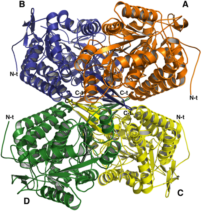
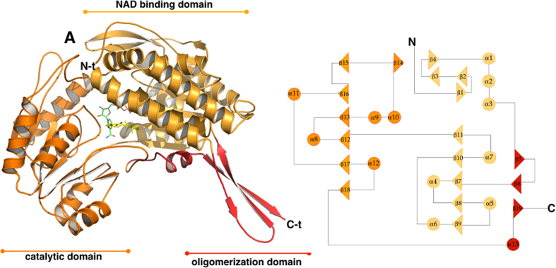
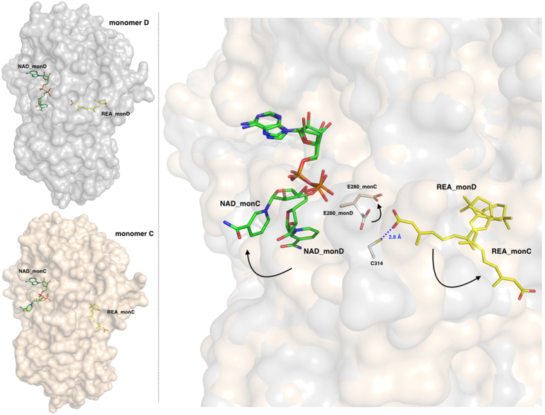
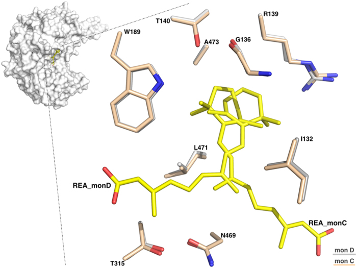
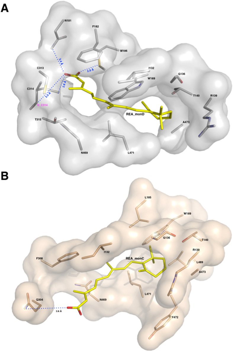
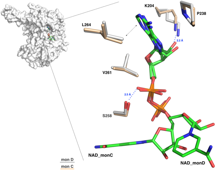
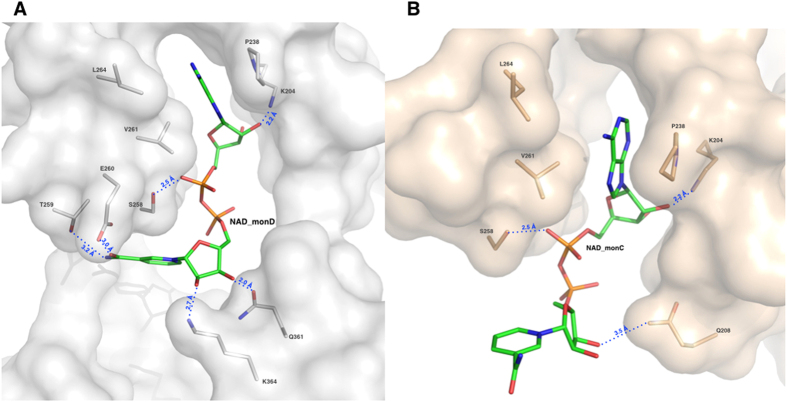
The aldehyde dehydrogenase family 1 member A3 (ALDH1A3) catalyzes the oxidation of retinal to the pleiotropic factor retinoic acid using NAD+. The level of ALDHs enzymatic activity has been used as a cancer stem cell marker and seems to correlate with tumour aggressiveness. Elevated ALDH1A3 expression in mesenchymal glioma stem cells highlights the potential of this isozyme as a prognosis marker and drug target. Here we report the first crystal structure of human ALDH1A3 complexed with NAD+ and the product all-trans retinoic acid (REA). The tetrameric ALDH1A3 folds into a three domain-based architecture highly conserved along the ALDHs family. The structural analysis revealed two different and coupled conformations for NAD+ and REA that we propose to represent two snapshots along the catalytic cycle. Indeed, the isoprenic moiety of REA points either toward the active site cysteine, or moves away adopting the product release conformation. Although ALDH1A3 shares high sequence identity with other members of the ALDH1A family, our structural analysis revealed few peculiar residues in the 1A3 isozyme active site. Our data provide information into the ALDH1As catalytic process and can be used for the structure-based design of selective inhibitors of potential medical interest.


Figures

Figure 1
Figure 1. The oligomeric structure of human ALDH1A3.
Ribbon representation of the hALDH1A3 tetramer with chains (A–D) coloured in orange, blue, yellow and green, respectively. The N-terminals are highly mobile and exposed on the surface of the tetramer while the C-terminals point toward its core. N-t →N-terminus; C-t →C-terminus.
Figure 2
Figure 2. The overall structure of human ALDH1A3 monomer.
Left hand side: ribbon representation of the hALDH1A3 monomer with its three domains coloured differently: the NAD binding-domain is shown in light orange, the catalytic-domain in orange and the oligomerization domain in red. The ligands NAD+ and REA are shown as green and yellow sticks, respectively. Right hand side: topology diagram of hALDH1A3.
Figure 3
Figure 3. The two different and coupled conformations adopted by REA and NAD+.
Left-hand side: surface representation of monomers D (upper, in grey) and C (lower, in light-pink) with bound NAD+ and REA shown as green and yellow sticks, respectively. Right-hand side: a zoom-in showing the REA and NAD+ conformations as observed in monomers D and C, shown with a surface representation in the background, after optimal superposition; the catalytic C314 from monomer D is depicted as grey stick and the E280 is shown in the two conformations adopted in monomer D and C. The two conformations observed for REA and NAD+ are coupled as indicated by the arrows, one couple representing the closed state (in monomer D) and the open state (in monomer C) that represents the product-release conformation. The residue E280 also oscillates between two conformations following those observed for the ligands.
Figure 4
Figure 4. The REA binding site.
Zoom-in of the REA binding site showing the two conformations adopted by REA in monomers D and C, after optimal superposition. Residues from the two monomers are represented as sticks, and coloured in light-pink for monomer C and in grey for monomer D. The two REA molecules (REA_C and REA_D) are represented as sticks and shown in yellow.
Figure 5
Figure 5. The REA binding site and major interactions established by REA with the protein milieu in its two conformations.
Surface representation of REA binding sites: the monomers D in grey and the monomer C in light-pink. Protein residues are represented as sticks and coloured in light-pink for monomer C and in grey for monomer D; the REA ligands (REA_C and REA_D) are shown as sticks and depicted in yellow. (A) The REA binding mode as observed in monomer D. The β-ionone ring establishes hydrophobic interactions with I132, G136, R139, T140, W189, L471 and A473. Its carboxyl group makes hydrogen bonds with C314, M186 and N181, and hydrophobic interactions with F182, C313, T315 and N469. (B) The REA binding mode as observed in monomer C. The β-ionone ring maintains the same interactions as in monomer D. On the contrary its carboxyl group significantly moves and makes hydrogen bonds with Q304 and hydrophobic interactions with F305 and N469.
Figure 6
Figure 6. The NAD+ binding site.
Zoom-in of the NAD+ binding site showing the two conformations adopted by the cofactor in monomers D and C, after optimal superposition. Residues from the two monomers are represented as sticks, and coloured in light-pink for monomer C and in grey for monomer D. The two NAD+ molecules (NAD_C and NAD_D) are represented as sticks and shown in green.
Figure 7
Figure 7. The NAD+ binding site and major interactions established by REA with the protein milieu in its two conformations.
Surface representation of NAD+ binding sites: the monomers D in grey and the monomer C in light-pink. Protein residues are represented as sticks and coloured in light-pink for monomer C and in grey for monomer D; the NAD+ ligands (NAD_C and NAD_D) are shown as sticks and depicted in green. (A) The NAD+ binding mode as observed in monomer D with P238, L264 and V261 contacting the NAD+ adenine moiety and K204 and S258 the adenine ribose. The NAD+ nicotinamide moiety is stabilized through hydrogen bonds established with four amino-acids: K364, Q361, T259 and E260. (B) The NAD+ binding mode as observed in monomer C. The ADP moiety maintains same orientation as in monomer D and establishes same contacts with protein residues. On the contrary, the nicotinamide changes orientation and loses the network of interactions observed in its closed conformation in monomer D and the nicotinamide ribose makes a new contact with Q208.

Similar articles

Cited by

References

    1. Vasiliou V., Pappa A. & Petersen D. R. Role of aldehyde dehydrogenases in endogenous and xenobiotic metabolism. Chem Biol Interact. 129, 1–19 (2000). - PubMed
    1. Vasiliou V., Pappa A. & Estey T. Role of human aldehyde dehydrogenases in endobiotic and xenobiotic metabolism. Drug Metab Rev. 36, 279–299 (2004). - PubMed
    1. Yokoyama A. et al. Alcohol and aldehyde dehydrogenase gene polymorphisms and oropharyngolaryngeal, esophageal and stomach cancers in Japanese alcoholics. Carcinogenesis 22, 433–439 (2001). - PubMed
    1. Vasiliou V. & Nebert D. W. Analysis and update of the human aldehyde dehydrogenase (ALDH) gene family. Hum Genomics. 2, 138–143 (2005). - PMC - PubMed
    1. Sladek N. E. Human aldehyde dehydrogenases: potential pathological, pharmacological, and toxicological impact. J Biochem Mol Toxicol. 17, 7–23 (2003). - PubMed

Publication types