. 2016 Oct 19:6:35581.
doi: 10.1038/srep35581.

Plasmodium vivax GPI-anchored micronemal antigen (PvGAMA) binds human erythrocytes independent of Duffy antigen status

Affiliations

Plasmodium vivax GPI-anchored micronemal antigen (PvGAMA) binds human erythrocytes independent of Duffy antigen status

Yang Cheng et al. Sci Rep. .

Abstract

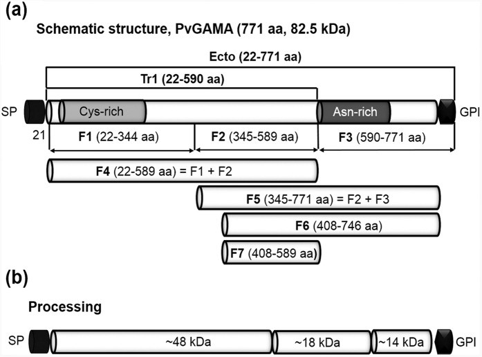
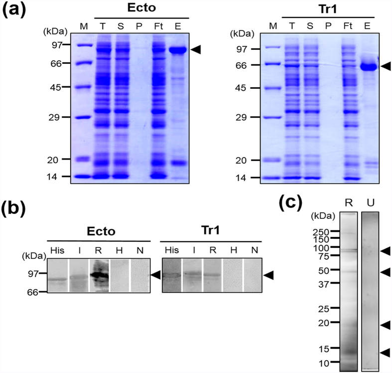
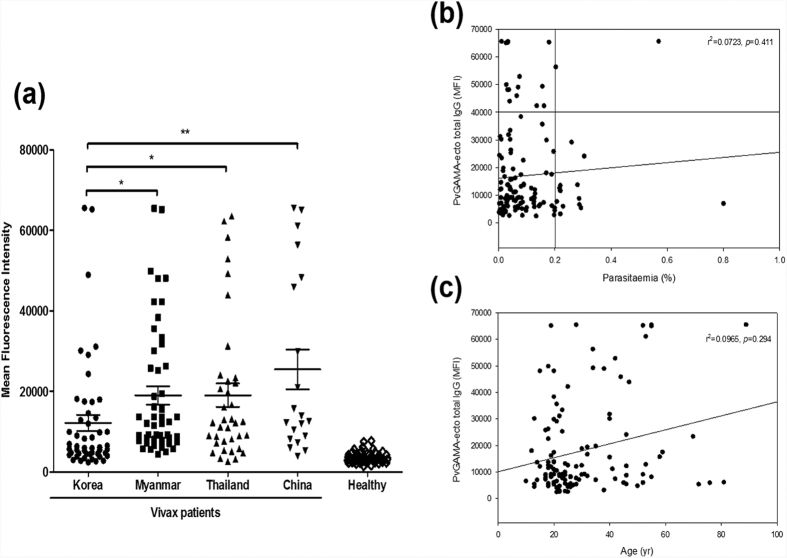
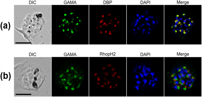
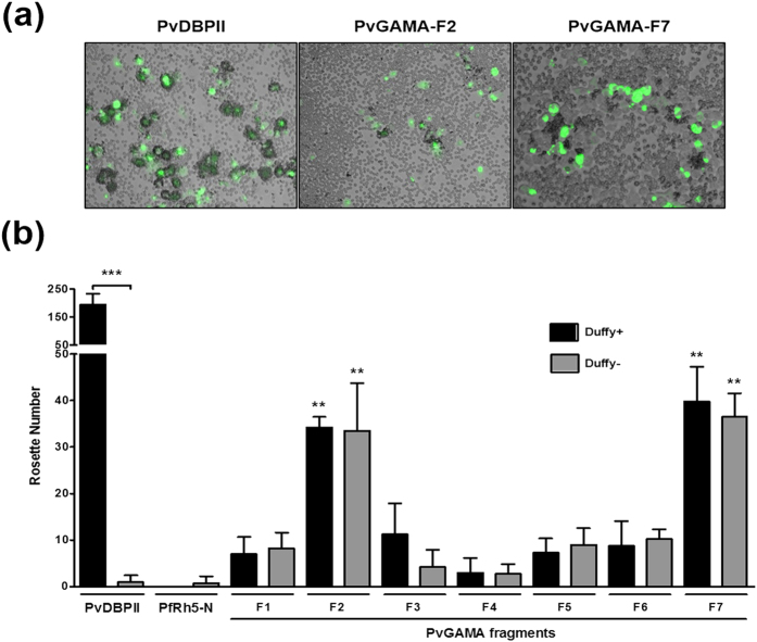
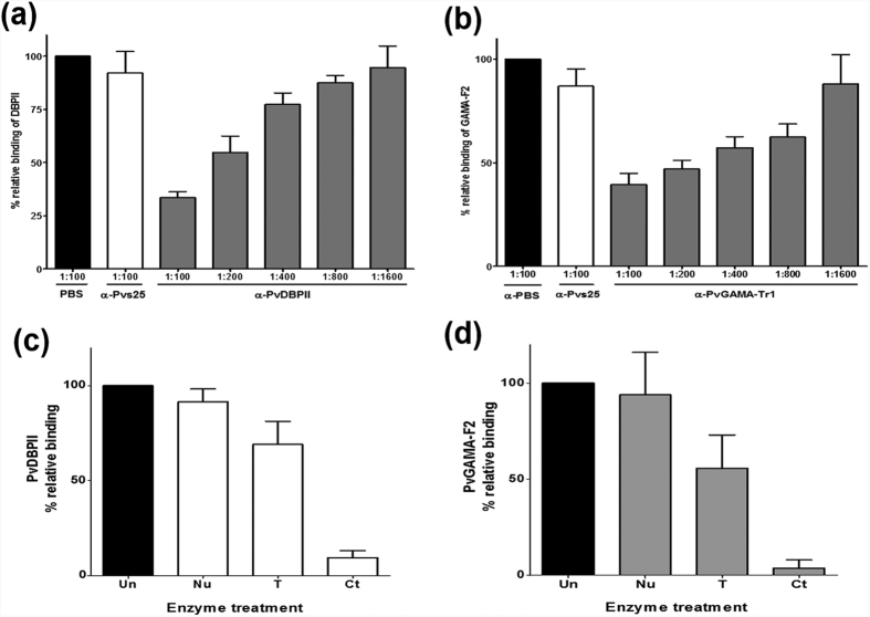
Plasmodium vivax, a major agent of malaria in both temperate and tropical climates, has been thought to be unable to infect humans lacking the Duffy (Fy) blood group antigen because this receptor is critical for erythrocyte invasion. Recent surveys in various endemic regions, however, have reported P. vivax infections in Duffy-negative individuals, suggesting that the parasite may utilize alternative receptor-ligand pairs to complete the erythrocyte invasion. Here, we identified and characterized a novel parasite ligand, Plasmodium vivax GPI-anchored micronemal antigen (PvGAMA), that bound human erythrocytes regardless of Duffy antigen status. PvGAMA was localized at the microneme in the mature schizont-stage parasites. The antibodies against PvGAMA fragments inhibited PvGAMA binding to erythrocytes in a dose-dependent manner. The erythrocyte-specific binding activities of PvGAMA were significantly reduced by chymotrypsin treatment. Thus, PvGAMA may be an adhesion molecule for the invasion of Duffy-positive and -negative human erythrocytes.


Figures

Figure 1
Figure 1. Schematic diagram of PvGAMA, fragments of PvGAMA for recombinant protein expression, erythrocyte binding assay and processing of PvGAMA.
(a) Schematic diagram of PvGAMA. The PvGAMA protein comprises 771 amino acids with a calculated molecular mass of 82.5 kDa. Two regions of PvGAMA, PvGAMA-Ecto (Ecto; amino acid (aa) position 22–771) and -Tr1 (Tr1; aa 22-590), were designed for protein expression, and 7 fragments (F1, aa 22–344; F2, aa 345–589; F3, aa 590–771; F4, aa 22–589; F5, aa 345–771; F6, aa 408–746 and F7, aa 408–589) were designed for the erythrocyte binding assay. Indicated are the predicted signal peptide (SP; aa position 1–21) and glycosylphosphatidylinositol-anchor signal (GPI; aa 747–771). PvGAMA-Ecto and -Tr1 were used to raise specific antisera. (b) Processing of PvGAMA was predicted from western blot results of the parasite lysate probed with anti-PvGAMA-Ecto sera (Fig. 2c). aa, amino acid; kDa, kilodalton; SP, signal peptide; GPI, glycophosphatidylinositol-anchor signal; Cys, Cysteine; Asn, Asparagine.
Figure 2
Figure 2. Recombinant protein expression, purification, and analysis of PvGAMA fragments.
(a) PvGAMA-Ecto and PvGAMA-Tr1 were synthesized using the wheat germ cell-free protein expression system and purified on Ni-Sepharose columns. The purified fragments of PvGAMA were found in the soluble elution fractions. The arrowheads indicate specific bands for each recombinant protein. T, total translation mix; S, supernatant; P, pellet; Ft, flow through; E, elution. (b) The purified PvGAMA-Ecto and PvGAMA-Tr1 were resolved by SDS-PAGE, transferred to a PVDF membrane, and probed with an anti-His-tag antibody (His), vivax-infected human sera (I), or rabbit PvGAMA-Ecto immunized sera (R). Healthy human sera (H) and non-immunized rabbit sera were used as negative controls (N). (c) Schizont lysates under reducing condition probed with PvGAMA-Ecto immune sera (R). A pre-immunized rabbit serum was used as a negative control (U). The arrowheads indicate specific bands for each recombinant protein.
Figure 3
Figure 3. Natural acquired antibodies against the PvGAMA-Ecto antigen.
(a) Total IgG prevalence of PvGAMA-Ecto with the P. vivax patient and healthy individual sera samples. The bar indicates the mean ± standard error of the fluorescence intensity. The p-values were calculated using Student’s t-test. (b) The correlation between PvGAMA-Ecto with parasitaemia was evaluated using Spearman’s correlation test. An MFI >40,000 was considered high intensity and parasitaemia >0.2% was considered high parasitaemia. (c) The correlation between PvGAMA-Ecto and parasitaemia was evaluated using Spearman’s correlation test. MFI, mean fluorescence intensity.
Figure 4
Figure 4. Subcellular localization of PvGAMA protein in asexual blood-stage parasites of P. vivax.
(a) The acetone-fixed mature schizont of P. vivax was dual-labelled with rabbit immune sera against PvGAMA (green) and mouse immune sera against PvDBP (red). (b) Another mature schizont of P. vivax was also dual-labelled with rabbit immune sera against PvGAMA (green) and mouse immune sera against PvRhopH2 (PVX_099930, red). The nuclei are visualized with DAPI (blue); the bar represents 5 μm.
Figure 5
Figure 5. Binding specificity of PvGAMA fragments expressed on HEK 293T cells to Duffy-positive and -negative erythrocytes.
(a) Erythrocyte-binding rosettes formed on the surface of HEK 293T cells expressing PvDBPII or different fragments of PvGAMA were visualized under light microscopy. (b) The number of rosettes formed by the HEK 293T cells transfected with genes coding for either PvDBPII, the non-binding domain of PfRH5 (PfRH5-N) or different fragments of PvGAMA (see Fig. 1a). Detection of the transfection efficiency of all constructs into HEK 293T cells by counting green signalling cells within 30 microscope fields (×200 magnification). Positive was defined as more than half the surface of the transfected cells covered with attached erythrocytes, and the total number of HEK 293T cells per coverslip was recorded. The data are shown as the mean number of rosettes of four independent experiments at different days, and the error bar represents ± standard deviation. The p-values were calculated using Student’s t-test. Significant differences are shown as double asterisks, p < 0.01, and triple asterisks, p < 0.0001.
Figure 6
Figure 6. Inhibition of erythrocyte rosettes to PvGAMA-F2 by anti-PvGAMA-Tr1 antibody and receptor specificities.
(a) HEK 293T cells were transfected with pEGFP-HSVgD1_DBPII plasmid DNA expressing a GFP-DBPII fusion protein. The cells were subsequently incubated with anti-PvDBPII rabbit sera at various dilutions before the addition of human erythrocytes. Binding was scored by counting the number of rosettes bound to HEK 293T cells in 30 microscope fields (×200 magnification). Rabbits immunized with PBS as a non-immunized control (PBS) and anti-Pvs25 (no erythrocyte-binding activity) immune rabbit sera diluted 1:100 were included as negative controls (Pvs25). (b) Inhibition of erythrocyte binding to PvGAMA-F2 expressed on HEK 293T cells by PvGAMA-Tr1 immune rabbit sera. The cells were incubated with PvGAMA-Tr1 immune rabbit sera at various dilutions before the addition of human erythrocytes. Error bars represent ± standard deviations. The erythrocyte binding abilities of HEK 293T cells transfected with pEGFP-HSVgD1_PvDBPII and PvGAMA-F2 plasmid DNA expressing a GFP-PvDBPII (c) and GFP-PvGAMA-F2 (d) were tested by incubation with untreated (Un), neuraminidase-treated (Nu), trypsin-treated (T), and chymotrypsin-treated (Ct) erythrocytes. The bars represent the standard deviation of the means of the three independent experiments.

References

    1. Miller L. H., Mason S. J., Dvorak J. A., McGinniss M. H. & Rothman I. K. Erythrocyte receptors for (Plasmodium knowlesi) malaria: Duffy blood group determinants. Science 189, 561–563 (1975). - PubMed
    1. Miller L. H., Mason S. J., Clyde D. F. & McGinniss M. H. The resistance factor to Plasmodium vivax in blacks. The Duffy-blood-group genotype, FyFy. N Engl J Med 295, 302–304, 10.1056/NEJM197608052950602 (1976). - DOI - PubMed
    1. Adams J. H. et al.. The Duffy receptor family of Plasmodium knowlesi is located within the micronemes of invasive malaria merozoites. Cell 63, 141–153 (1990). - PubMed
    1. Horuk R. et al.. A receptor for the malarial parasite Plasmodium vivax: the erythrocyte chemokine receptor. Science 261, 1182–1184 (1993). - PubMed
    1. Fraser T. et al.. Expression and serologic activity of a soluble recombinant Plasmodium vivax Duffy binding protein. Infect Immun 65, 2772–2777 (1997). - PMC - PubMed

Publication types

MeSH terms

Substances

LinkOut - more resources