

Optical Coherence Tomography Features in Diabetic Macular Edema and the Impact on Anti-VEGF Response
- PMID: 27759856
- PMCID: PMC5512288
- DOI: 10.3928/23258160-20161004-03
Optical Coherence Tomography Features in Diabetic Macular Edema and the Impact on Anti-VEGF Response
Abstract
Background and objective: To assess the relationship between spectral-domain optical coherence tomography (SD-OCT) features and functional outcomes for diabetic macular edema (DME) undergoing treatment with intravitreal bevacizumab (Avastin; Genentech, South San Francisco, CA).
Patients and methods: Institutional review board-approved, retrospective, consecutive case series of eyes receiving intravitreal bevacizumab (1.25 mg) for DME. SD-OCT features were evaluated and correlated with functional response to anti-vascular endothelial growth factor (VEGF) therapy.
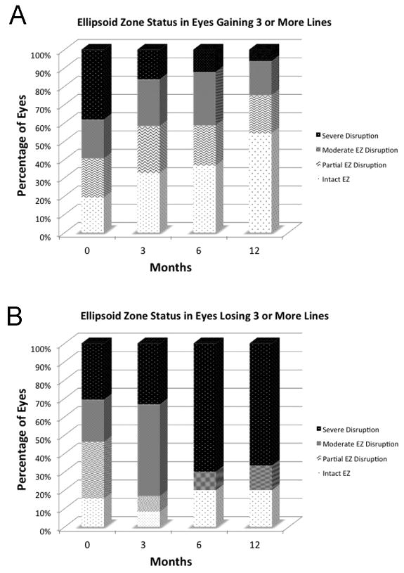
Results: One hundred fifty-nine eyes of 159 subjects were included in this study. Mean visual acuity improved from 20/76 to 20/58. The proportion of eyes with 20/40 or greater visual acuity increased with treatment (35% at initial visit, 51% at final visit). SD-OCT factors that were associated with functional outcomes to anti-VEGF therapy include ellipsoid zone integrity and severity of intraretinal fluid.
Conclusion: SD-OCT features appear to provide important markers for functional response to anti-VEGF therapy in DME. [Ophthalmic Surg Lasers Imaging Retina. 2016;47:908-913.].
Copyright 2016, SLACK Incorporated.
Figures
References
-
- Rogers SL, Tikellis G, Cheung N, et al. Retinal arteriolar caliber predicts incident retinopathy: the Australian Diabetes, Obesity and Lifestyle (AusDiab) study. Diabetes Care. 2008 Apr;31(4):761–3. - PubMed
-
- Soheilian M, Ramezani A, Bijanzadeh B, et al. Intravitreal bevacizumab (avastin) injection alone or combined with triamcinolone versus macular photocoagulation as primary treatment of diabetic macular edema. Retina. 2007 Nov-Dec;27(9):1187–95. - PubMed
-
- Solaiman KA, Diab MM, Abo-Elenin M. Intravitreal bevacizumab and/or macular photocoagulation as a primary treatment for diffuse diabetic macular edema. Retina. 2010 Nov-Dec;30(10):1638–45. - PubMed
-
- Focal photocoagulation treatment of diabetic macular edema. Relationship of treatment effect to fluorescein angiographic and other retinal characteristics at baseline: ETDRS report no. 19. Early Treatment Diabetic Retinopathy Study Research Group. Arch Ophthalmol. 1995 Sep;113(9):1144–55. - PubMed
MeSH terms
Substances
Grants and funding
LinkOut - more resources
Full Text Sources
Other Literature Sources
Medical
