

Analysis of Scleral Feeder Vessel in Myopic Choroidal Neovascularization Using Optical Coherence Tomography Angiography
- PMID: 27759864
- PMCID: PMC5279865
- DOI: 10.3928/23258160-20161004-11
Analysis of Scleral Feeder Vessel in Myopic Choroidal Neovascularization Using Optical Coherence Tomography Angiography
Abstract
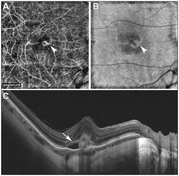
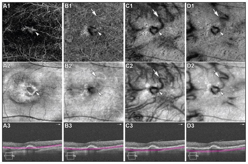
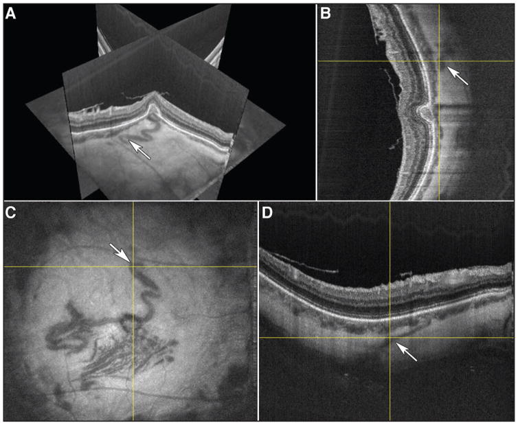
To describe the appearance of a scleral-derived feeder vessel in a highly myopic eye with secondary choroidal neovascularization (CNV) as visualized on both en face high-speed swept-source (SS) optical coherence tomography angiography (OCTA) prototype, and a commercially available spectral-domain (SD) OCTA, with the corresponding en face and cross-sectional structural OCT images. In this case report, a 60-year-old white male presented with high myopia and secondary CNV in the right eye, previously treated with anti-vascular endothelial growth factor, and was imaged on both SD-OCT and SS-OCT. The neovascular complex could be visualized on both devices. Structural en face SS-OCT images demonstrated a large choroidal-scleral feeder vessel that was not visualized with SD-OCT. The authors concluded that structural en face SS-OCT better visualizes scleral feeder vessel compared to SD-OCT due to the longer wavelength (∼1,050 nm) with increased choroidal penetration and decreased sensitivity roll-off in the SS-OCT system. [Ophthalmic Surg Lasers Imaging Retina. 2016;47:960-964.].
Copyright 2016, SLACK Incorporated.
Figures
References
-
- Kotsolis AI, Killian FA, Ladas ID, Yannuzzi LA. Fluorescein angiography and optical coherence tomography concordance for choroidal neovascularisation in multifocal choroidtis. Br J Ophthalmol. 2010;94(11):1506–1508. - PubMed
-
- Makita S, Hong Y, Yamanari M, Yatagai T, Yasuno Y. Optical coherence angiography. Opt Express. 2006;14(17):7821–7840. - PubMed
-
- Fingler J, Schwartz D, Yang C, Fraser SE. Mobility and transverse flow visualization using phase variance contrast with spectral domain optical coherence tomography. Opt Express. 2007;15(20):12636–12653. - PubMed
-
- Tao YK, Davis AM, Izatt JA. Single-pass volumetric bidirectional blood flow imaging spectral domain optical coherence tomography using a modified Hilbert transform. Opt Express. 2008;16(16):12350–12361. - PubMed
-
- An L, Wang RK. In vivo volumetric imaging of vascular perfusion within human retina and choroids with optical microangiography. Opt Express. 2008;16(15):11438–11452. - PubMed
Publication types
MeSH terms
Grants and funding
LinkOut - more resources
Full Text Sources
Other Literature Sources
Research Materials
