. 2016 Nov 1;126(11):4237-4249.
doi: 10.1172/JCI85057. Epub 2016 Oct 17.

Helicase CHD4 is an epigenetic coregulator of PAX3-FOXO1 in alveolar rhabdomyosarcoma

Helicase CHD4 is an epigenetic coregulator of PAX3-FOXO1 in alveolar rhabdomyosarcoma

Maria Böhm et al. J Clin Invest. .

Abstract

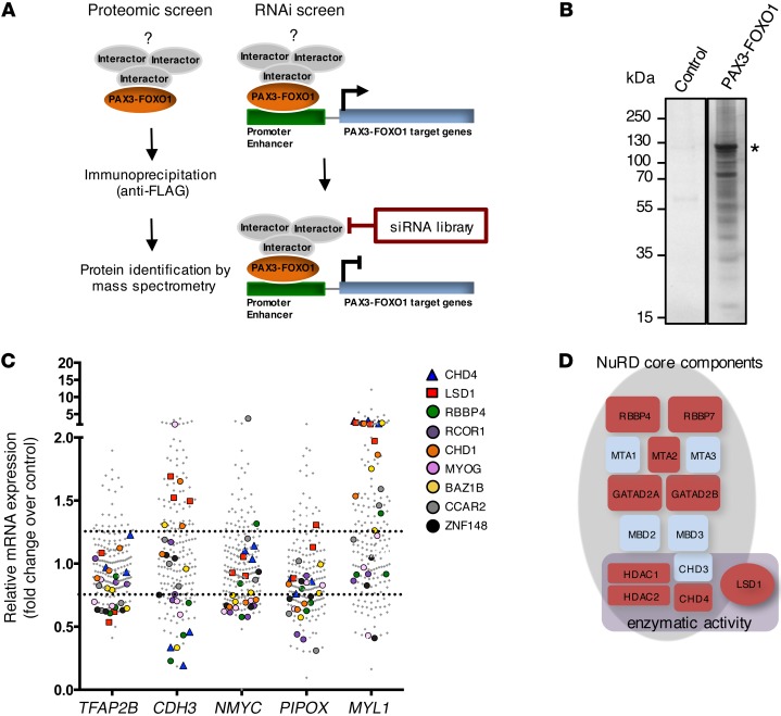
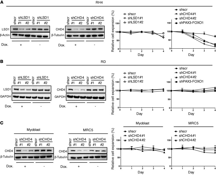
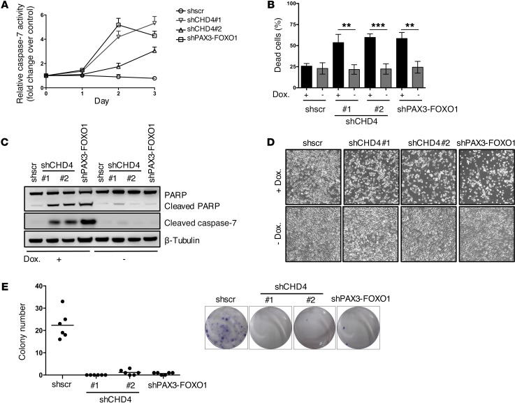
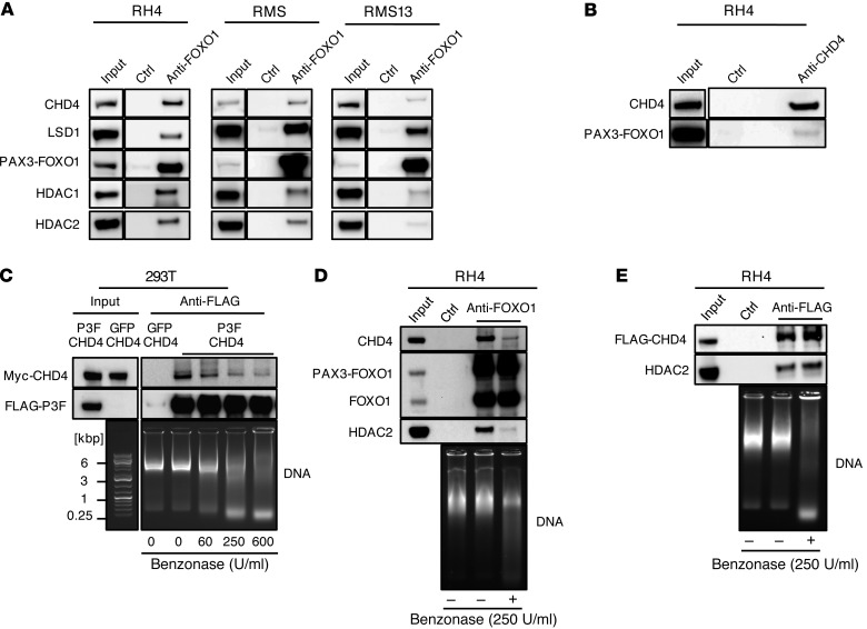
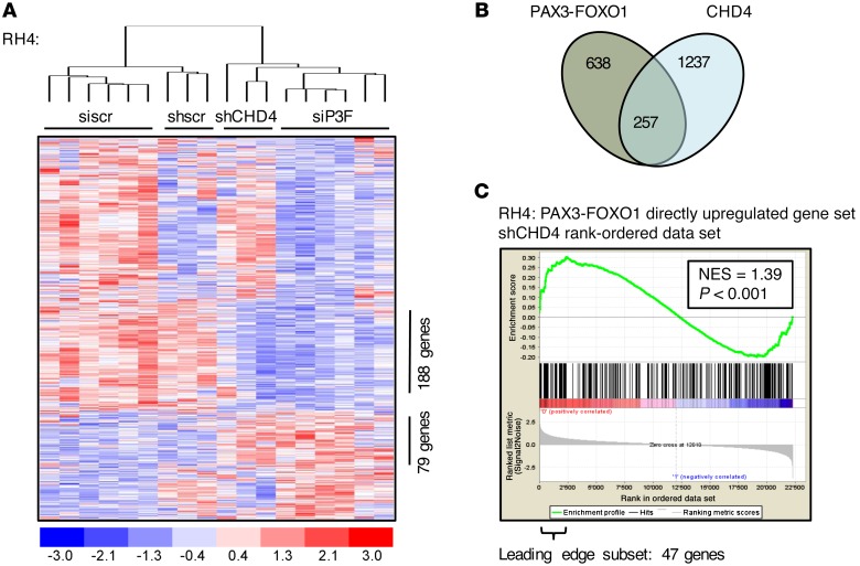
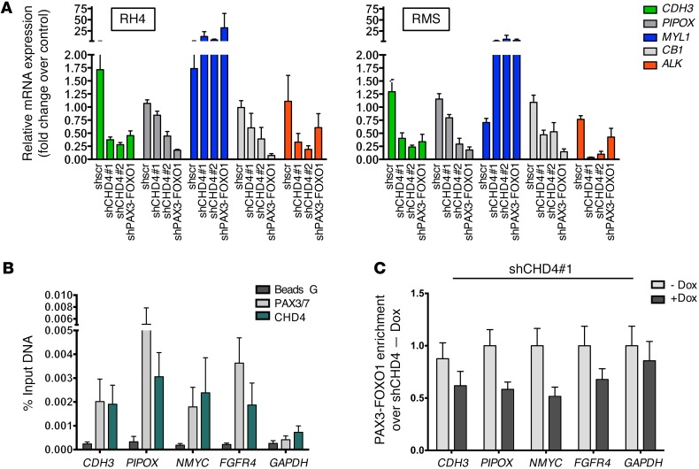
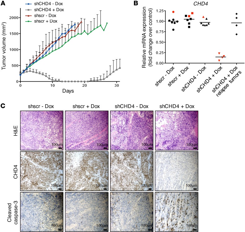
A vast number of cancer genes are transcription factors that drive tumorigenesis as oncogenic fusion proteins. Although the direct targeting of transcription factors remains challenging, therapies aimed at oncogenic fusion proteins are attractive as potential treatments for cancer. There is particular interest in targeting the oncogenic PAX3-FOXO1 fusion transcription factor, which induces alveolar rhabdomyosarcoma (aRMS), an aggressive cancer of skeletal muscle cells for which patient outcomes remain dismal. In this work, we have defined the interactome of PAX3-FOXO1 and screened 60 candidate interactors using siRNA-mediated depletion to identify candidates that affect fusion protein activity in aRMS cells. We report that chromodomain helicase DNA binding protein 4 (CHD4), an ATP-dependent chromatin remodeler, acts as crucial coregulator of PAX3-FOXO1 activity. CHD4 interacts with PAX3-FOXO1 via short DNA fragments. Together, they bind to regulatory regions of PAX3-FOXO1 target genes. Gene expression analysis suggested that CHD4 coregulatory activity is essential for a subset of PAX3-FOXO1 target genes. Depletion of CHD4 reduced cell viability of fusion-positive but not of fusion-negative RMS in vitro, which resembled loss of PAX3-FOXO1. It also caused specific regression of fusion-positive xenograft tumors in vivo. Therefore, this work identifies CHD4 as an epigenetic coregulator of PAX3-FOXO1 activity, providing rational evidence for CHD4 as a potential therapeutic target in aRMS.


Figures

Figure 1
Figure 1. Two-step proteomic and siRNA screen identifies putative PAX3-FOXO1 interaction partners.
(A) Schematic representation of the 2-step screening approach. (B) Representative silver-stained gel of FLAG immunoprecipitates from RMS13 cells transfected with FLAG-tagged PAX3-FOXO1. The asterisk marks the band corresponding to FLAG-PAX3-FOXO1. RMS13 cells expressing only FLAG-tag served as negative control. (C) SiRNA screening results for 60 candidate interactors. Each candidate was silenced using 3 different siRNAs per target. Fold change of expression of PAX3-FOXO1 target genes was measured by quantitative real-time PCR relative to RH4 cells treated with scrambled control. Values for the 9 candidate interactors resulting from the siRNA screen are indicated. (D) Schematic representation of the NuRD complex. General subunit composition and components with enzymatic activity are displayed. NuRD subunits identified by MS are marked in red.
Figure 2
Figure 2. Silencing of CHD4 affects specifically FP-RMS cell expansion.
(A) Left panels: Western blots after knockdown of CHD4 or LSD1 in RH4 cell extracts 72 hours after induction with doxycycline (Dox.). Uninduced cells served as negative control, and actin or tubulin was used as loading control. Right panels: RH4 cell expansion was measured by WST assay at indicated time points after silencing of LSD1 or CHD4 by shRNA induction with doxycycline. Data represent the mean ± SD of 3 independent experiments and were normalized to uninduced cells. PAX3-FOXO1 knockdown served as positive control. (B) Western blots and cell expansion after LSD1 or CHD4 knockdown in RD cells treated and analyzed as in A. (C) Western blots and cell proliferation after CHD4 knockdown in human myoblast or MRC5 cells treated and analyzed as in A.
Figure 3
Figure 3. CHD4 knockdown decreases viability of FP-RMS cells.
(A) Caspase-3/7 activity was measured in RH4 cells 72 hours after induction of CHD4 silencing. Fold change of caspase-3/7 activity was normalized to uninduced cells. Shscr-treated cells served as negative and PAX3-FOXO1 knockdown cells as positive controls. Values represent the mean ± SD of 3 independent biological experiments. (B) Percentage of dead cells 96 hours after induction of CHD4 silencing. Cells were stained with NucView Caspase-3 Substrate and 7AAD and analyzed by flow cytometry. Values represent the mean ± SD of 4 independent experiments (**P < 0.01; ***P < 0.001; uncorrected Fisher’s LSD). (C) Western blot of PARP and cleaved caspase-7 from extracts of RH4 cells 72 hours after induction of CHD4 or PAX3-FOXO1 silencing and uninduced or shscr-treated control cells. (D) Representative phase-contrast images of RH4 cells 72 hours after induction of silencing with doxycycline (Dox.) transduced with indicated constructs; original magnification, ×100. (E) Clonogenic assays of RH4 cells 12 days after induction of CHD4 silencing. Quantitation of number of colonies with black lines representing the mean values. Representative images of crystal violet–stained colonies are shown in the right panel.
Figure 4
Figure 4. CHD4 and PAX3-FOXO1 mainly interact via short DNA fragments.
(A) Representative Western blots of endogenous PAX3-FOXO1 immunoprecipitates and lysates from 3 different FP-RMS cell lines. PAX3-FOXO1 was immunoprecipitated by the anti-FOXO1 antibody, and uncoated beads served as negative control (ctrl). (B) Representative Western blot of reciprocal CHD4 immunoprecipitate from RH4 cells. Endogenous CHD4 was immunoprecipitated by an anti-CHD4 antibody, and uncoated beads served as negative control (ctrl). (C) Western blot detection of indicated proteins in anti-FLAG immunoprecipitates and lysates from 293T cells transfected with FLAG-tagged P3F and Myc-tagged CHD4. Cell lysate was treated with indicated amounts of Benzonase during immunoprecipitation to digest the DNA, and DNA digestion was evaluated by agarose gel electrophoresis. (D) Western blot detection of indicated endogenous proteins in anti-FOXO1 immunoprecipitates from RH4 cells. Uncoated beads served as negative control (ctrl). Lysates were digested or not with 250 U/ml Benzonase, and DNA digestion was evaluated by agarose gel electrophoresis. (E) Western blot detection of indicated endogenous proteins in anti-FLAG immunoprecipitates from RH4 cells with stable knock-in of 3X FLAG at C-terminus of CHD4. Uncoated beads served as negative control (Ctrl). Lysates were digested or not with 250 U/ml Benzonase, and DNA digestion was evaluated by agarose gel electrophoresis.
Figure 5
Figure 5. CHD4 coregulates PAX3-FOXO1–activated target genes.
(A) Heat map of unsupervised hierarchical clustering of PAX3-FOXO1 (siP3F) and CHD4 (shCHD4#1) knockdown profiles using the gene set directly regulated by PAX3-FOXO1 (638 genes differently expressed between siP3F-treated and siscr-treated [24 and 48 hours after transfection] or untreated RH4 cells; fold change >1.5; P < 0.05). Each column represents a different time point for shRNA-treated cells in singlicate and for siRNA-treated cells in duplicate (24, 48, 72 hours from left to right for each condition). Numbers of coexpressed genes between these transcriptional profiles are displayed. (B) Schematic representation of genes directly regulated by PAX3-FOXO1 and CHD4 (fold change >1.5; P < 0.05 between siP3F-treated and siscr-treated [24 and 48 hours after transfection] or untreated RH4 cells and between shCHD4-expressing and shscr-expressing RH4 cells [24 and 48 hours after doxycycline induction] or uninduced control cells). The coregulated subset as read out from the heatmap clustering in A is displayed. (C) GSEA using CHD4-regulated genes in RH4 cells as the rank-ordered data set and targets directly upregulated by PAX3-FOXO1 as the gene set (259 genes differently expressed between siP3F-treated and siscr-treated or untreated RH4 cells 24 and 48 hours after transfection; fold change >1.7; P < 0.05). Normalized enrichment score (NES) and P value are shown.
Figure 6
Figure 6. CHD4 colocalizes to PAX3-FOXO1 binding sites.
(A) Expression levels of indicated PAX3-FOXO1 target genes were quantified by quantitative real-time PCR after CHD4 knockdown in 2 FP-RMS cell lines. Bar charts are geometric means from 4 independent experiments with 95% CI (P < 0.05; Dunnett’s multiple comparison test). Fold change of mRNA expression was normalized to uninduced cells with PAX3-FOXO1 knockdown serving as positive control. (B) ChIP was performed in RH4 cells using PAX3/7 and CHD4 antibodies on known PAX3-FOXO1 DNA binding sites in target genes, and only beads without antibody served as negative control (Beads G). Bar charts indicate the mean ± SEM of at least 3 independent biological replicates. Abundance of precipitated DNA fragments was measured by quantitative PCR, and results are presented as the percentage of input. The GAPDH promoter region served as negative control. (C) Effect on PAX3-FOXO1 binding upon CHD4 silencing (48 hours of incubation with doxycycline) was determined by ChIP as described in B and is shown as fold enrichment over PAX3-FOXO1 ChIP signal in the presence of CHD4 (no doxycycline control).
Figure 7
Figure 7. CHD4 inhibition causes tumor regression in mouse xenografts.
In vivo treatment of NOD/SCID mice engrafted with RH4 cells containing stably integrated doxycycline-inducible shCHD4#1 or shscr expression vectors. Mice bearing palpable tumors were treated i.p. for 2 days with either vehicle control or doxycycline at a dose of 53.3 mg/kg. Additionally, they were fed with doxycycline-supplemented or control food. (A) Absolute tumor volumes of FP-RMS xenografts as measured by caliper. Vehicle-treated control groups consisted of 6 mice, and doxycycline-treated groups consisted of 5 (shscr) and 7 (shCHD4#1) mice. (B) CHD4 mRNA expression level in indicated tumors of doxycycline- or vehicle control–treated mice analyzed by quantitative real-time PCR. Tumors were isolated at the end of treatment (black; tumor set shown in A) or after 4 days of treatment (red; separate tumor set). Fold changes of mRNA expression are normalized to the mean of CHD4 expression in shscr vehicle control–treated tumors. (C) Immunohistochemical staining of tumors with indicated antibodies. Tumors were isolated 4 days after the start of treatment.

References

    1. Breneman JC, et al. Prognostic factors and clinical outcomes in children and adolescents with metastatic rhabdomyosarcoma — a report from the Intergroup Rhabdomyosarcoma Study IV. J Clin Oncol. 2003;21(1):78–84. doi: 10.1200/JCO.2003.06.129. - DOI - PubMed
    1. Van Gaal JC, De Bont ES, Kaal SE, Versleijen-Jonkers Y, van der Graaf WT. Building the bridge between rhabdomyosarcoma in children, adolescents and young adults: the road ahead. Crit Rev Oncol Hematol. 2012;82(3):259–279. doi: 10.1016/j.critrevonc.2011.06.005. - DOI - PubMed
    1. Barr FG. Gene fusions involving PAX and FOX family members in alveolar rhabdomyosarcoma. Oncogene. 2001;20(40):5736–5746. doi: 10.1038/sj.onc.1204599. - DOI - PubMed
    1. Skapek SX, et al. PAX-FOXO1 fusion status drives unfavorable outcome for children with rhabdomyosarcoma: a children’s oncology group report. Pediatr Blood Cancer. 2013;60(9):1411–1417. doi: 10.1002/pbc.24532. - DOI - PMC - PubMed
    1. Missiaglia E, et al. Genomic imbalances in rhabdomyosarcoma cell lines affect expression of genes frequently altered in primary tumors: an approach to identify candidate genes involved in tumor development. Genes Chromosomes Cancer. 2009;48(6):455–467. doi: 10.1002/gcc.20655. - DOI - PubMed

Publication types

MeSH terms

Substances