. 2016 Oct 19;12(10):e1006393.
doi: 10.1371/journal.pgen.1006393. eCollection 2016 Oct.

Alignment of Homologous Chromosomes and Effective Repair of Programmed DNA Double-Strand Breaks during Mouse Meiosis Require the Minichromosome Maintenance Domain Containing 2 (MCMDC2) Protein

Affiliations

Alignment of Homologous Chromosomes and Effective Repair of Programmed DNA Double-Strand Breaks during Mouse Meiosis Require the Minichromosome Maintenance Domain Containing 2 (MCMDC2) Protein

Friederike Finsterbusch et al. PLoS Genet. .

Abstract

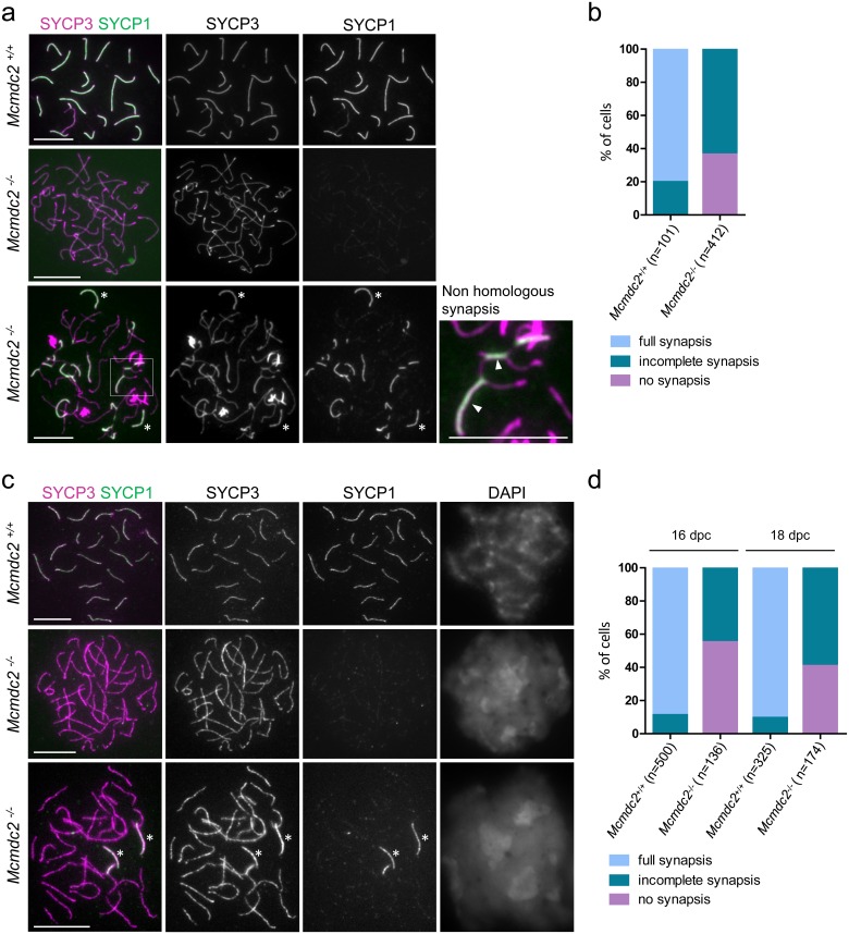
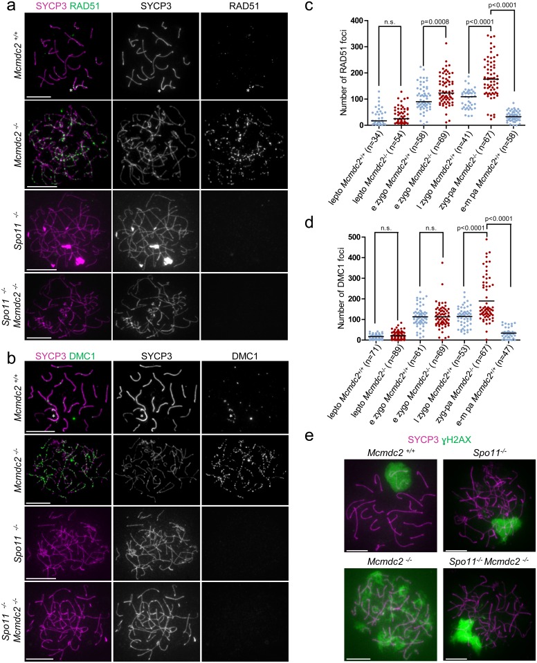
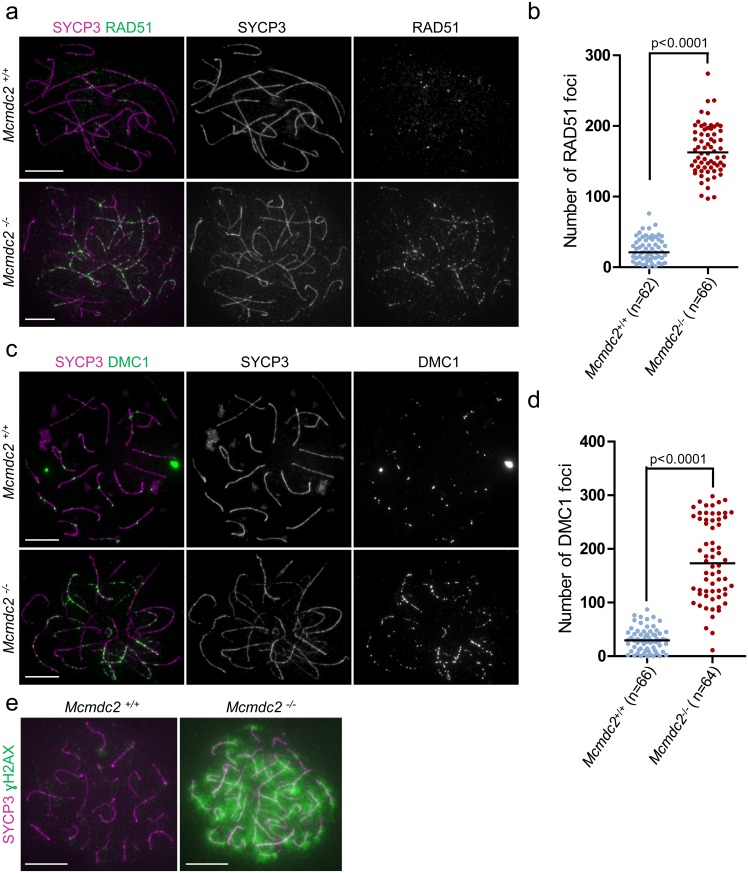
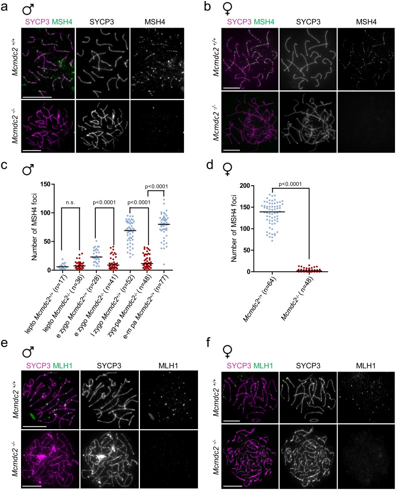
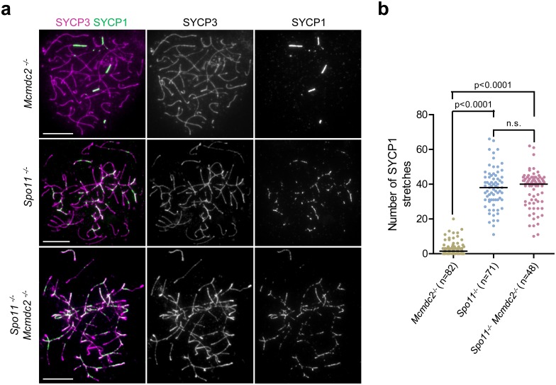
Orderly chromosome segregation during the first meiotic division requires meiotic recombination to form crossovers between homologous chromosomes (homologues). Members of the minichromosome maintenance (MCM) helicase family have been implicated in meiotic recombination. In addition, they have roles in initiation of DNA replication, DNA mismatch repair and mitotic DNA double-strand break repair. Here, we addressed the function of MCMDC2, an atypical yet conserved MCM protein, whose function in vertebrates has not been reported. While we did not find an important role for MCMDC2 in mitotically dividing cells, our work revealed that MCMDC2 is essential for fertility in both sexes due to a crucial function in meiotic recombination. Meiotic recombination begins with the introduction of DNA double-strand breaks into the genome. DNA ends at break sites are resected. The resultant 3-prime single-stranded DNA overhangs recruit RAD51 and DMC1 recombinases that promote the invasion of homologous duplex DNAs by the resected DNA ends. Multiple strand invasions on each chromosome promote the alignment of homologous chromosomes, which is a prerequisite for inter-homologue crossover formation during meiosis. We found that although DNA ends at break sites were evidently resected, and they recruited RAD51 and DMC1 recombinases, these recombinases were ineffective in promoting alignment of homologous chromosomes in the absence of MCMDC2. Consequently, RAD51 and DMC1 foci, which are thought to mark early recombination intermediates, were abnormally persistent in Mcmdc2-/- meiocytes. Importantly, the strand invasion stabilizing MSH4 protein, which marks more advanced recombination intermediates, did not efficiently form foci in Mcmdc2-/- meiocytes. Thus, our work suggests that MCMDC2 plays an important role in either the formation, or the stabilization, of DNA strand invasion events that promote homologue alignment and provide the basis for inter-homologue crossover formation during meiotic recombination.


Conflict of interest statement

The authors have declared that no competing interests exist.

Figures

Fig 1
Fig 1. Preferential expression of Mcmdc2 in the gonads, and Mcmdc2 targeting in mice.
(a) Expression of Mcmdc2 and a “house-keeping” gene (S9) in testis and a somatic tissue mix measured by RT-PCR. cDNAs were prepared from four RNA mixtures: (1) Equal amounts of RNAs from 17 somatic tissues (see Materials and Methods for the tissue list) were mixed and 1μg of the resulting mixture was used for RT (17 somatic tissues). (2) Mixture “1” supplemented with testis RNA at a concentration equal to that of the individual somatic RNAs (17 somatic tissues + 1x testis). (3) Mixture “1” supplemented with testis RNA at a concentration equal to five times that of the individual somatic RNAs (17 somatic tissues + 5 x testis) (4) Mixture “3” with no RT (17 somatic tissues + 5xtestis noRT). Mcmdc2-specific PCR-products were amplified preferentially from templates that contained testis cDNA. (b) Mcmdc2 targeting strategy. Schematics of the targeting construct, the wild-type (WT) and the modified Mcmdc2 genomic locus. Black boxes represent exons (not to scale). Recombination at the homology arms (HA) of the targeting construct modifies intron 4 by introducing: 1) an additional exon (SA-IRES-LacZ) that contains a strong splice acceptor site (SA) and poly-adenylation site (left grey box), 2) a transcriptional unit that contains the strong housekeeping human ß-Actin promotor (hBactP) driving the neomycin (Neo) resistance gene as a selection marker. This modification of intron 4 also disrupts the Mcmdc2 open reading frame after the 95th codon (Mcmdc2insertion allele). Recombination catalyzed by FLPe at FRT sites removes the SA-IRES-LacZ exon and the hBactP-Neo gene, and restores the MCMDC2 ORF (Mcmdc2restored). Mcmdc2restored is a functional allele that can be disrupted by Cre-mediated recombination between loxP sites (Mcmdc2deletion). Excision of exon 5–7 causes a frameshift after the 80th codon. Cre-mediated recombination between loxP sites of a Mcmdc2insertion allele results in Mcmdc2insertion-deletion allele. The positions of PCR-genotyping primers are indicated. Red bars mark the 3`and the internal Southern blot probes; the predicted length of restriction fragments is indicated. (c) Southern blot of DNA from wild-type (+/+) and targeted Mcmdc2+/insertion (+/i) embryonic stem cell clones (C6 and F7) that were used to derive two independent mouse lines. DNA was digested with Eco31I and hybridized with an internal probe for LacZ (left panel), or DNA was digested with BclI and hybridized with a 3’ probe (right panel). The blots indicate a single integration of the targeting cassette in the Mcmdc2 locus. (d) RT-PCR was used to detect Mcmdc2 and "house-keeping" Rps9 (S9) transcripts in testes of wild-type and Mcmdc2-/- (insertion-deletion) mice. Oligo-pairs specific to Mcmdc2 exon 3 and 4, 5 and 6, 6 and 7, 8 and 9, or 10 and 11 were used.
Fig 2
Fig 2. Mcmdc2-/- mice are deficient in germ cells from late meiotic prophase onwards in both sexes.
(a, b) Growth curves of five (a) or three (b) independent lines of Mcmdc2+/+ (+/+) and Mcmdc2-/- mouse embryonic fibroblasts. Cells were grown either without aphidicolin treatment (a) or with aphidicolin treatment for the first 24 hours (b), where 1μM aphidicolin was added at day 0. (a, b) Cell numbers were determined at the indicated time points in three technical replicates of each fibroblast line. Means and standard deviations of the medians of technical triplicates are shown. Growth curves of Mcmdc2+/+ and Mcmdc2-/- mouse embryonic fibroblasts are not significantly different (a: p = 0.8201, b: p = 0.9932, two-way ANOVA test). (c) Images of Mcmdc2+/+ (+/+) and Mcmdc2-/- (-/-) testes (upper panel) and ovaries (lower panel). Scale bars; 500μm. (d) Cryosections of testes from adult Mcmdc2+/+ and Mcmdc2-/- mice. DNA was detected by DAPI, histone H1T (marker of spermatocytes after mid-pachytene) and nuclear cleaved PARP1 (marker of apoptotic cells) were detected by immunostaining. Outlines of testis tubules are marked by dashed lines. The upper panels of d show stage V-VI and VII-VIII wild-type testis tubules, which contain several layers of germ cells at distinct spermatogenic stages: Sertoli cells (Se), spermatogonia B (SgB, stage V-VI), preleptotene (pl, stage VII-VIII), mid-pachytene (pa, stage V-VI), late-pachytene (pa, stage VII-VIII) spermatocytes, post-meiotic spermatids (sd) and spermatozoa (sp). Lower panels of d show that Mcmdc2-/- meiocytes underwent apoptosis at a stage corresponding to wild-type mid-pachytene in stage IV tubules. Consequently, spermatocytes were not found in the inner layers of testis tubules beyond stage IV, and post-meiotic spermatids and spermatozoa were also missing from Mcmdc2-/- testes. To illustrate this, stage IV, V-VI and VII-VIII tubules of Mcmdc2-/- mice are shown. Apoptotic (ap) and non-apoptotic early-mid pachytene (pa) spermatocytes are shown in the stage IV tubule, which was identified by the presence of mitotic intermediate spermatogonia (m) and intermediate spermatogonia (Int). Stage V-VI and VII-VIII tubules contain somatic Sertoli cells (Se) and spermatogonia B (SgB) or preleptotene (pl) spermatocytes, respectively, but more advanced spermatogenic cells are missing. Due to elimination at mid-pachytene, histone H1T positive cells are missing from Mcmdc2-/- testis tubules. (e) NOBOX (oocyte marker) was detected by immunofluorescence on cryosections of ovaries from 6-week-old mice. DNA was stained by DAPI. Oocytes in primordial (pd) and secondary (s) follicles are shown in the section of a wild-type ovary. In contrast, oocytes are not detected in the shown Mcmdc2-/- ovary section. (d, e) Scale bars; 50μm.
Fig 3
Fig 3. Synaptonemal complex formation is defective in Mcmdc2-/- mice.
(a, c) SYCP3 (axis marker) and SYCP1 (synaptonemal complex marker) were detected by immunofluorescence on nuclear surface spreads of Mcmdc2+/+ pachytene and Mcmdc2-/- zygotene-pachytene spermatocytes (a) and oocytes (c). Two distinct categories of Mcmdc2-/- zygotene-pachytene meiocytes were found. They either had no synapsis with no or weak punctate SYCP1 along unsynapsed axes (middle rows, a and c), or stretches of SYCP1 formed between some chromosomes that have managed to synapse (bottom panels, a and c). The spermatocyte shown in the bottom panels of a illustrates the maximum extent of synaptonemal complex formation observed in Mcmdc2-/- spermatocytes. Although some chromosomes evidently managed to fully synapse (asterisks in a and c) most synaptonemal complexes are incomplete and often form between apparently non-homologous axes (a inset, arrowheads mark synaptonemal complex stretches between apparently non-homologous chromosomes that have different lengths). Note that X and Y sex chromosomes are synapsed only in their short PAR regions in spermatocytes (a, top). Scale bars; 10μm. (b, d) Quantification of synaptonemal complex formation in Mcmdc2+/+ and Mcmdc2-/- spermatocytes (b) or oocytes (d). Spermatocytes were collected from testes of adult mice, and oocytes were collected from the ovaries of 16 or 18 days post coitum (dpc) fetuses. Synaptonemal complex development was assessed by detecting SYCP3 (axis marker) and SYCP1 (synaptonemal complex marker) on nuclear spreads of meiocytes from littermate pairs of wild-type and Mcmdc2-/- mice. Three categories of synaptonemal complex formation were distinguished in cells with fully formed continuous axes: 1) No synapsis (characterized by either no SYCP1 stain or punctate weak SYCP1 stain along unsynapsed axes), 2) incomplete synapses (stretches of axes have formed but there is at least one chromosome without fully completed synaptonemal complex) or 3) full synapsis (all chromosomes are fully synapsed except the male sex chromosomes). Counted numbers of cells are indicated (n).
Fig 4
Fig 4. RAD51 and DMC1 foci persist in Mcmdc2-/- spermatocytes.
(a, b, e) Immunostaining showing SYCP3 together with RAD51 (a), DMC1 (b) or γH2AX (e) on nuclear surface spreads of pachytene Mcmdc2+/+, late zygotene-pachytene Mcmdc2-/-, Spo11-/-, and Spo11-/- Mcmdc2-/—spermatocytes. RAD51 and DMC1 foci are present at comparatively high density along the axes of unsynapsed sex chromosomes (a, b, asterisk), and are largely absent from synapsed autosomes of Mcmdc2+/+ spermatocytes. Both RAD51 and DMC1 foci are present in high numbers along the unpaired axes of Mcmdc2-/- spermatocytes. Absence of RAD51 and DMC1 foci is shown in Spo11-/- and Spo11-/- Mcmdc2-/—spermatocytes. (e) γH2AX preferentially accumulates on the partially synapsed sex chromosomes of the Mcmdc2+/+ spermatocyte. γH2AX associates with chromatin throughout the nucleus in the Mcmdc2-/- spermatocytes. γH2AX is largely restricted to the sex chromatin in wild-type pachytene spermatocytes, and to pseudo-sex bodies in Spo11-/- and Spo11-/- Mcmdc2-/—spermatocytes. Scale bars; 10μm. (c, d) Numbers of RAD51 (c) or DMC1 (d) foci are shown in leptotene (lepto), early zygotene (e zygo) in Mcmdc2+/+ and Mcmdc2-/-, late zygotene (l zygo) and early-mid pachytene (e-m pa) in Mcmdc2+/+ and zygotene-pachytene (zyg-pa) in Mcmdc2-/- spermatocytes. Median numbers of foci are marked, and n corresponds to the number of analyzed spermatocytes in three pooled experiments. DMC1 and RAD51 foci numbers are significantly higher in zygotene-pachytene Mcmdc2-/- spermatocytes than in late-zygotene or early-mid-pachytene Mcmdc2+/+ spermatocytes (Mann Whitney test).
Fig 5
Fig 5. RAD51 and DMC1 foci persist in Mcmdc2-/- oocytes.
(a, c, e) Immunostaining of SYCP3 along with RAD51 (a), DMC1 (c) or γH2AX (e) on nuclear surface spreads of pachytene Mcmdc2+/+, or zygotene-pachytene Mcmdc2-/- oocytes. Oocytes were collected from the ovaries of littermate fetuses at 18dpc, which is a time point when most wild-type oocytes are in the late pachytene stage. RAD51 and DMC1 foci are largely absent from synapsed chromosomes in Mcmdc2+/+ oocytes. Both RAD51 and DMC1 foci are present in high numbers along the unpaired axes of Mcmdc2-/- oocytes. (e) γH2AX is largely absent from the synapsed chromosomes of the Mcmdc2+/+ oocyte. γH2AX associates with chromatin throughout the nucleus in the Mcmdc2-/- oocyte. Scale bars; 10μm. (b, d) Numbers of RAD51 (b) or DMC1 (d) foci are shown in Mcmdc2+/+ and Mcmdc2-/- oocytes at 18dpc. Median numbers of foci are marked, and n corresponds to the number of analyzed oocytes in two pooled experiments. DMC1 and RAD51 foci numbers are significantly higher in Mcmdc2-/- than in Mcmdc2+/+ oocytes (Mann Whitney test).
Fig 6
Fig 6. MutSγ and MutLγ foci formation are defective in Mcmdc2-/- meiocytes.
(a, b, e, f) Immunostaining of SYCP3 together with MSH4 (a, b) or MLH1 (e, f) on nuclear surface spreads of pachytene Mcmdc2+/+ or zygotene-pachytene Mcmdc2-/- meiocytes. (a, b) MSH4 foci are readily detected along synapsed axes of pachytene spermatocytes and oocytes (16dpc). MSH4 foci numbers are much lower in Mcmdc2-/- meiocytes. (e, f) Typically, a single MLH1 focus is detected along each synapsed axis pair of Mcmdc2+/+ pachytene spermatocytes and oocytes (from ovaries of newborn mice). MLH1 foci are not present along the unsynapsed axes of Mcmdc2-/- meiocytes. Scale bars; 10μm. (c, d) Numbers of MSH4 foci in Mcmdc2+/+ and Mcmdc2-/- spermatocytes and oocytes. (c) Spermatocytes were examined at leptotene (lepto), early zygotene (e zygo) in Mcmdc2+/+ and Mcmdc2-/-, late zygotene (l zygo) and early-mid pachytene (e-m pa) in Mcmdc2+/+ and zygotene-pachytene (zyg-pa) in Mcmdc2-/- mice. MSH4 foci numbers are significantly lower in Mcmdc2-/- than in Mcmdc2+/+ spermatocytes from early-zygotene stage onwards (Mann Whitney test). (d) Oocytes with fully formed axes (late zygotene and early pachytene) were examined from fetal ovaries at the 16dpc developmental time point. MSH4 foci numbers are significantly lower in Mcmdc2-/- than in Mcmdc2+/+ oocytes (Mann Whitney test). (c, d) Median numbers of foci are marked, and n corresponds to the number of analyzed meiocytes in two pooled experiments.
Fig 7
Fig 7. MCMDC2 is not required for extensive non-homologous synaptonemal complex formation in the Spo11-/- background.
(a) SYCP3 (axis marker) and SYCP1 (synaptonemal complex marker) were detected by immunofluorescence on nuclear surface spreads of zygotene-pachytene Mcmdc2-/-, Spo11-/- or Spo11-/- Mcmdc2-/—spermatocytes. Whereas comparatively few synaptonemal complex stretches are detected in the Mcmdc2-/-spermatocyte, extensive non-homologous synaptonemal complex formation is seen in the Spo11-/- or Spo11-/- Mcmdc2-/—spermatocytes. Scale bars; 10μm (b) Quantification of SYCP1 stretch numbers in zygotene-pachytene spermatocytes with fully condensed chromosome axes of the indicated genotypes. The numbers of synaptonemal complex stretches is significantly higher in Spo11-/- or Spo11-/- Mcmdc2-/—spermatocytes than in Mcmdc2-/- (Mann Whitney test). The numbers of synaptonemal complex stretches are not significantly different in Spo11-/- or Spo11-/- Mcmdc2-/—spermatocytes (p = 0.8639, Mann Whitney test). Median numbers of foci are marked, and n corresponds to the number of analyzed spermatocytes in two (Spo11-/- or Spo11-/- Mcmdc2-/-) or three (Mcmdc2-/-) pooled experiments.

Similar articles

Cited by

References

    1. Page SL, Hawley RS (2003) Chromosome choreography: the meiotic ballet. Science 301: 785–789. 10.1126/science.1086605 - DOI - PubMed
    1. Petronczki M, Siomos MF, Nasmyth K (2003) Un menage a quatre: the molecular biology of chromosome segregation in meiosis. Cell 112: 423–440. 10.1016/S0092-8674(03)00083-7 - DOI - PubMed
    1. Keeney S, Giroux CN, Kleckner N (1997) Meiosis-specific DNA double-strand breaks are catalyzed by Spo11, a member of a widely conserved protein family. Cell 88: 375–384. 10.1016/S0092-8674(00)81876-0 - DOI - PubMed
    1. Bergerat A, de Massy B, Gadelle D, Varoutas PC, Nicolas A, et al. (1997) An atypical topoisomerase II from Archaea with implications for meiotic recombination. Nature 386: 414–417. 10.1038/386414a0 - DOI - PubMed
    1. Baudat F, de Massy B (2007) Regulating double-stranded DNA break repair towards crossover or non-crossover during mammalian meiosis. Chromosome Res 15: 565–577. 10.1007/s10577-007-1140-3 - DOI - PubMed