. 2016 Oct 19;11(10):e0164087.
doi: 10.1371/journal.pone.0164087. eCollection 2016.

Role of PXR in Hepatic Cancer: Its Influences on Liver Detoxification Capacity and Cancer Progression

Affiliations

Role of PXR in Hepatic Cancer: Its Influences on Liver Detoxification Capacity and Cancer Progression

Deepak Kotiya et al. PLoS One. .

Abstract

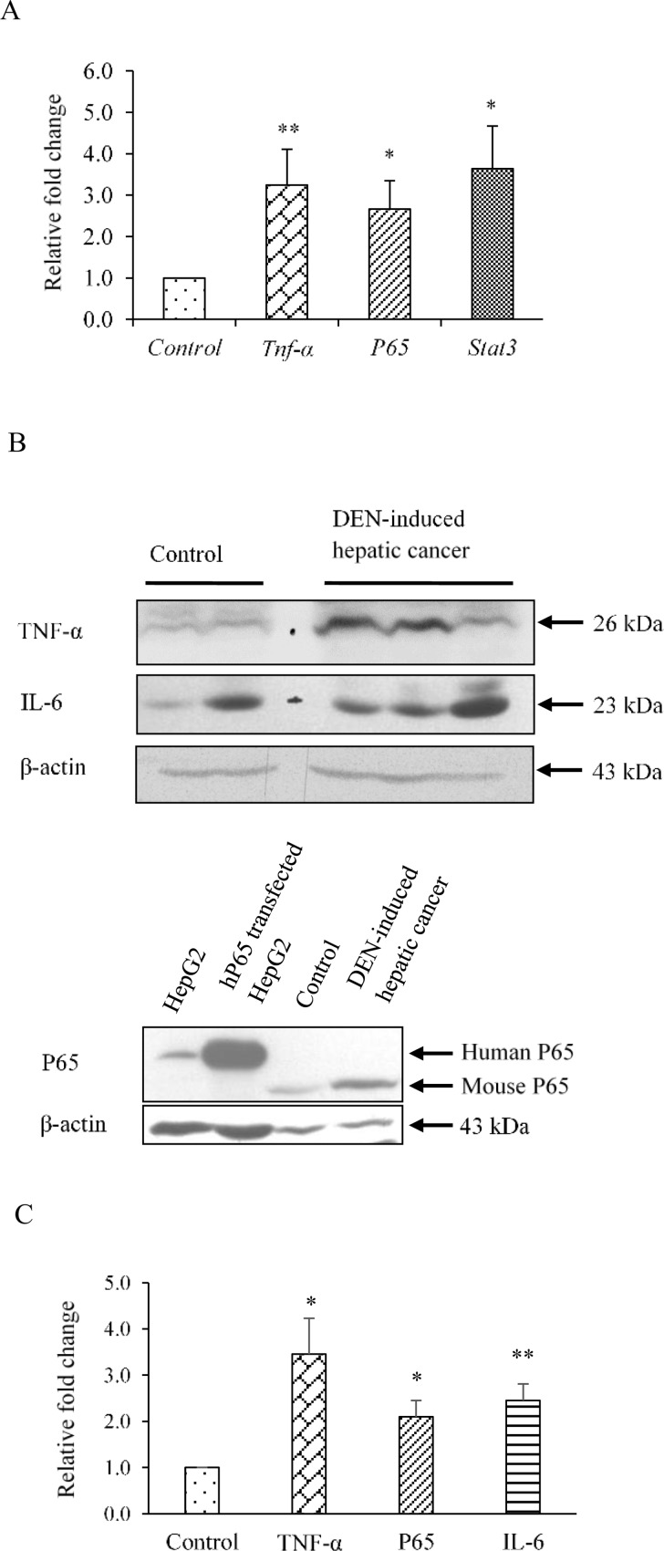
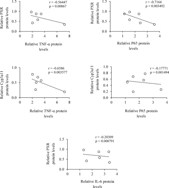
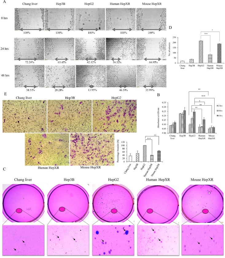
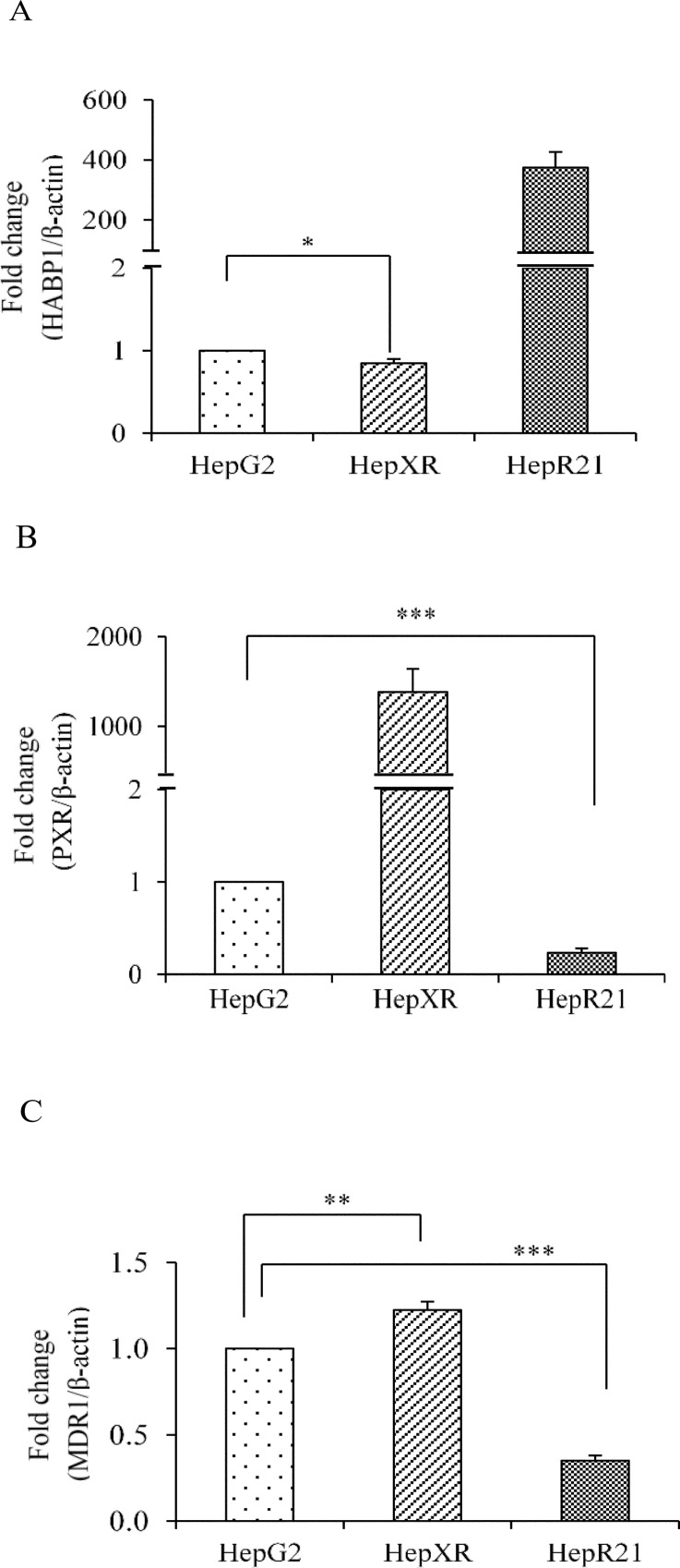
The role of nuclear receptor PXR in detoxification and clearance of xenobiotics and endobiotics is well-established. However, its projected role in hepatic cancer is rather illusive where its expression is reported altered in different cancers depending on the tissue-type and microenvironment. The expression of PXR, its target genes and their biological or clinical significance have not been examined in hepatic cancer. In the present study, by generating DEN-induced hepatic cancer in mice, we report that the expression of PXR and its target genes CYP3A11 and GSTa2 are down-regulated implying impairment of hepatic detoxification capacity. A higher state of inflammation was observed in liver cancer tissues as evident from upregulation of inflammatory cytokines IL-6 and TNF-α along with NF-κB and STAT3. Our data in mouse model suggested a negative correlation between down-regulation of PXR and its target genes with that of higher expression of inflammatory proteins (like IL-6, TNF-α, NF-κB). In conjunction, our findings with relevant cell culture based assays showed that higher expression of PXR is involved in reduction of tumorigenic potential in hepatic cancer. Overall, the findings suggest that inflammation influences the expression of hepatic proteins important in drug metabolism while higher PXR level reduces tumorigenic potential in hepatic cancer.


Conflict of interest statement

The authors declare that there is no conflict of interest that could be perceived as prejudicing the impartiality of the research reported.

Figures

Fig 1
Fig 1. Assessment of expression levels of nuclear receptor PXR, CAR and RXR-α in DEN-induced hepatic cancer.
A. Real-time PCR analysis was performed for PXR, CAR and RXR-α genes with the total RNA extracted from mouse liver tissues of saline-treated control and DEN-induced hepatic cancer mice. GAPDH served as an internal control. B. Cell lysate of control and hepatic cancerous mice liver tissues were electrophoresed (50 μg per sample) on a 10% SDS-PAGE. The proteins were transferred onto the methanol-activated PVDF membrane and probed with anti-mouse PXR antibody, anti-mouse CAR antibody and anti-mouse RXR-α antibody. β-Actin antibody served as control; C. The relative endogenous protein expression levels of PXR, CAR and RXR-α in control and DEN-induced hepatic cancer were quantified by densitometry. The experiments were performed with six samples of each control and DEN-induced cancerous mice. The values are represented the mean ±SE. The P-value represents the significance in DEN-induced hepatic cancer mice as compared to the control mice. P-value <0.05 represented by a single asterisk (*), P-value <0.005 represented by a double asterisk (**), P-value <0.0005 represented by a triple asterisk (***).
Fig 2
Fig 2. Altered expression of PXR-regulated genes in DEN-induced hepatic cancer.
A. Total RNA was extracted from mouse liver tissues of saline-treated control and DEN-induced hepatic cancer mice. Real-time PCR analysis was performed for Cyp3A11, Gsta2 and Mrp3. For Mdr1 semi-quantitative PCR was performed. GAPDH served as an internal control. B. Cell lysates of control and hepatic cancerous mice liver tissues were electrophoresed (50 μg per sample) on a 10% SDS-PAGE. The proteins were transferred onto the methanol-activated PVDF membrane and probed with anti-mouse Cyp3a11 antibody (upper panel). β-Actin antibody served as control (lower panel); C. The relative endogenous Cyp3a11 protein expression in control and DEN-induced hepatic cancer was quantified by densitometry. The experiments were performed with six samples of each control and DEN-induced cancerous mice, and the values are represented as the mean ±SE. The P-value represents the significance in DEN-induced hepatic cancer mice as compared to the control mice. The P-value <0.05 represented by a single asterisk (*), P-value <0.005 represented by a double asterisk (**), P-value <0.0005 represented by a triple asterisk (***).
Fig 3
Fig 3. Levels of inflammatory proteins are enhanced in DEN-induced hepatic cancer.
A. Real-time PCR analysis was performed for Tnf-α, p65, and Stat3 with total RNA extracted from mouse liver tissues of saline-treated control and DEN-induced hepatic cancer mice. GAPDH served as an internal control. B. Cell extracts of control and DEN-induced hepatic cancerous mice liver tissues were electrophoresed (50 μg per sample) on a 10% SDS-PAGE. The proteins were transferred onto the methanol-activated PVDF membrane and probed with anti-mouse TNF-α, anti-mouse P65, and anti-mouse IL-6 antibody. Bands of human and mouse P65 were detected at 65 KDa and 60 KDa respectively. β-Actin antibody served as control; C. The relative endogenous TNF-α, P65 and IL-6 protein expressions in control and DEN-induced hepatic cancer were quantified by densitometry. The experiments were performed with seven samples of each control and DEN-induced cancerous mice, and the values are represented as the mean ±SE. The P-value represents the significance in DEN-induced hepatic cancer mice as compared to the control mice. P-value <0.05 represented by a single asterisk (*), P-value <0.005 represented by a double asterisk (**), P-value <0.0005 represented by a triple asterisk (***).
Fig 4
Fig 4. Correlations between the reduced levels of PXR and PXR-regulated CYP3A11 DME with enhanced levels of inflammatory proteins in hepatic cancer.
Relative protein levels of TNF-α, P65, and IL-6 were correlated with PXR and CYP3A11 protein levels using Pearson’s correlation coefficient (r) in scattered plot. Respective p-value (p) represents the significance between the correlations.
Fig 5
Fig 5. Human HepXR and mouse HepXR cells stably expressing higher levels of PXR exhibit reduced tumorigenic potential.
All the experiments were performed with cell lines that included Chang liver, Hep3B, HepG2, human HepXR and mouse HepXR cells as described under ‘Materials and Methods’. A. In ‘wound healing assay’ cell lines were cultured to achieve 70–80% confluency. A scratch was inflicted with a 200 μl of pipette tip to create the wound. The width of the cell-free zone in the wound was monitored at 0 hrs, 24 hrs and 48 hrs and the migration ability of the cells to fill the wound gap was calculated using Image J software. Values presented are percentage of the wound gap as compared to control (100%). B. In ‘adhesion assay’ cell lines were seeded at 4 × 10⁴ cells per well in a 24-well plate. Cells were harvested at 2 hrs, 4 hrs and 6 hrs after fixing with 4% para-formaldehyde for 10 minutes followed by staining with 0.1% (W/V) crystal violet for 20 minutes. This was followed by solubilization in 1% Triton X 100 for 24 hrs and the adhesion ability of the cells was calculated by taking the spectrophotometric readings at 620 nm. Values presented are absorbance at 620 nm. The measured colour intensity was proportional to the number of adhered cells. C. In ‘colony formation assay’ the experimental cell lines were seeded with 5,000 cells with 0.35% agarose-DMEM per 35 mm plate after plating the 0.5% agarose-DMEM bottom layer as described under ‘Materials and Methods’. Plates were incubated at 37°C in humidified incubator and intermittently observed for 3 weeks. Plates were stained with 0.005% (W/V) crystal violet for 3–4 hrs followed by washing with PBS. Stained colony images were recorded with Sony BionZ X camera. Anchorage-independent growth of the cells was calculated by manually counting the colonies (indicated by arrows) using Microsoft office picture manager software at 200% zoom after randomly choosing the areas on the plate. D. In ‘cell invasion assay’ after seeding cell lines as mentioned in ‘Materials and Methods’, cultures were allowed to invade through the matrigel up to 36 hrs. Invaded cells were stained with crystal violet and counted to quantify. Values presented in the graph are number of colonies of cell-specific type in each plate. All the values are represented as the mean ±SE. The P-value represents the significance in human HepXR and mouse HepXR cell lines as compared to the control HepG2 cell line in all experiments. P-value <0.05 represented by a single asterisk (*), P-value <0.005 represented by a double asterisk (**), P-value <0.0005 represented by a triple asterisk (***).
Fig 6
Fig 6. PXR reduces cell-ECM interactions in hepatic cancer cells.
Total RNA was extracted from HepG2, human HepXR and HepR21 cell lines and used for real-time PCR analysis of A. human HABP1; B. human PXR; and C. human MDR1. β-Actin served as an internal control. The values are represented as the mean ±SE. The P-value represents the significance in human HepXR and HepR21 cell lines as compared to the control HepG2 cell line. P-value <0.05 represented by a single asterisk (*), P-value <0.005 represented by a double asterisk (**), P-value <0.0005 represented by a triple asterisk (***).
Fig 7
Fig 7. PXR enhances expression of apoptotic genes while reduces expression of cell-cycle regulatory genes in hepatic cancer cells A.
Real-time PCR analysis was performed for Bcl-xl, Bcl-2, Cdk2 and Cdk4 with total RNA extracted from HepG2, human HepXR and mouse HepXR cell lines for various cell cycle and apoptosis regulatory genes. β-Actin served as an internal control. B. Higher cellular expression of PXR increased the doubling time in human HepXR and mouse HepXR cells in comparison to HepG2 cells. The experiments were performed three times with three samples of each cell lines and the values are represented as the mean ±SE. The P-value represents the significance in human HepXR and mouse HepXR cell lines as compared to the control HepG2 cell line. P-value <0.05 represented by a single asterisk (*), P-value <0.005 represented by a double asterisk (**), P-value <0.0005 represented by a triple asterisk (***).
Fig 8
Fig 8. Overexpression of FLAG-tagged mPXR gene at protein level and its effect on tissue histology.
A. Liver tissue extracts of Control and transgenic mice were electrophoresed (50 μg per sample) on a 10% SDS-PAGE. The proteins were transferred onto the methanol-activated PVDF membrane and probed with anti-FLAG antibody and anti-mouse PXR antibody (upper panels). β-Actin antibody served as control (lower panel). B. Control and transgenic mice were sacrificed and their livers were removed. Paraffin sections were prepared from control and transgenic mice livers harvested in the experiment and immunodetection were performed with anti-FLAG antibody (indicated by arrows). The immunodetected tissues were viewed and recorded at 20X magnification using a light microscope. The experiments were performed with three samples of each of the controls and the transgenic mice.

References

    1. Pondugula SR, Mani S. Pregnane xenobiotic receptor in cancer pathogenesis and therapeutic response. Cancer Lett. 2013;328:1–9. 10.1016/j.canlet.2012.08.030 - DOI - PMC - PubMed
    1. Ihunnah CA, Jiang M, Xie W. Nuclear receptor PXR, transcriptional circuits and metabolic relevance. BBA-Mol Basis Dis. 2011;1812:956–63. - PMC - PubMed
    1. Wang H, Venkatesh M, Li H, Goetz R, Mukherjee S, Biswas A, et al. Pregnane X receptor activation induces FGF19-dependent tumor aggressiveness in humans and mice. The J Cli Invest. 2011;121:3220–32. - PMC - PubMed
    1. Heng Jiang KC, He Jianming, Pan Feng, Li Jianjun, Chen Jianfang,Chen Wensheng, Liang Houjie. Association of Pregnane X Receptor with multidrug resistance-related protein 3 and its role in human colon cancer chemoresistance. J Gastrointest Surg. 2009. 13:1831–8. 10.1007/s11605-009-0964-x - DOI - PubMed
    1. Ouyang N, Ke S, Eagleton N, Xie Y, Chen G, Laffins B, et al. Pregnane X receptor suppresses proliferation and tumourigenicity of colon cancer cells. Br J Cancer. 2010;102:1753–61. 10.1038/sj.bjc.6605677 - DOI - PMC - PubMed

MeSH terms