. 2016 Oct 18;17(4):1128-1140.
doi: 10.1016/j.celrep.2016.09.076.

HMGB1 Activates Proinflammatory Signaling via TLR5 Leading to Allodynia

Affiliations

HMGB1 Activates Proinflammatory Signaling via TLR5 Leading to Allodynia

Nabanita Das et al. Cell Rep. .

Abstract

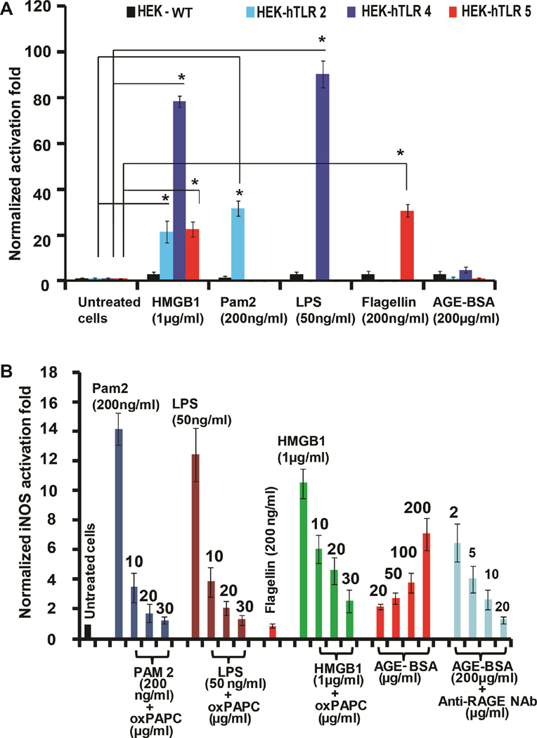
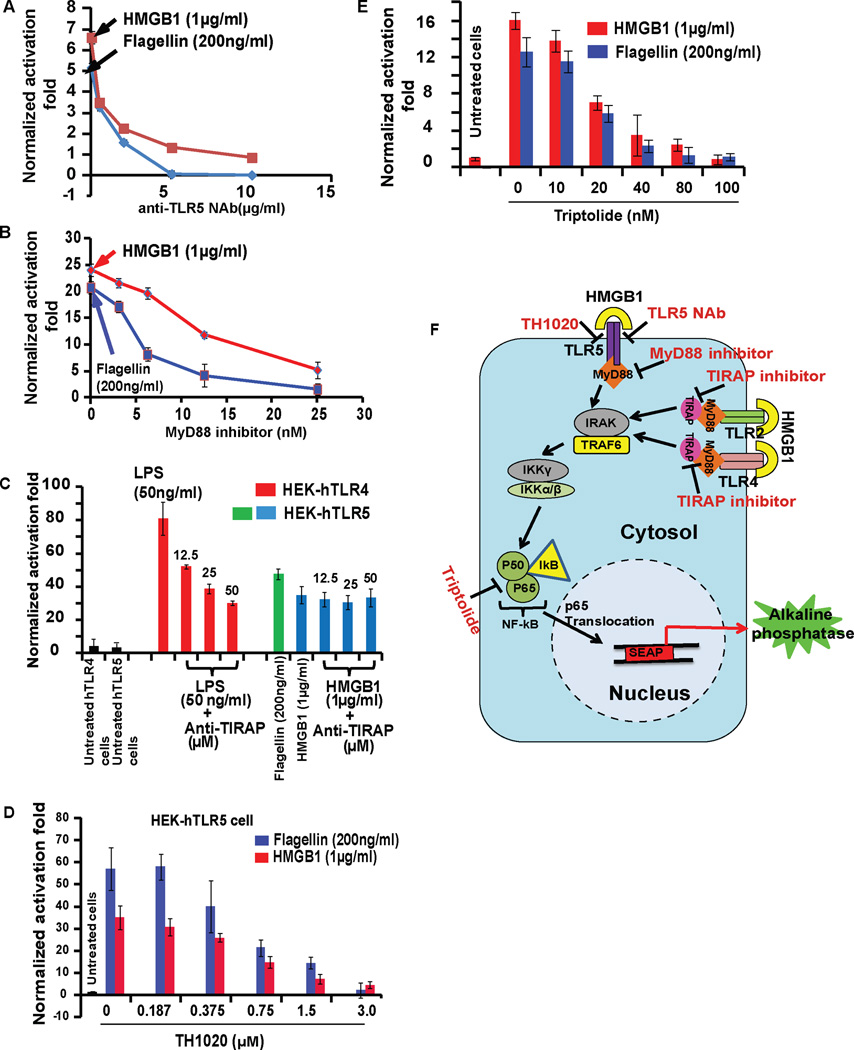
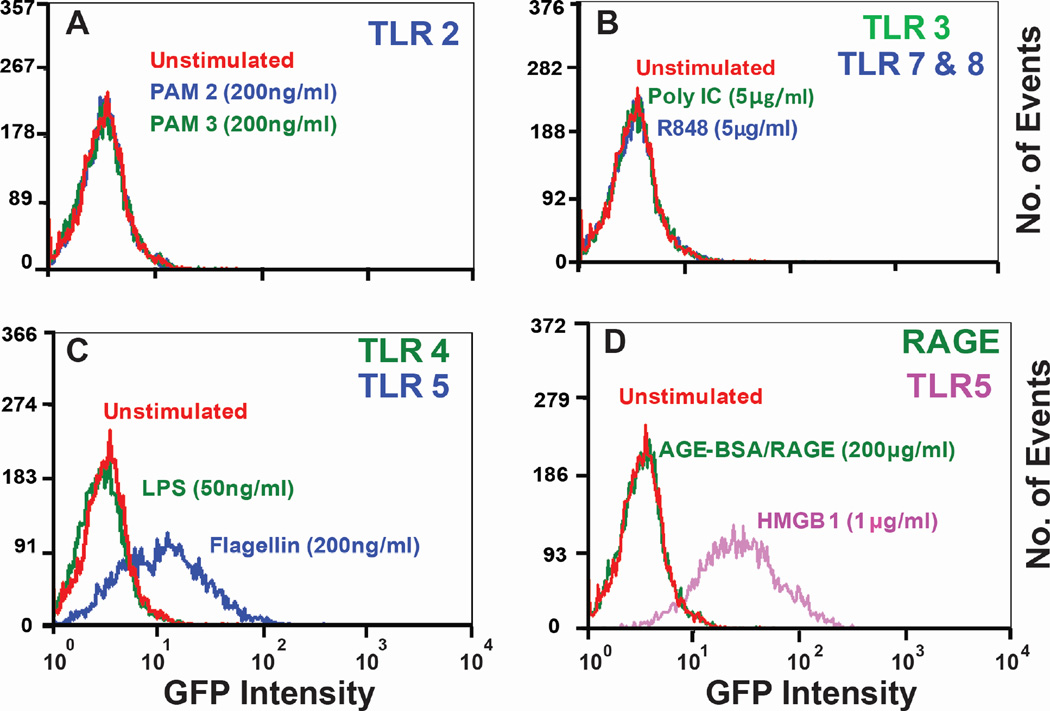
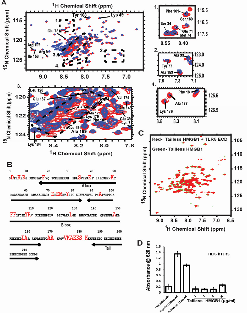
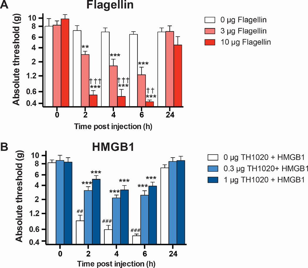
Infectious and sterile inflammatory diseases are correlated with increased levels of high mobility group box 1 (HMGB1) in tissues and serum. Extracellular HMGB1 is known to activate Toll-like receptors (TLRs) 2 and 4 and RAGE (receptor for advanced glycation endproducts) in inflammatory conditions. Here, we find that TLR5 is also an HMGB1 receptor that was previously overlooked due to lack of functional expression in the cell lines usually used for studying TLR signaling. HMGB1 binding to TLR5 initiates the activation of NF-κB signaling pathway in a MyD88-dependent manner, resulting in proinflammatory cytokine production and pain enhancement in vivo. Biophysical and in vitro results highlight an essential role for the C-terminal tail region of HMGB1 in facilitating interactions with TLR5. These results suggest that HMGB1-modulated TLR5 signaling is responsible for pain hypersensitivity.

Keywords: HMGB1; Toll-like receptors; cytokines; inflammation; pain.


Figures

Figure 1
Figure 1. Activation of TLR5 by HMGB1
(A) TLR ligands activate SEAP signaling in HEK cells. WT, TLR2-, TLR4-, and TLR5-overexpressing HEK cells were incubated with TLR ligands, HMGB1 or AGE-BSA for 24 h. NF-κB activation was evaluated by SEAP secretion in the culture supernatant by the QUANTI-Blue SEAP reporter assay. See also Figure S1A. (B) RAW 264.7 cells were incubated with TLR ligands with HMGB1, AGE-BSA and anti-RAGE NAb for 24 h. oxPAPC was added at varying doses. NF-κB-driven iNOS activation was evaluated by the NO produced. All data are represented as mean ± SD, n=3. *P< 0.05 (unpaired student’s t-test) relative to untreated cells.
Figure 2
Figure 2. HMGB1 activates TLR5 signaling through MyD88 pathway
(A) HEK-hTLR5 cells were treated with HMGB1 or flagellin followed by addition of anti-TLR5 neutralizing Ab (anti-TLR5 NAb), (B) MyD88 inhibitor, (C) TIRAP inhibitor peptide, (D) TH1020, TLR5 inhibitor or (E) Triptolide, an NF-κB inhibitor for 24 h. (F) Schematic overview of the treatments of anti-TLR5 NAb, MyD88 inhibitor, TH1020 and Triptolide in the TLR5 signaling cascade. NF-κB activation was evaluated by SEAP secretion in the culture supernatant by the QUANTI-Blue SEAP reporter assay. Both HMGB1 (1 µg/ml) and flagellin (200 ng/ml) strongly activated NF-κB-mediated SEAP signaling, followed by dose-dependent inhibition by anti-TLR5 NAb, MyD88 inhibitor, TH1020 and Triptolide, but not the TIRAP inhibitor peptide. For the TIRAP inhibitor assay, we confirmed that the peptide decreased SEAP secretion from LPS-treated HEK-hTLR4 cells in a dose-dependent manner. All data are represented as mean ± SD, n=3.
Figure 3
Figure 3. HMGB1 activates downstream NF-κB signaling through TLR5
Flow cytometric analysis of NF-κB activation in human Jurkat TLR5 sensitive T cells with various TLR ligands: (A) PAM2 and PAM3 for TLR2, (B) Poly I:C for TLR3, R848 for TLR7/8, (C) LPS for TLR4, flagellin for TLR5 and (D) AGE-BSA for RAGE, HMGB1 for TLR5. HMGB1 and flagellin both triggered NF-κB activation upon binding to TLR5 while other ligands failed to activate NF-κB. Human Jurkat-T cell line was stably transfected with a GFP-labeled NF-κB reporter gene. Cells sensitive to TLR5 activation were sorted using a MoFlo cytomation fluorescence-activated cell sorter. 10% of activated cells were collected and used for this experiment.
Figure 4
Figure 4. TLR5-HMGB1 interaction produces NO in primary cells and releases proinflammatory cytokines in THP-1 cells
(A) Normalized iNOS activation folds of the inhibitory effect of TH1020 on flagellin- and HMGB1-induced TLR5 activation in rat primary PBMCs. *P<0.05 for TH1020 relative to the positive controls. Values are the mean ±SD, n=2. (B) Western blot analysis of HEK-hTLR5 cells treated with flagellin (200ng/ml) or HMGB1 (1µg/ml) for 4 h at 37°C, 5% CO 2, shows significant translocation of p65 (NF-ΚB subunit) from the cytosolic to nuclear fraction. GAPDH and LaminB are shown as internal controls. (C) In HEK-hTLR5 cells, HMGB1 and flagellin induced IL-8 and TNF-α mRNA expression at 16 h post-treatment. Changes in gene expression levels are shown relative to untreated controls. IL-8 and TNF-α gene expression levels were determined using the expression ratio of the gene of interest to GAPDH. (D) ELISA assays conducted by inhibition of TLR2 and TLR4 in THP-1 cells by oxPAPC. oxPAPC displayed significant inhibition of TNF-α release in THP-1 cells upon activation by PAM2, LPS and HMGB1, whereas no inhibition was observed for flagellin-induced TNF-α release in comparison to uninhibited cells. (E) Treatment of THP-1 cells by AGE-BSA and HMGB1 induced significant TNF-α secretion. Inhibition of TLR2 and TLR4 by oxPAPC along with HMGB1 treatment displayed significant inhibition of TNF-α release. To elucidate the roles of TLR5 or RAGE in HMGB1-mediated NF-κB signaling, we combined anti-TLR5 NAb alone/or with anti-RAGE NAb with oxPAPC and HMGB1 samples. Significant inhibition of TNF-α release was observed with anti-TLR5 NAb, suggesting the role of TLR5 in mediating TNF-α release via HMGB1. See also Figure S2H. (F) oxPAPC (inhibitor of TLR2 and TLR4) and oxPAPC/TLR5-NAb displayed significant inhibition of IL-8 release in THP-1 cells upon activation by HMGB1. qPCR and ELISA data are represented as mean ± SD, n=3. *P<0.05-unpaired student’s t-test.
Figure 5
Figure 5. Characterization of binding sites of HMGB1 with TLR5
(A) Two-dimensional 1H-15N HSQC spectra of 15N-uniformly labeled CBP-HMGB1 (blue) superimposed on 15N-uniformly labeled CBP-HMGB1 bound with unlabeled TLR5 ECD (red). Changes in chemical shifts, peak broadening and sharpening upon TLR5 addition are marked as dotted boxes, subsets 1–4. The subsets include HMGB1 N-terminal residues, the basic linker region (residue 170–187), and A-and B-box helices. The “random coil region” of the spectra (subset 3: 1H: 8.1–8.6 ppm, 15N: 120.5 ppm-123.5 ppm) has the largest chemical shift changes upon TLR5 ECD addition. All spectra were collected in 800 MHz proton frequency at 25°C, using 84 µM of each protein at pH 7.8. See al so Figure S4. (B) HMGB1 sequence highlighting chemical shift changes upon TLR5 addition. Different segments of the protein are shown by arrows. (C) HSQC spectra of the 1:1 complex of TLR5 ECD and 15N tailless HMGB1 (red) superimposed onto the 15N tailless HMGB1 spectra (green) confirms that the HMGB1 tail is the primary TLR5 ECD interaction site. See also Figure S4. (D) HEK-TLR5 cells treated with flagellin (200ng/ml), full length HMGB1 (1µg/ml) showed TLR5-induced SEAP activation while the tailless HMGB1 mutant (1–10 µg/ml) could not do so. Data are represented as mean ± SD, n=3. *P<0.05 (unpaired student’s t-test) relative to untreated cells.
Figure 6
Figure 6. TLR5-HMGB1 interaction causes allodynia in vivo
(A) Dose-dependent effect of subcutaneous flagellin on hindpaw withdrawal thresholds to von Frey hairs (allodynia). N = 4/group, mean ± SEM. (B) Dose-dependent effect of subcutaneous TLR5 antagonist TH1020 on HMGB1-dependent allodynia (10 µg, subcutaneous). N = 6/group, mean ± SEM. **P < 0.01, ***P < 0.001, relative to 0 µg; ††P < 0.01, †††P < 0.001, relative to 3 µg; ##P < 0.01, ###P < 0.001, relative to baseline (BL).

References

    1. Akira S, Takeda K. Toll-like receptor signalling. Nat Rev Immunol. 2004;4:499–511. - PubMed
    1. Aravalli RN, Peterson PK, Lokensgard JR. Toll-like receptors in defense and damage of the central nervous system. J Neuroimmune Pharmacol. 2007;2:297–312. - PubMed
    1. Banerjee S, Friggeri A, Liu G, Abraham E. The C-terminal acidic tail is responsible for the inhibitory effects of HMGB1 on efferocytosis. J Leukoc Biol. 2010;88:973–979. - PMC - PubMed
    1. Bradford MM. A rapid and sensitive method for the quantitation of microgram quantities of protein utilizing the principle of protein-dye binding. Anal Biochem. 1976;72:248–254. - PubMed
    1. Brown GT, Mcintyre TM. Lipopolysaccharide signaling without a nucleus: kinase cascades stimulate platelet shedding of proinflammatory IL-1beta-rich microparticles. J Immunol. 2011;186:5489–5496. - PMC - PubMed

Publication types

MeSH terms