. 2016 Oct 18;17(4):1171-1183.
doi: 10.1016/j.celrep.2016.09.061.

Phenotypic Characterization of a Comprehensive Set of MAPK1/ERK2 Missense Mutants

Affiliations

Phenotypic Characterization of a Comprehensive Set of MAPK1/ERK2 Missense Mutants

Lisa Brenan et al. Cell Rep. .

Abstract

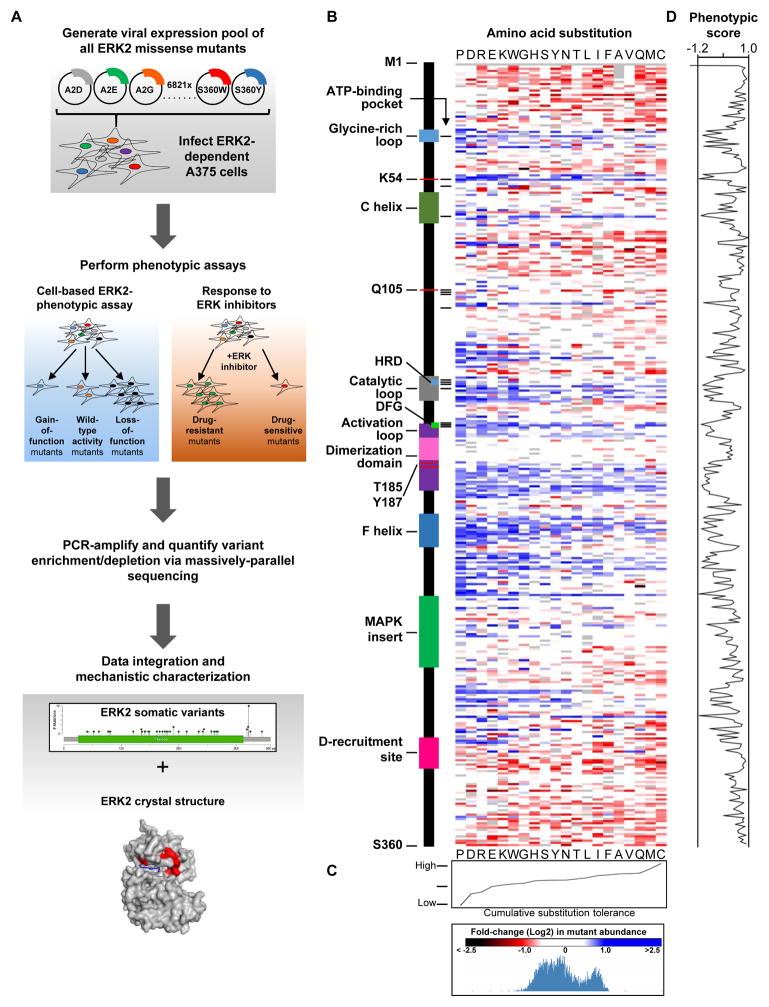
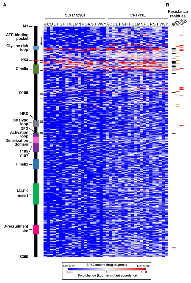
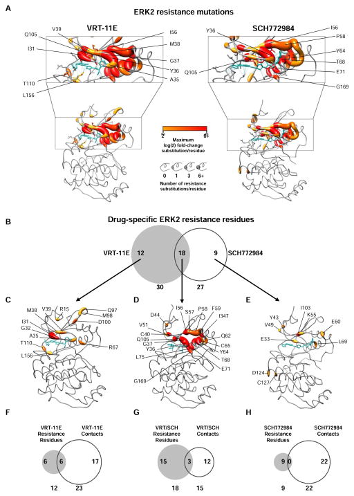
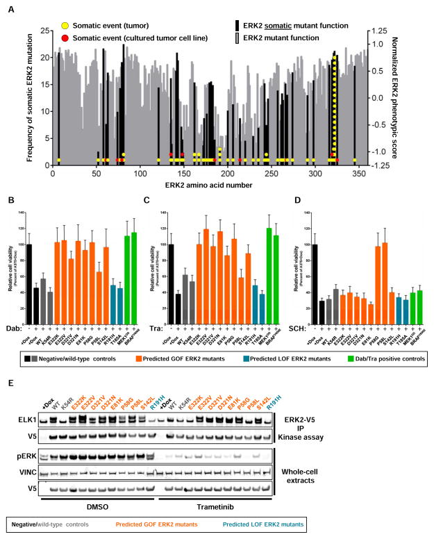
Tumor-specific genomic information has the potential to guide therapeutic strategies and revolutionize patient treatment. Currently, this approach is limited by an abundance of disease-associated mutants whose biological functions and impacts on therapeutic response are uncharacterized. To begin to address this limitation, we functionally characterized nearly all (99.84%) missense mutants of MAPK1/ERK2, an essential effector of oncogenic RAS and RAF. Using this approach, we discovered rare gain- and loss-of-function ERK2 mutants found in human tumors, revealing that, in the context of this assay, mutational frequency alone cannot identify all functionally impactful mutants. Gain-of-function ERK2 mutants induced variable responses to RAF-, MEK-, and ERK-directed therapies, providing a reference for future treatment decisions. Tumor-associated mutations spatially clustered in two ERK2 effector-recruitment domains yet produced mutants with opposite phenotypes. This approach articulates an allele-characterization framework that can be scaled to meet the goals of genome-guided oncology.

Keywords: ERK; MAPK; cancer; functional biology; precision medicine; precision oncology; rare mutants.


Figures

Figure 1
Figure 1. Functional characterization of 6,810 ERK2 mutants
(A) A pooled-format proliferation screen in A375 cells to measure the functional impact of 6,810 MAPK1/ERK2 mutants. (B) Heat map of the enrichment/depletion of ERK2 mutants in a pooled assay in which the proliferation of A375 cells serves as an indirect measure of ERK2 function. Data points represent the mean of 4 biological replicates. Grey boxes identify wild-type residues (see also Table S1, S2). (C) The aggregate impact of amino acid substitutions on ERK2 function in A375 cells. (D) Composite metric of ERK2 function when mutated at each amino acid, based on mutant enrichment/depletion in A375 cells. See also Figure S1, S2 and Table S1, S2 and S3.
Figure 2
Figure 2. Comprehensive identification of ERK2 mutants that induce resistance to ERK-directed kinase inhibitors
(A) Heat map displaying the normalized enrichment/depletion of individual ERK2 mutants expressed in A375 cells in the presence of the ERK-inhibitors SCH772984 or VRT-11E. Data points represent the mean of 4–6 biological replicates. Grey boxes identify wild-type residues. (B) Annotation of ERK2 amino acids that when mutated confer resistance to VRT-11E (VRT, red), SCH772984 (SCH, orange) or both inhibitors (S+V, black) when expressed in A375 cells. (C) Composite metric of ERK2 function when mutated at each amino acid, based on mutant enrichment/depletion in A375 cells. See also Figure S3 and Table S1, S3, S4, S5.
Figure 3
Figure 3. Spatial mapping of ERK2 mutants that induce resistance to ERK-directed kinase inhibitors
(A) Projection of ERK2 mutant enrichment in A375 cells in the presence of VRT-11E or SCH772984 on the structure of drug-bound ERK2 (PDB ID: 4QTE and 4QTA, respectively). All mutants displaying a log(2) fold-change of ≥2 are represented. Blue lines identify intramolecular contacts between ERK2 and inhibitor. Inhibitors are shown in blue. (B) Venn diagram highlighting the overlap of ERK2 residues that, when mutated, confer resistance to either VRT-11E only, SCH772984 only, or both VRT-11E and SCH772984. (C) Structural representation of ERK2 residues that, when mutated, confer resistance to VRT-11E (PDB ID: 4QTE). VRT-11E is shown in blue, residues are colored and sized as in (A). (D) Structural representation of ERK2 residues that, when mutated, confer resistance to VRT-11E and SCH772984 (PDB ID: 4FMQ). ANP is shown in blue, MAPK-docking peptide is not shown, residues are colored and sized as in (A). (E) Structural representation of ERK2 residues that, when mutated, confer resistance to SCH772984 (PDB ID: 4QTE). SCH772984 is shown in blue, residues are colored and sized as in (A). (F) Venn diagram highlighting the overlap of VRT-11E-specific residues with residues that form direct contacts with VRT-11E. (G) Venn diagram highlighting the overlap of VRT-11E/SCH772984-specific residues with residues that form direct contacts with both inhibitors. (H) Venn diagram highlighting the overlap of VRT-11E-specific residues with residues that form direct contacts with SCH772984. See also Figure S3, S4 and Table S1, S3, S4 and S5.
Figure 4
Figure 4. Identification of somatic ERK2 mutants with non-wild-type function
(A) Bars represent the composite ERK2 function score for each residue of ERK2. Each circle represents an annotated ERK2 mutant found in human tumors. Data bars represent the mean of 4 biological replicates. (B) Suppression of proliferation in A375 cells expressing ERK2 mutants after treatment with 10 nM dabrafenib (Dab, 96 hours of treatment). Data are represented as a mean (+/− S.D.) of at least 6 replicates. (C) Suppression of A375 proliferation with 2 nM trametinib (Tra, 96 hours of treatment) and proliferative rescue with GOF ERK2 mutants. Data are represented as a mean (+/− S.D.) of at least 6 replicates. (D) Suppression of A375 proliferation with 500 nM SCH772984 (SCH, 96 hours of treatment) and proliferative rescue with ERK2 resistance mutants. Data are represented as a mean (+/− S.D.) of at least 6 replicates. (E) In vitro kinase assay using V5-tagged ERK2 mutants immunoprecipitated from A375 cells treated with DMSO or 4 nM trametinib for 4 hours. Kinase activity is determined by the capacity to phosphorylate purified ELK1 (pERK, phosphorylated ERK; VINC, vinculin; V5 IP KA, immunoprecipitated V5 epitope followed by kinase assay). See also Figure S2, S4 and Table S1, S2, S3 and S7.
Figure 5
Figure 5. Somatic ERK2 mutants co-cluster in effector recruitment domains, but have opposite phenotypes
(A) ERK2 residues containing tumor-associated mutations are enriched and cluster in ERK2 effector recruitment domains (p = 4.303 × 10⁻⁵, Fishers exact test) (PDB ID: 2ERK). (B) Alteration of residues in the DRS of ERK2 are associated with GOF phenotypes, whereas alterations in FRS residues are associated with LOF phenotypes (p = 2.13 × 10⁷, Students T-test). Data points represent the mean of 4 biological replicates in A375 cells. (C) Western blot of lysates from A375 cells showing that ERK2 DRS mutants induce DUSP6 expression, whereas FRS mutants behave like kinase impaired ERK2K54R or the phosphorylation site (p-site) mutants ERK2T185A ERK2T187A (pERK, phosphorylated ERK; VINC, vinculin). (D) ERK2 DRS mutants rescue the anti-proliferative effects of trametinib in A375 cells (Tra, 96 hours of treatment), whereas FRS mutants do not. Data are represented as a mean (+/− S.D.) of at least 6 replicates. (E) Compound mutation of the FRS (Y233A) in ERK2 DRS mutants abrogate their capacity to rescue the anti-proliferative effects of trametinib (Tra, 96 hours of treatment) in A375 cells. Data are represented as a mean (+/− S.D.) of at least 6 replicates. (F) Western blot showing that compound mutation of the FRS (Y233A) in DRS ERK2 mutants abrogate their capacity to signal to downstream effectors in A375 cells. See also Figure S5, Table S3.
Figure 6
Figure 6. Somatic DRS ERK2 mutants are refractory to de-phosphorylation by the DUSP6 phosphatase
(A) Somatically mutated residues in the DRS of ERK2 (blue) form direct contacts with ERK2 effectors, including RSK1 (green, PDB ID: 4NIF). (B) Somatic DRS ERK2 mutants are refractory to dephosphorylation by the DUSP6 phosphatase in transfected HEK293T cells (pERK, phosphorylated ERK; VINC, vinculin). (C) Quantification of phosphorylated mutant ERK2 when co-expressed with DUSP6, as in (B). Data are represented as the mean (+/− S.D.) from 3 independent experiments in HEK293T cells. Values are normalized to wild-type ERK2.

Similar articles

Cited by

References

    1. Alexa A, Gogl G, Glatz G, Garai A, Zeke A, Varga J, Dudas E, Jeszenoi N, Bodor A, Hetenyi C, Remenyi A. Structural assembly of the signaling competent ERK2-RSK1 heterodimeric protein kinase complex. Proc Natl Acad Sci U S A. 2015;112:2711–2716. - PMC - PubMed
    1. Aronov AM, Tang Q, Martinez-Botella G, Bemis GW, Cao J, Chen G, Ewing NP, Ford PJ, Germann UA, Green J, et al. Structure-guided design of potent and selective pyrimidylpyrrole inhibitors of extracellular signal-regulated kinase (ERK) using conformational control. J Med Chem. 2009;52:6362–6368. - PubMed
    1. Berger Alice H, Brooks Angela N, Wu X, Shrestha Y, Chouinard C, Piccioni F, Bagul M, Kamburov A, Imielinski M, Hogstrom L, et al. High-throughput Phenotyping of Lung Cancer Somatic Mutations. Cancer Cell - PMC - PubMed
    1. Bott CM, Thorneycroft SG, Marshall CJ. The sevenmaker gain-of-function mutation in p42 MAP kinase leads to enhanced signalling and reduced sensitivity to dual specificity phosphatase action. FEBS Lett. 1994;352:201–205. - PubMed
    1. Cancer Genome Atlas Research N. Weinstein JN, Collisson EA, Mills GB, Shaw KR, Ozenberger BA, Ellrott K, Shmulevich I, Sander C, Stuart JM. The Cancer Genome Atlas Pan-Cancer analysis project. Nat Genet. 2013;45:1113–1120. - PMC - PubMed

Publication types

MeSH terms

Substances