. 2016 Oct 18;111(8):1750-1760.
doi: 10.1016/j.bpj.2016.09.001.

Bilayer Properties of Lipid A from Various Gram-Negative Bacteria

Affiliations

Bilayer Properties of Lipid A from Various Gram-Negative Bacteria

Seonghoon Kim et al. Biophys J. .

Abstract

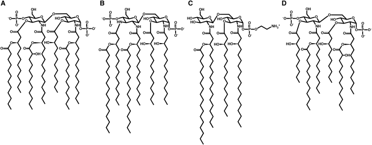
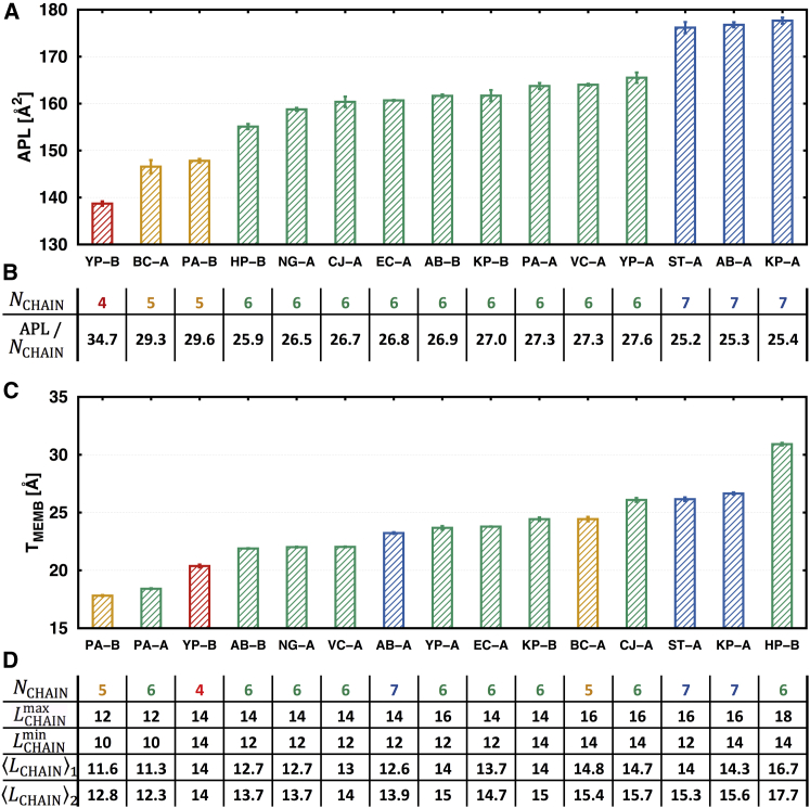
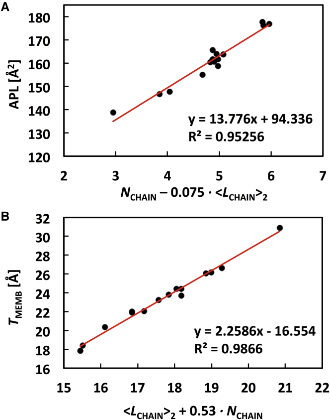
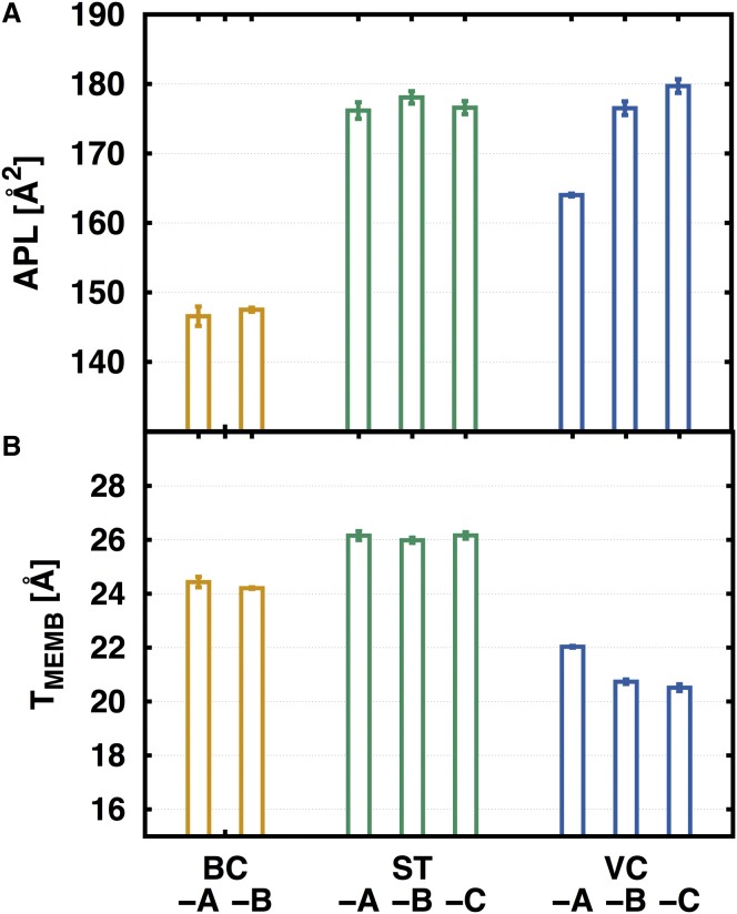
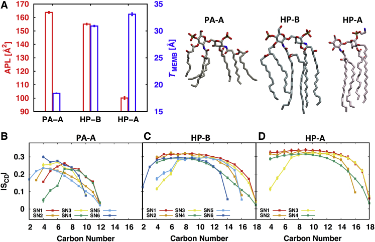
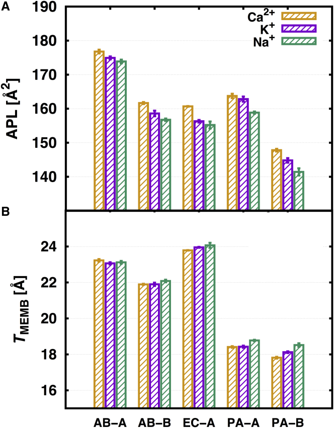
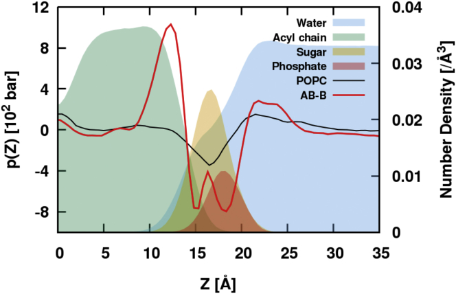
Lipid A is the lipid anchor of a lipopolysaccharide in the outer leaflet of the outer membrane of Gram-negative bacteria. In general, lipid A consists of two phosphorylated N-acetyl glucosamine and several acyl chains that are directly linked to the two sugars. Depending on the bacterial species and environments, the acyl chain number and length vary, and lipid A can be chemically modified with phosphoethanolamine, aminoarabinose, or glycine residues, which are key to bacterial pathogenesis. In this work, homogeneous lipid bilayers of 21 distinct lipid A types from 12 bacterial species are modeled and simulated to investigate the differences and similarities of their membrane properties. In addition, different neutralizing ion types (Ca2+, K+, and Na+) are considered to examine the ion's influence on the membrane properties. The trajectory analysis shows that (1) the area per lipid is mostly correlated to the acyl chain number, and the area per lipid increases as a function of the acyl chain number; (2) the hydrophobic thickness is mainly determined by the average acyl chain length with slight dependence on the acyl chain number, and the hydrophobic thickness generally increases with the average acyl chain length; (3) a good correlation is observed among the area per lipid, hydrophobic thickness, and acyl chain order; and (4) although the influence of neutralizing ion types on the area per lipid and hydrophobic thickness is minimal, Ca2+ stays longer on the membrane surface than K+ or Na+, consequently leading to lower lateral diffusion and a higher compressibility modulus, which agrees well with available experiments.


Figures

Figure 1
Figure 1
Selected lipid A chemical structures: (A) A. baumannii, (B) C. jejuni, (C) H. pylori, and (D) P. aeruginosa; see Fig. S1 for all lipid A structures in Table 1 with the position numbers and acyl chain terminal carbon numbers.
Figure 2
Figure 2
(A) APL and (C) TMEMB of 1-, 4′-phosphorylated lipid A with the standard errors over three replicas. (B) APL/LCHAIN of each lipid A system in the order of (A). (D) LCHAINmin, LCHAINmax, LCHAIN1, and LCHAIN2 of each lipid A system in the order of (C). LCHAIN1 and LCHAIN2 are the average LCHAIN with different weight schemes (see the main text). To see this figure in color, go online.
Figure 3
Figure 3
Correlation (A) between APL and [NCHAIN0.075×LCHAIN2] and (B) between TMEMB and [LCHAIN2+0.53×NCHAIN]. To see this figure in color, go online.
Figure 4
Figure 4
(A) APL and (B) TMEMB of the eight chemically modified lipid A bilayers. To see this figure in color, go online.
Figure 5
Figure 5
(A) APL and TMEMB of PA-A, HP-B, and HP-A with the representative lipid A structure. |SCD| of each acyl chain for (B) PA-A, (C) HP-B, and (D) HP-A. To see this figure in color, go online.
Figure 6
Figure 6
(A) Results of APL and (B) TMEMB on different neutralizing ion types with color as yellow, Ca2+; purple, K+; and green, Na+. To see this figure in color, go online.
Figure 7
Figure 7
Lateral pressure profile of AB-B (red line) and POPC (black line) bilayers with the lateral density profiles (solid layers) of AB-B system with sky-blue for water, green for acyl tail, orange for disaccharide, and red for phosphate. Note that, as the magnitude of the lateral pressure depends on the system size, its variation along the z axis is the important feature. To see this figure in color, go online.

References

    1. Thomson J.M., Bonomo R.A. The threat of antibiotic resistance in Gram-negative pathogenic bacteria: β-lactams in peril! Curr. Opin. Microbiol. 2005;8:518–524. - PubMed
    1. Silhavy T.J., Kahne D., Walker S. The bacterial cell envelope. Cold Spring Harb. Perspect. Biol. 2010;2:a000414. - PMC - PubMed
    1. Gessmann D., Chung Y.H., Fleming K.G. Outer membrane β-barrel protein folding is physically controlled by periplasmic lipid head groups and BamA. Proc. Natl. Acad. Sci. USA. 2014;111:5878–5883. - PMC - PubMed
    1. Fontana C., Conde-Álvarez R., Widmalm G. Structural studies of lipopolysaccharide-defective mutants from Brucella melitensis identify a core oligosaccharide critical in virulence. J. Biol. Chem. 2016;291:7727–7741. - PMC - PubMed
    1. Peleg A.Y., Seifert H., Paterson D.L. Acinetobacter baumannii: emergence of a successful pathogen. Clin. Microbiol. Rev. 2008;21:538–582. - PMC - PubMed

LinkOut - more resources