. 2017 Jan 1;312(1):F109-F120.
doi: 10.1152/ajprenal.00115.2016. Epub 2016 Oct 19.

Deletion of protein kinase C-ε attenuates mitochondrial dysfunction and ameliorates ischemic renal injury

Affiliations

Deletion of protein kinase C-ε attenuates mitochondrial dysfunction and ameliorates ischemic renal injury

Grazyna Nowak et al. Am J Physiol Renal Physiol. .

Abstract

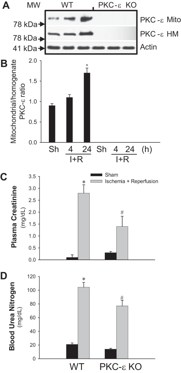
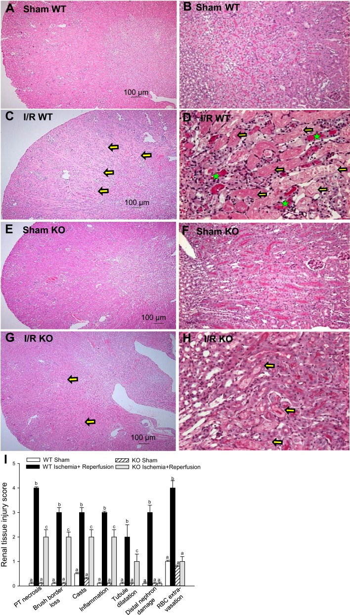
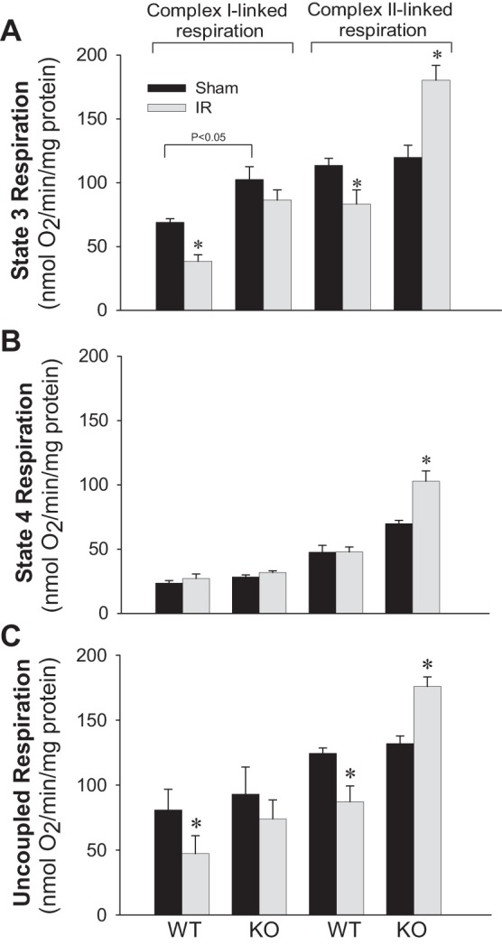
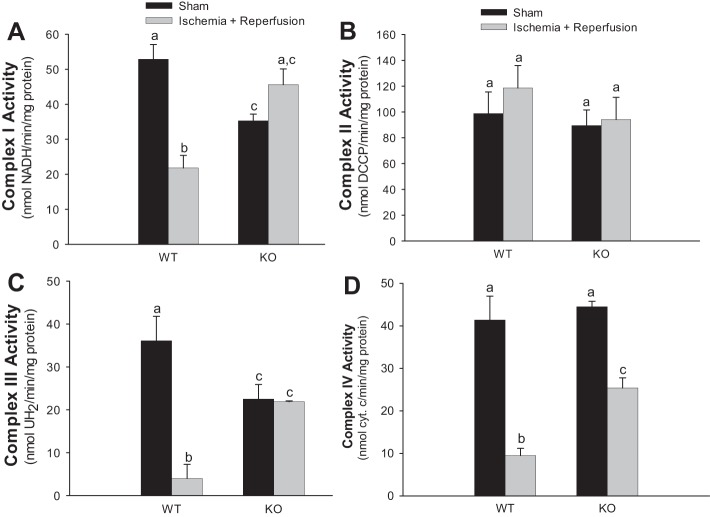
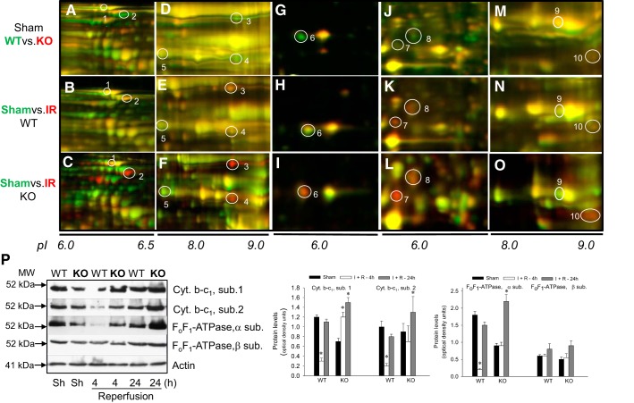
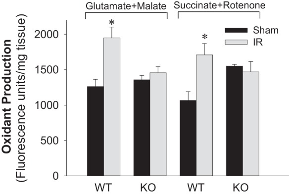
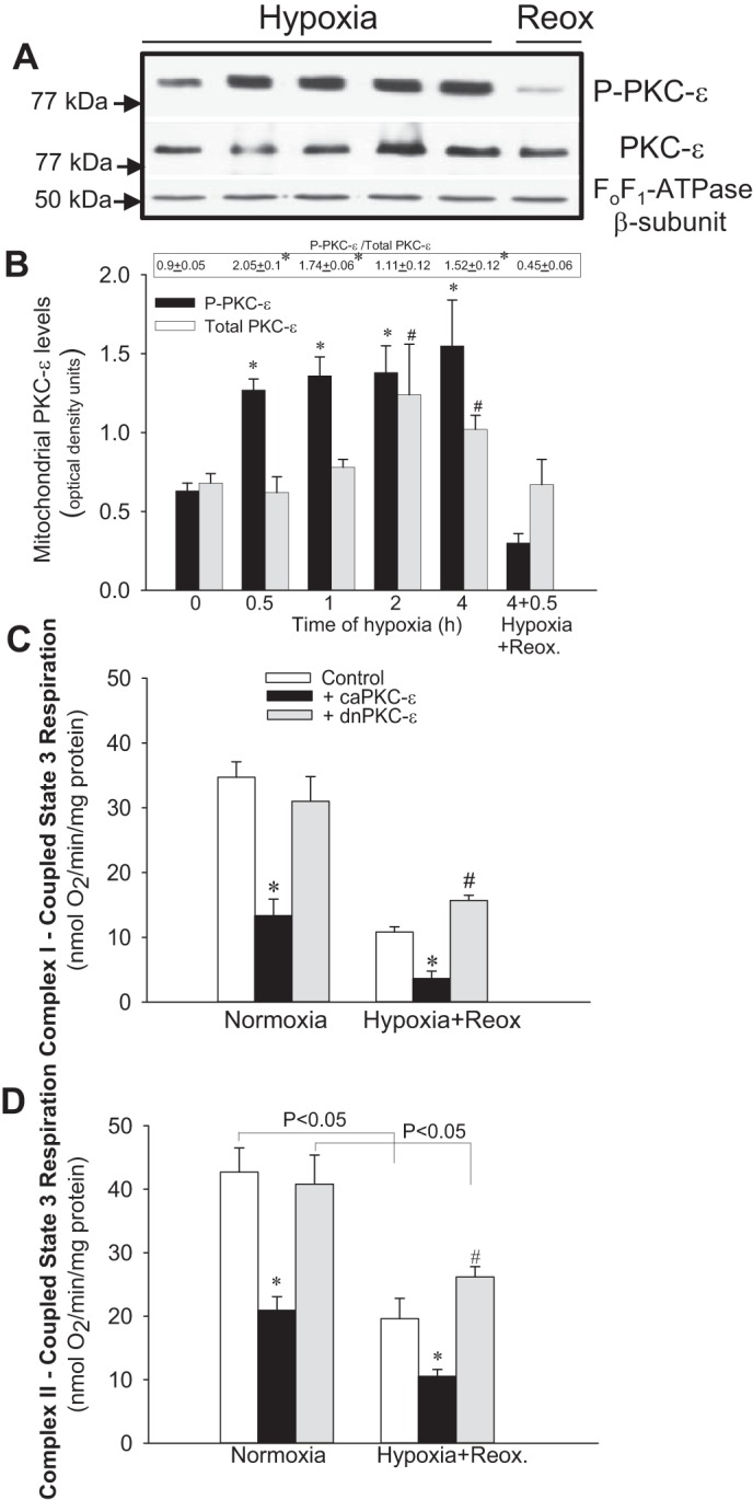
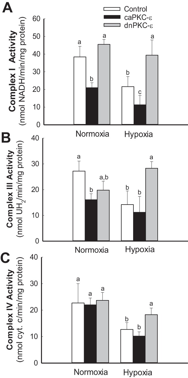
Previously, we documented that activation of protein kinase C-ε (PKC-ε) mediates mitochondrial dysfunction in cultured renal proximal tubule cells (RPTC). This study tested whether deletion of PKC-ε decreases dysfunction of renal cortical mitochondria and improves kidney function after renal ischemia. PKC-ε levels in mitochondria of ischemic kidneys increased 24 h after ischemia. Complex I- and complex II-coupled state 3 respirations were reduced 44 and 27%, respectively, in wild-type (WT) but unchanged and increased in PKC-ε-deficient (KO) mice after ischemia. Respiratory control ratio coupled to glutamate/malate oxidation decreased 50% in WT but not in KO mice. Activities of complexes I, III, and IV were decreased 59, 89, and 61%, respectively, in WT but not in KO ischemic kidneys. Proteomics revealed increases in levels of ATP synthase (α-subunit), complexes I and III, cytochrome oxidase, α-ketoglutarate dehydrogenase, and thioredoxin-dependent peroxide reductase after ischemia in KO but not in WT animals. PKC-ε deletion prevented ischemia-induced increases in oxidant production. Plasma creatinine levels increased 12-fold in WT and 3-fold in KO ischemic mice. PKC-ε deletion reduced tubular necrosis, brush border loss, and distal segment damage in ischemic kidneys. PKC-ε activation in hypoxic RPTC in primary culture exacerbated, whereas PKC-ε inhibition reduced, decreases in: 1) complex I- and complex II-coupled state 3 respirations and 2) activities of complexes I, III, and IV. We conclude that PKC-ε activation mediates 1) dysfunction of complexes I and III of the respiratory chain, 2) oxidant production, 3) morphological damage to the kidney, and 4) decreases in renal functions after ischemia.

Keywords: acute kidney injury; electron transport chain; hypoxia; ischemia and reperfusion; mitochondria; proteomics; renal proximal tubular cells.


Figures

Fig. 1.
Fig. 1.
Deletion of protein kinase C-ε (PKC-ε) reduces decreases in renal functions after ischemia. A: PKC-ε levels in renal cortical mitochondria (Mito) and homogenates (HM) at 4 and 24 h after renal ischemia in wild-type (WT) and PKC-ε-deficient (PKC-ε KO) male mice. B: ratio of mitochondrial/total (homogenate) PKC-ε levels in renal cortexes at 4 and 24 h after renal ischemia in WT and PKC-ε KO male mice. Quantification of the PKC-ε levels was carried out by densitometric analysis of immunoblots. Renal function was evaluated using creatinine (C) and urea nitrogen (D) levels in mouse plasma collected at 24 h of reperfusion after renal ischemia. Results are averages ± SE of data obtained from 7–11 animals. *, #Values significantly different (P < 0.05) from respective controls. The immunoblot results are representative of 3 different experiments.
Fig. 2.
Fig. 2.
Deletion of PKC-ε improves renal morphology during reperfusion after ischemia. Renal histology of the cortical-medullary junction assessed in periodic acid-Schiff (PAS)-stained kidney sections from WT (A–D) and KO (E–H) mice. Sections represent kidneys from sham-operated mice (A, B, E, and F) and mice killed at 24 h of reperfusion after 50 min of renal ischemia (C, D, G, and H). Magnification: ×48.8 (A, C, E, and G) and ×244 (B, D, F, and H). The sections are representative of WT animals (n = 5) and KO animals (n = 4). I: quantitative evaluation of morphological kidney injury at 24 h of reperfusion after 50 min of renal ischemia expressed relatively according to materials and methods. The images were captured using a Nikon Eclipse E800 microscope using objectives Nikon Plan Apo ×4 (A, C, E, and G) and Nikon Plan Apo ×20 (B, D, F, and H). Morphology was scored according to proximal tubule necrosis, brush border loss, cast formation within tubules, inflammation (infiltration of the inflammatory cells), tubule dilatation, distal nephron damage, and red blood cell extravasation. Yellow arrows show severe confluent necrosis in the proximal tubules, and green stars indicate tubular cast formation. Results are averages ± SE of data obtained from kidney sections from WT (n = 5) and KO (n = 4) mice. Values with dissimilar superscripts (a, b, c) within a group are significantly different (P < 0.05) from each other.
Fig. 3.
Fig. 3.
Deletion of PKC-ε restores mitochondrial respiration in renal cortical tissue injured by ischemia. State 3 (A), state 4 (B), and uncoupled (C) respirations coupled to the oxidation of electron donors to complex I (5 mM glutamate + 5 mM malate) and complex II (10 mM succinate + 0.1 μM rotenone) in renal cortical mitochondria isolated at 24 h of reperfusion after 50 min of renal ischemia in WT and KO mice. Results are averages ± SE of data obtained from 4–8 animals. *Values significantly different (P < 0.05) from respective controls.
Fig. 4.
Fig. 4.
Deletion of PKC-ε restores activities of complexes of the electron transport chain in mitochondria of renal cortical tissue injured by ischemia. Activities of NADH-ubiquinone oxidoreductase (complex I) (A), succinate-ubiquinone oxidoreductase (complex II) (B), ubiquinol-cytochrome c oxidoreductase (C), and cytochrome oxidase (complex IV) (D) in mitochondria isolated from renal cortexes of WT and KO mice at 24 h of reperfusion after 50 min renal ischemia. Results are averages ± SE of data obtained from 4–8 animals. Values with dissimilar superscripts (a, b, c) are significantly different (P < 0.05) from each other.
Fig. 5.
Fig. 5.
Deletion of PKC-ε upregulates the levels of crucial proteins associated with oxidative phosphorylation in mitochondria of renal cortical tissue injured by ischemia and reperfusion. Mitochondria were isolated from sham-operated and ischemic WT and KO mice at 24 h of reperfusion after 50 min renal ischemia as described in materials and methods. Proteins present in mitochondria were labeled using Cy3 and Cy5 dyes, resolved using 2-dimensional differential in-gel electrophoresis (2D-DIGE: horizontally, by isoelectric focusing point from pH 3 to 10; vertically, by molecular weight from 150 to 10 kDa), excised and eluted from the gel, and identified by mass spectrometry (MS)/MS analysis. Top (WT vs. KO, sham), proteins from WT mice were labeled green (Cy5), whereas proteins from KO mice were labeled red (Cy3). Middle [WT, sham vs. ischemia-reperfusion (IR)], proteins from sham-operated mice were labeled green (Cy5), whereas proteins from ischemic mice were labeled red (Cy3). Bottom (KO, sham vs. IR), proteins from sham-operated mice were labeled green (Cy5), whereas proteins from ischemic mice were labeled red (Cy3). A–C: mitochondrial pyruvate carboxylase (spot 1) and 2-oxoglutarate dehydrogenase (spot 2). D–F: α-subunit of ATP synthase (spot 3), subunit 2 of cytochrome b-c1 complex (complex III, spot 4), and mitochondrial medium-chain specific acyl-CoA dehydrogenase (spot 5). G–I: subunit 5B of cytochrome oxidase (spot 6). J–L: thioredoxin-dependent peroxide reductase (spot 7) and NADH dehydrogenase (ubiquinone) iron-sulfur protein 3 (complex I, spot 8). M–O: lipoamide acyltransferase component of branched-chain α-keto acid dehydrogenase (spot 9) and succinate dehydrogenase (ubiquinone) iron-sulfur subunit (complex II, spot 10). P: left, protein levels of subunits (core proteins) 1 and 2 of cytochrome b-c1 complex and subunits-α and -β of ATP synthase (FoF1-ATPase) in renal cortical tissues of sham-operated and ischemic WT and PKC-ε KO mice at 4 and 24 h of reperfusion after 50 min renal ischemia. Immunoblot results are representative of 3 different experiments involving 3 WT mice and 3 KO mice each. Right, densitometric analysis of immunoblot bands shown on left. *Values significantly different (P < 0.05) from respective sham controls.
Fig. 6.
Fig. 6.
Deletion of PKC-ε decreases oxidant production in renal tissue after ischemia. Oxidant production was assessed using 5 μM 5-(and-6)-carboxy-2′,7′-dichlorodihydro-fluorescein (carboxy-H2DCFDA) in renal tissue slices energized with electron donors to the respiratory complex I (5 mM glutamate + 5 mM malate) and complex II (10 mM succinate + 0.1 μM rotenone) in WT and KO mice at 24 h of reperfusion after 50 min renal ischemia. Data are expressed as %sham controls. Results are averages ± SE of data obtained from 4–12 animals. *Values significantly different (P < 0.05) from respective sham controls.
Fig. 7.
Fig. 7.
Activation of PKC-ε exacerbates hypoxia-induced decreases in mitochondrial respiration in renal proximal tubule cells (RPTC). A: protein levels of phosphorylated PKC-ε (P-PKC-ε) and total PKC-ε (PKC-ε) in RPTC mitochondria at different time points of hypoxia and reoxygenation. Levels of the β-subunit of FoF1-ATPase were used as loading controls. B: densitometric analysis of the phosphorylated and total PKC-ε levels at different time points of hypoxia (0.5–4 h) and at 0.5 h of reoxygenation after 4 h of hypoxia in mitochondria isolated from renal proximal tubular cells in primary culture. Quantification of the PKC-ε levels was carried out by densitometric analysis of immunoblots. The ratio of phosphorylated and total PKC-ε levels is shown above the bar graph in B. C and D: state 3 respiration coupled to the oxidation of electron donors to complex I (5 mM glutamate + 5 mM malate) and complex II (10 mM succinate + 0.1 μM rotenone) in cultured RPTC overexpressing the constitutively active (caPKC-ε) and inactive [dominant-negative (dn) PKC-ε] mutants of PKC-ε. RPTC were subjected to hypoxia for 4 h, and state 3 respiration was assessed at 0.5 h of reoxygenation after hypoxia. PKC-ε was activated in RPTC by expressing caPKC-ε mutant [multiplicity of infection (MOI) = 50] or inhibited by expressing dnPKC-ε (MOI = 100). Results are averages ± SE of data obtained from 5–7 independent experiments (RPTC isolations). *, #Values significantly different (P < 0.05) from respective controls.
Fig. 8.
Fig. 8.
Activation of PKC-ε exacerbates hypoxia-induced decreases in activities of complexes of the electron transport chain in RPTC mitochondria. Activities of NADH-ubiquinone oxidoreductase (complex I) (A), ubiquinol-cytochrome c oxidoreductase (B), and cytochrome oxidase (complex IV) (C) in mitochondria isolated from cultured RPTC overexpressing caPKC-ε and dnPKC-ε mutants of PKC-ε. PKC-ε was activated in RPTC by expressing caPKC-ε mutant (MOI = 50) or inhibited by expressing dnPKC-ε mutant (MOI = 100) at 48 h before the exposure to hypoxia. RPTC were subjected to hypoxia for 4 h, and mitochondria were isolated at 0.5 h of reoxygenation. Results are averages ± SE of data obtained from 4–8 independent experiments (RPTC isolations). Values with dissimilar superscripts (a, b, c) are significantly different (P < 0.05) from each other.

Comment in

References

    1. Baines CP, Song CX, Zheng YT, Wang GW, Zhang J, Wang OL, Guo Y, Bolli R, Cardwell EM, Ping P. Protein kinase Cε interacts with and inhibits the permeability transition pore in cardiac mitochondria. Circ Res 92: 873–880, 2003. - PMC - PubMed
    1. Baines CP, Zhang J, Wang GW, Zheng YT, Xiu JX, Cardwell EM, Bolli R, Ping P. Mitochondrial PKCε and MAPK form signaling modules in the murine heart: enhanced mitochondrial PKCε-MAPK interactions and differential MAPK activation in PKCε-induced cardioprotection. Circ Res 90: 390–397, 2002. - PubMed
    1. Bak S, León I, Jensen O, Højlund K. Tissue specific phosphorylation of mitochondrial proteins isolated from rat liver, heart muscle, and skeletal muscle. J Proteome Res 12: 4327–4339, 2013. - PubMed
    1. Bender E, Kadenbach B. The allosteric ATP-inhibition of cytochrome c oxidase activity is reversibly switched on by cAMP-dependent phosphorylation. FEBS Lett 466: 130–134, 2000. - PubMed
    1. Bonventre JV, Weinberg JM. Recent advances in the pathophysiology of ischemic acute renal failure. J Am Soc Nephrol 14: 2199–2210, 2003. - PubMed

Publication types

MeSH terms

Substances

LinkOut - more resources