. 2017 Jan 1;312(1):F157-F171.
doi: 10.1152/ajprenal.00386.2016. Epub 2016 Oct 19.

A small molecule screening to detect potential therapeutic targets in human podocytes

Affiliations

A small molecule screening to detect potential therapeutic targets in human podocytes

Eugen Widmeier et al. Am J Physiol Renal Physiol. .

Abstract

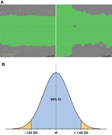
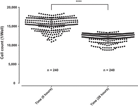
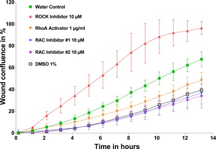
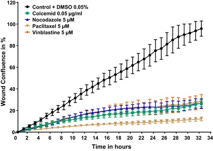
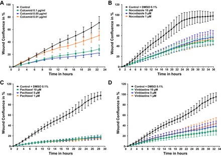
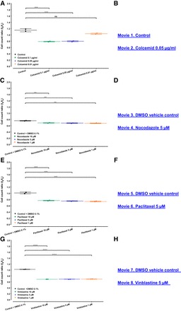
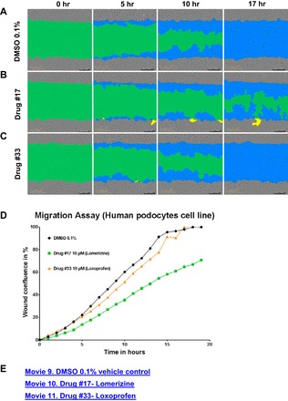
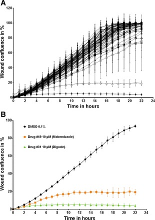
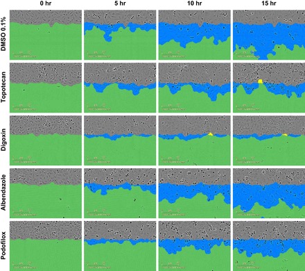
A small molecule screening to detect potential therapeutic targets in human podocytes. Am J Physiol Renal Physiol 312: F157-F171, 2017. First published October 19, 2016; doi:10.1152/ajprenal.00386.2016. Steroid-resistant nephrotic syndrome (SRNS) inevitably progresses to end-stage kidney disease, requiring dialysis or transplantation for survival. However, treatment modalities and drug discovery remain limited. Mutations in over 30 genes have been discovered as monogenic causes of SRNS. Most of these genes are predominantly expressed in the glomerular epithelial cell, the podocyte, placing it at the center of the pathogenesis of SRNS. Podocyte migration rate (PMR) represents a relevant intermediate phenotype of disease in monogenic causes of SRNS. We therefore adapted PMR in a high-throughput manner to screen small molecules as potential therapeutic targets for SRNS. We performed a high-throughput drug screening of a National Institutes of Health Clinical Collection (NCC) library (n = 725 compounds) measuring PMR by videomicroscopy. We used the Woundmaker to perform individual 96-well scratch wounds and screened compounds using a quantitative kinetic live cell imaging migration assay using IncuCyte ZOOM technology. Using a normal distribution for the average PMR in wild-type podocytes with a vehicle control (DMSO), we applied a 90% confidence interval to define "distinct" compounds (5% faster/slower PMR) and found that 12 of 725 compounds (at 10 μM) reduced PMR. Clusters of drugs that alter PMR included actin/tubulin modulators such as the azole class of antifungals and antineoplastic vinca-alkaloids. We hereby identify compounds that alter PMR. The PMR assay provides a new avenue to test therapeutics for nephrotic syndrome. Positive results may reveal novel pathways in the study of glomerular diseases such as SRNS.

Keywords: podocyte; small molecule screen; steroid-resistant nephrotic syndrome.


Figures

Fig. 1.
Fig. 1.
Methods to determine positive hits with videomicroscopy based scratch wound assay. Wells for the podocyte migration rate (PMR) assay were chosen for quality depending on wound width, confluence (A), and characteristics of a normal distribution curve (B). A: 2 representative images are shown of human podocytes at time point t0, after a scratch-wound. Left: adequate scratch with a confluent monolayer above and below the wound with no debris/cellular remains within the wound to confound analysis. Right: inadequate wound width and cell confluency due to poor podocyte coverage above and below the scratch wound. B: normal distribution curve is displayed with demarcations at 1.65 SD from the mean [90% confidence interval (CI)]. In the first part of our evaluation, outliers (outside the 90% CI) in wound width were deemed inadequate and discarded from analysis. In the second part of our evaluation, wound confluence was used to identify wells with “distinct” PMR outside the 90% CI and kept as positive results.
Fig. 2.
Fig. 2.
Proliferation assay in immortalized undifferentiated human podocytes. Stably nuclear mKate2 expressing wild-type podocytes from an immortalized human podocytes cell line were seeded 12 h before the beginning of the experiment on a 96-well image-lock plate. Images were captured at the beginning and at the end of the experiment. A significant reduction of cell count in average of 23.2% on 240 replicates is shown over a period of 26 h in cultured media containing DMSO vehicle control (0.1%). Data are expressed as means ± SD for 3 independent experiments, 80 replicates each.
Fig. 3.
Fig. 3.
Proof of principle experiment with RhoA and Rac1 effectors. Wild-type podocytes were seeded 12 h before scratch wound analysis on a 96-well image-lock plate. After scratch wound was made in the confluent podocyte monolayer, podocytes were exposed to different compounds including ROCK inhibitor (CN06; Cytoskeleton), RhoA activator (CN03; Cytoskeleton), and Rac1 inhibitor #1 (553502; Millipore), and Rac1 inhibitor #2 (553511; Millipore). Concentrations are as follows: RhoA inhibitor: 10 μM; RhoA activator: 1 μg/ml; Rac1 inhibitor #1: 10 μM; and Rac1 inhibitor #2: 10 μM, as per dosing instructions. RhoA effectors were diluted in water. Rac1 effectors were diluted in DMSO. The volume of total media including drug in each well is 100 μl. Note that, whereas ROCK inhibitors increased PMR, RhoA activators as well as Rac1 inhibitors #1 and #2 decreased PMR. Data are expressed as means ± SD for 2 independent experiments.
Fig. 4.
Fig. 4.
Proof-of-principle experiment with established modulators of microtubule polymerization/depolymerization such as colcemid, nocodazole, paclitaxel, and vinblastine. Stably nuclear mKate2 expressing wild-type podocytes were seeded 12 h before scratch wound analysis on a 96 well image-lock plate. After scratch wound was made in the confluent podocyte monolayer, podocytes were exposed to compounds including colcemid (no. 10295892001; Roche), nocodazole (M1404; Sigma), paclitaxel (T7402; Sigma), vinblastine (V1377; Sigma), and DMSO vehicle control (0.05%). Concentrations are as follows: 0.05 μg/ml colcemid, 5 μM nocodazole, 5 μM paclitaxel, and 5 μM vinblastine. Colcemid was delivered as ready to use solution, DMSO was added accordingly. Nocodazole, paclitaxel, and vinblastine were diluted in DMSO. All drugs significantly decreased PMR at the established concentrations.
Fig. 5.
Fig. 5.
Dose response of established modulators of microtubule demonstrates decreased PMR. Stably nuclear mKate2 expressing wild-type podocytes were seeded 12 h before scratch wound analysis on a 96 well image-lock plate. After scratch wound was made in the confluent podocyte monolayer, podocytes were exposed to compounds in different concentration as indicated. A: colcemid (control, n = 47; 0.1 µg/ml colcemid, n = 47; 0.05 µg/ml colcemid, n = 32; and 0.01 µg/ml colcemid, n = 31) alters the PMR partially in a concentration-response manner. B and C: nocodazole (0.1% DMSO vehicle control, n = 45; 10 µM nocodazole, n = 48; 5 µM nocodazole, n = 3; and 1 µM nocodazole, n = 32) and paclitaxel (0.1% DMSO vehicle control, n = 46; 10 µM paclitaxel, n = 48; 5 µM paclitaxel, n = 31, 1 µM paclitaxel, n = 32) reduce PMR in a concentration-independent manner. D: whereas vinblastine (0.1% DMSO vehicle control, n = 47; 10 µM vinblastine, n = 48; 5 µM vinblastine, n = 32; and 1 µM vinblastine, n = 32) reduces PMR in a concentration-dependent manner.
Fig. 6.
Fig. 6.
Toxicity of established microtubule modulators reduce podocytes cell count without compromizing the PMR. Wild-type podocytes from an immortalized human podocytes cell line stably expressing nuclear mKate2 were seeded 12 h before the beginning of the experiment on a 96-well image-lock plate. Whole well images were captured after drug exposure at the beginning (t0) and at the end (tx) of the experiment to perform the quantitative analysis of total cell count. Live cell imaging captured immortalized human podocytes on an hourly basis to characterize the cellular morphology and viability of podocytes, while undergoing migration in a scratch wound assay. A, C, E, and G: all compounds at indicated concentration reduce the cell count ratio and show substantial cell morphology changes vs. control condition. B, D, F, and H: all compounds significantly affected PMR as well, however, without compromising the migratory behavior (see Fig. 5; also see Supplemental Movies 1-8; Supplemental material for this article is available at the Journal website). Data are expressed as a ratio of means ± SD for 2 independent experiments. NS = not significant; **P < 0.01, ***P < 0.001, and ****P < 0.0001 by one-way ANOVA Bonferroni’s multiple comparison test.
Fig. 7.
Fig. 7.
Positive drug screening hits determined by PMR as compared with vehicle control. Examples of positive and negative results in National Institutes of Health Clinical Collection (NIHCC) Plate #4. A: DMSO 0.1% vehicle control at time 0, 5, 10, and 15 h, respectively. B: Drug #17 (10 μM), lomerizine, at time 0, 5, 10, and 15 h, respectively. C: Drug #33 (10 μM), loxoprofen, at time 0 and 5, 10, and 15 h, respectively. D: graphical representation of wound confluence (y-axis) over time (x-axis) for DMSO, Drug #17 (lomerizine), and Drug #33 (loxoprofen) over an average of 3 replicates. E: see Supplemental Movies 9-11 for movies of wound confluence over time of vehicle control DMSO 0.1%, Drug #17 (lomerizine), and Drug #33 (loxoprofen) for 1 replicate.
Fig. 8.
Fig. 8.
Representative example of PMR assay (NIHCC Plate #8) demonstrating wound confluence over time and positive hits. Podocytes from an immortalized human podocyte cell line was seeded 12 h before scratch wound analysis on a 96-well image-lock plate. After a scratch wound was made in the confluent podocyte monolayer, podocytes were exposed to 80 different compounds from NIHCC Drug Plate #8. Each individual drug (80 compounds) was placed in 1 individual well. DMSO vehicle control (0.1%) was seeded in the 16 remaining wells of the 96-well plate. A: wound confluence over time demonstrates podocyte migration over a period of 24 h after creation of scratch wound for all 80 individual compounds. B: 2 specific compounds with PMR outside the <90% CI are shown as positive hits. Drug #49 (mobendazole) and Drug #31 (digoxin) are highlighted in comparison to DMSO vehicle control as they are significantly outside the 90% confidence interval for Δwound confluence/Δtime, a measurement of PMR. Data are expressed as means ± SD for 3 independent experiments.
Fig. 9.
Fig. 9.
Podocyte cellular morphology at sequential time points after drug exposure. Human podocytes were qualitatively examined for podocyte morphology changes during the podocyte migration assay. Live cell imaging captured immortalized human podocytes at different time points (0, 5, 10, and 15 h) after drug exposure, while undergoing migration in a scratch wound assay. Compounds shown include DMSO vehicle control (0.1%), topotecan (10 μM), digoxin (10 μM), albendazole (10 μM), and podofilox (10 μM). All compounds significantly affected PMR as well (see Table 2).

Comment in

  • Stop that podocyte!
    Gupta V, Reiser J. Gupta V, et al. Am J Physiol Renal Physiol. 2017 Feb 1;312(2):F373-F374. doi: 10.1152/ajprenal.00499.2016. Epub 2016 Oct 19. Am J Physiol Renal Physiol. 2017. PMID: 27760773 Free PMC article. No abstract available.

References

    1. Aizman O, Uhlén P, Lal M, Brismar H, Aperia A. Ouabain, a steroid hormone that signals with slow calcium oscillations. Proc Natl Acad Sci USA 98: 13420–13424, 2001. doi:10.1073/pnas.221315298. - DOI - PMC - PubMed
    1. Altmann SW, Davis HR Jr, Zhu LJ, Yao X, Hoos LM, Tetzloff G, Iyer SP, Maguire M, Golovko A, Zeng M, Wang L, Murgolo N, Graziano MP. Niemann-Pick C1 Like 1 protein is critical for intestinal cholesterol absorption. Science 303: 1201–1204, 2004. doi:10.1126/science.1093131. - DOI - PubMed
    1. Alvarez J, Montero M, Garcia-Sancho J. High affinity inhibition of Ca(2+)-dependent K+ channels by cytochrome P-450 inhibitors. J Biol Chem 267: 11789–11793, 1992. - PubMed
    1. Anderson-Beck R, Wilson L, Brazier S, Hughes IE, Peers C. Doxapram stimulates dopamine release from the intact rat carotid body in vitro. Neurosci Lett 187: 25–28, 1995. doi:10.1016/0304-3940(95)11328-T. - DOI - PubMed
    1. Andrews PM. The effect of vinblastine-induced microtubule loss on kidney podocyte morphology. Am J Anat 150: 53–61, 1977. doi:10.1002/aja.1001500104. - DOI - PubMed

Publication types

MeSH terms

Substances

LinkOut - more resources